|
|
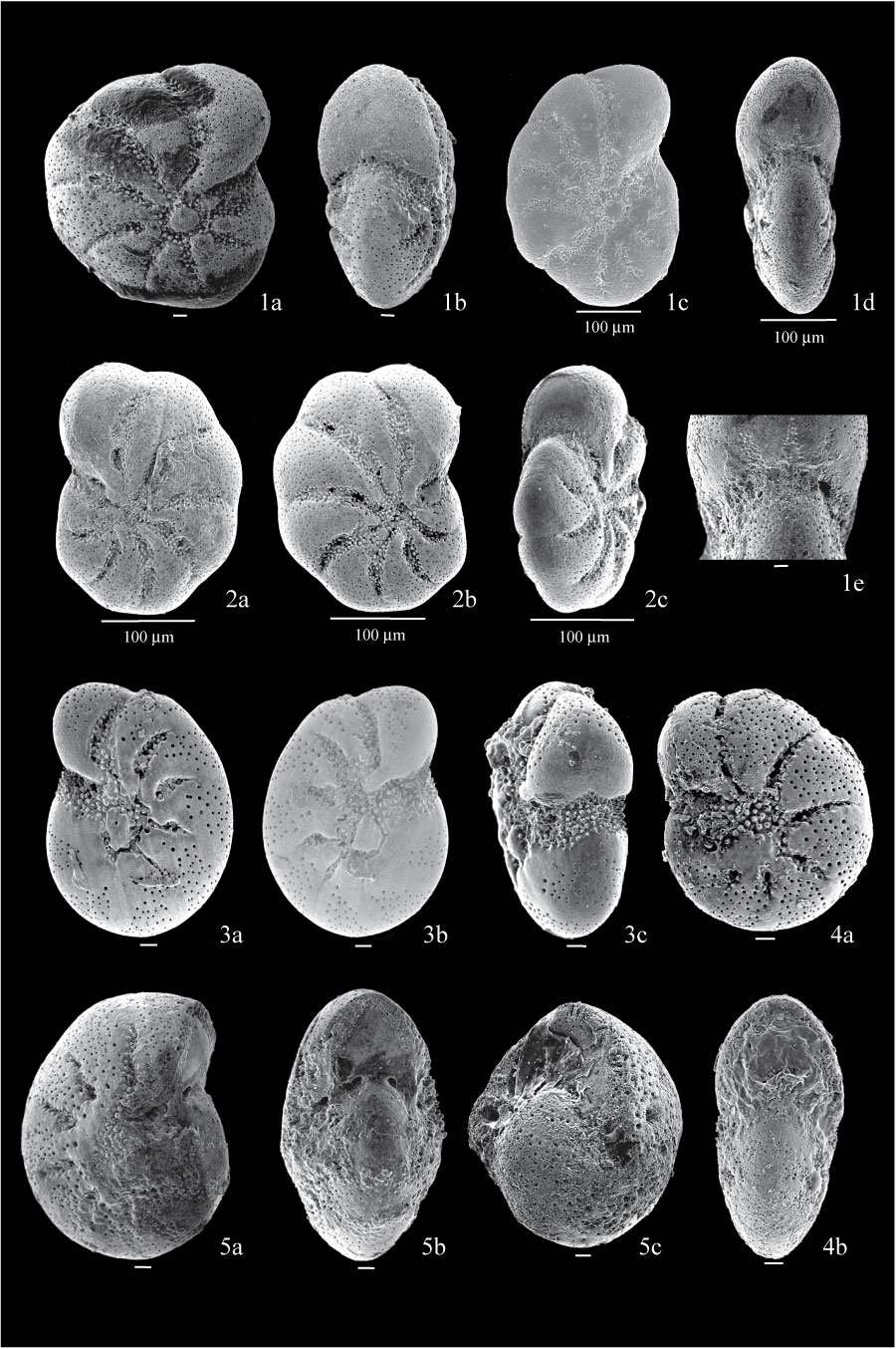
Figure 14. 1. Cribroelphidium excavatum forma excavata ; 1a, side view of specimen from sample BE5, showing straight sutures extending unconstricted to the umbilicus; 1b, apertural view of specimen 1a; 1c, side view of specimen from sample BE1; 1d, apertural view of specimen 1c; 1d, detail of aperture of specimen 1c, showing pustulose material arranged on triangular patterns on the apertural face. 2. Cribroelphidium excavatum forma lidoensis from sample BE1; 2a, side view showing sutures curved backwards, broadening towards the umbilicus, filled with papillae; 2b, other side; 2c, apertural view. 3. Cribroelphidium excavatum forma clavata from sample BE1; 3a, side view showing umbilical boss and backwards-curved sutures; 3b, view of the other side; 3c, apertural view. 4. Cribroelphidium excavatum forma selseyensis from sample BE1; 4a, side view showing granular material filling the umbilicus; 4b, apertural view. 5. Cribroelphidium excavatum forma magna from sample FC04-62; 5a, side view showing large umbilicus filled with one knobby boss, and sutures constricted before reaching the umbilicus; 5b, apertural view showing strongly convex walls and raised umbilicus; 5c, view of another specimen from sample FC04-62, with very strongly convex walls that give it an almost circular cross-section. All scales are 10 µm unless otherwise indicated.
|