|
|
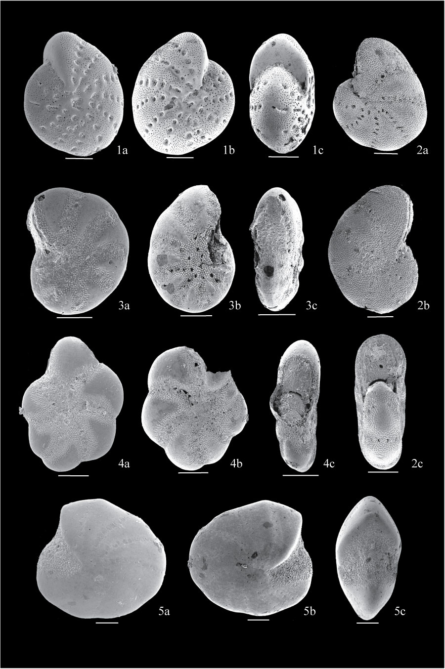
Figure 15. 1. Cribroelphidium foraminosum from sample BE1; 1a, side view showing coarse perforation, sutural pores and sutural bridges; 1b, view of the other side; 1c, apertural view showing imperforate apertural face and multiple aperture. 2. Cribroelphidium microgranulosum from sample BE1; 2a, side view showing round pores along sutures; 2b, view of other side showing wall completely covered with pustulose material; 2c, apertural view showing narrow curved aperture. 3. Cribroelphidium hallandense from sample BE1; 3a, side view showing bands of pustulose material covering umbilicus and along sutures; 3b, view of the other side; 3c, apertural view. 4. Cribroelphidium magellanicum from sample BE1; 4a, side view showing very lobulate periphery; 4b, view of the other side with last chamber broken; 4c, apertural view. 5. Elphidiella hannai from sample BE1; 5a, side view showing rounded periphery; 5b, view of the other side showing thickened flushed sutures with double row of fine pores; 5c, apertural view showing concentration of granular material. All scales are 100 µm.
|