|
|
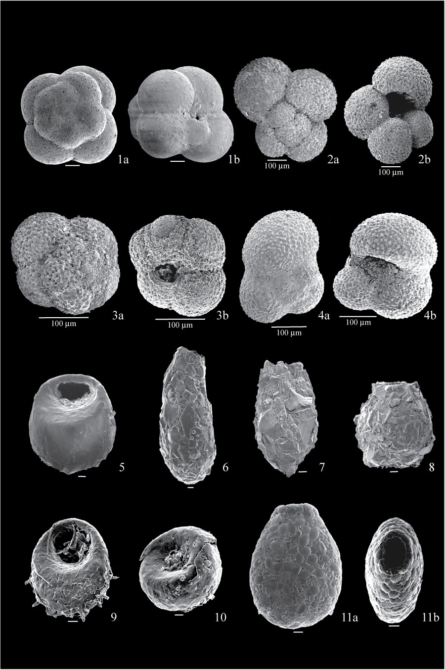
Figure 16. 1. Globigerinita uvula from sample FC04-64; 1a, dorsal view showing very smooth test; 1b, ventral view showing low-arched aperture. 2. Globigerina bulloides from sample FC04-77; 2a, dorsal view showing globular chambers; 2b, ventral view showing high-arched aperture. 3. Globigerina quinqueloba from sample FC04-77; 3a, dorsal view showing low trochospiral arrangement; 3b, ventral view showing lipped aperture. 4. Globigerinoides cyclostomus from sample BE1; 4a, dorsal view showing ovoid chambers; 4b, ventral view showing rectangular test outline. 5. Centropyxis constricta aerophila from sample BE3, side view of specimen covered with organic material. 6. Difflugia oblonga Ehrenberg, from sample FC04-85, side view showing pyriform test. 7. Difflugia protaeiformis Lamarck, from sample FC04-85, side view showing spine in fundus. 8. Difflugia urceolata from sample FC04-85, side view showing cauldron-like test. 9. Centropyxis constricta constricta from sample FC04-110, showing spines in fundus. 10. Cyclopyxis kahli from sample FC04-80, side view showing hemispherical test. 11. Heleopera sphagni from sample FC04-92; 11a, side view showing test entirely composed of idiosomes; 11b, apertural view showing compressed test and elliptical aperture. All scales are 10 µm unless otherwise indicated.
|