|
|
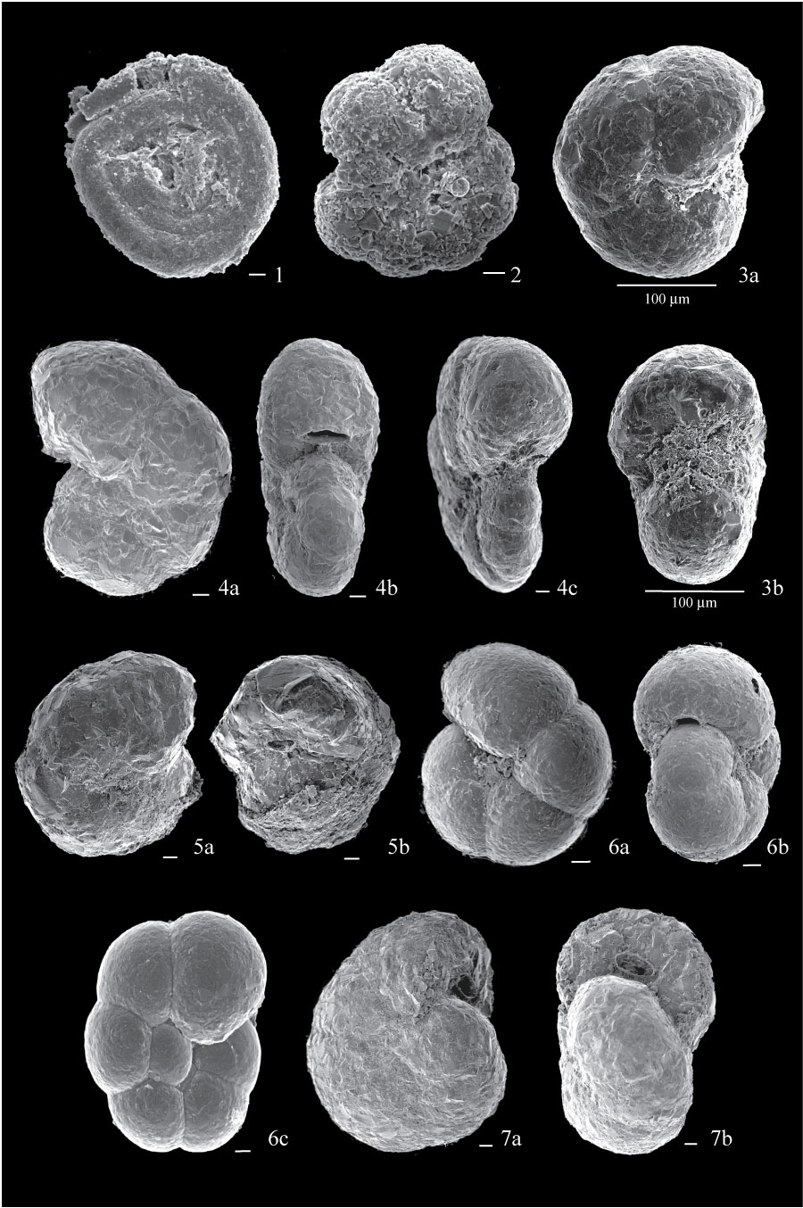
Figure 2. 1. Ammodiscus gullmarensis from sample FC04-86, side view. 2. Cribrostomoides crassimargo, from sample FC04-90, side view showing lobulate periphery and coarse agglutination. 3. Cribrostomoides sp. A, from sample FC04-77; 3a, side view, showing rounded periphery and inflated last chamber; 3b, apertural view, showing inflated last chamber. 4. Cribrostomoides jeffreysii, from sample BE5; 4a, side view, showing ovate periphery; 4b, apertural view, showing depressed test and typical aperture; 4c, apertural view from another specimen from sample BE7, showing leaf-like aperture. 5. Cribrostomoides cf. subglobosum, from sample ALS2; 5a, side view showing rounded periphery; 5b, apertural view showing globular test and characteristic aperture. 6. Haplophragmoides bradyi, from sample BE3; 6a, side view showing very smooth test; 6b, apertural view showing characteristic interiomarginal aperture; 6c, side view of a more evolute specimen from sample BE5. 7. Recurvoides turbinatus, from sample ALS1; 7a, side view; 7b, apertural view showing asymmetry of test and angled aperture. All scales are 10 µm unless otherwise indicated.
|