|
|
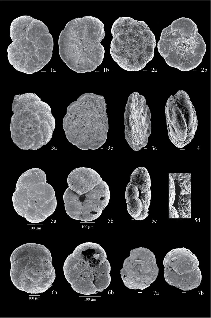
Figure 4. 1. Polystomammina nitida, from sample BE7; 1a, dorsal view, showing very straight sutures; 1b, umbilical view. 2. Trochammina squamata from sample FC04-96; 2a, dorsal view; 2b, umbilical view. 3. Lepidodeuterammina ochracea, from sample ALS1; 3a, dorsal view; 3b, umbilical view, with umbilical region filled with organic matter; 3c, side view, showing depressed concave test. 4. Two forms of D. discorbis attached to each other, from sample FC04-79. 5. Trochammina pacifica from sample BE5; 5a, dorsal view; 5b, umbilical view, showing inflated chambers; 5c, side view, showing aperture reaching the umbilicus; 5d, detail of aperture showing lip. 6. Deuterammina rotaliformis, both specimens from sample BE1; 6a, dorsal view; 6b, umbilical view, with debris partially obscuring star-shaped umbilicus. 7. Ammoglobigerina globigeriniformis from sample BE5; 7a, dorsal view of specimen with last chamber broken; 7b, umbilical view of another specimen. All scales are 10 µm unless otherwise indicated.
|