|
|
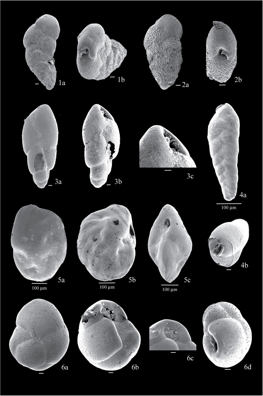
Figure 9. 1. Bolivina decussata from sample BE1; 1a, side view showing roughened surface; 1b, apertural view. 2. Bolivina minuta from sample BE1; 2a, side view showing nearly parallel sides; 2b, apertural view. 3. Stainforthia feylingi from sample BE1; 3a, side view showing smooth, finely perforated test; 3b, side view showing fusiform test; 3c, detail of aperture showing toothplate. 4. Bolivinellina pacifica from sample BE1; 4a, side view showing elongated test and biserial arrangement; 4b, apertural view, showing lip and internal toothplate. 5. Islandiella helenae from sample BE1; 5a, side view showing smooth test; 5b, view of the other side, showing position of aperture along periphery; 5c, apertural view showing biconvex test. 6. Cassidulina crassa from sample FC04-14; 6a, side view showing rectangular last chamber; 6b, view of the other side, showing slightly inflated chambers; 6c, detail of aperture; 6d, view of another specimen from sample BE3, showing rounded outline and depressed sutures, different from the more compressed test and flushed sutures of C. reniforme. All scales are 10 µm unless otherwise indicated.
|