|
|
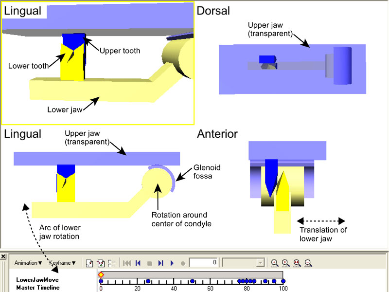
Figure 1. Setup and procedure for tooth movement reconstructions demonstrated using model upper and lower jaws, and model carnassial teeth from Evans and Sanson (2006). Four views of the reconstruction are given: a lingual view of the teeth; a dorsal view of the teeth; a lingual view showing both the teeth and the jaw joint surfaces; and an anterior view of the teeth and right jaw surfaces. Top two views are perspective views (which give a sense of depth and distance between objects), and bottom two views are orthogonal views (which aid in viewing the tooth and joint surfaces at the same time). Lower left shows arc of lower jaw rotation as the lower jaw rotates around the centre of the condyle. The condyle fits within the glenoid fossa of the upper jaw (size of condyle is enlarged for greater clarity). Lower right shows the path of translation of the lower jaw as it sits in the glenoid fossa. Upper teeth in dark blue, upper joint surfaces and jaw in light blue, lower teeth in yellow, lower joint surfaces and jaw in light yellow. Upper jaw is transparent to allow viewing teeth from dorsal.
|