|
 |
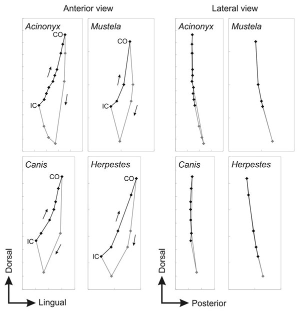
Figure 7. Trace plots of protoconid position for anterior (left) and lateral (right) views of four species for a single cycle of jaw movement. Occlusal path reconstructed when teeth are in occlusion is shown as a black line; jaw positions inserted merely to complete the chewing cycle are shown in grey. Small arrows indicate direction of movement for anterior views. Tick marks are 2 mm apart for all plots. Dorsal, lingual and posterior directions are indicated.
|