|
|
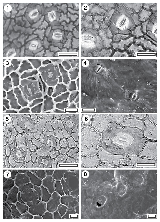
Figure 29. 1-8. Fossil Sapindaceae: CUT-Z-EDD and CUT-Z- FJI. 1. CUT-Z-EDD, TLM view showing stomatal complexes (SL1818, scale-bar = 50 Ám); 2. CUT-Z-EDD, TLM view showing three stomatal complexes. Note that the walls of the subsidiary cells abut or slightly bisect the outline of the guard cells (SL1818, scale-bar = 20 Ám); 3. CUT-Z-EDD, SEM view of inner cuticular surface showing two stomatal complexes. Note buttressing of epidermal cell walls (S-1036, scale-bar = 20 Ám); 4. CUT-Z-EDD, SEM view of outer cuticular surface showing two stomatal complexes. Note striae radiating from each complex (S-1036, scale-bar = 20 Ám); 5. CUT-Z- FJI, TLM view showing stomatal complexes (SL2539, scale-bar = 50 Ám); 6. CUT-Z- FJI, TLM detail of single stomatal complex (SL2539, scale-bar = 20 Ám); 7. CUT-Z- FJI, SEM view of inner cuticular surface showing a single stomatal complex (S-1393, scale-bar = 10 Ám); 8. CUT-Z- FJI, SEM view of outer cuticular surface showing stomatal complexes. Note guard cell pair are slightly sunken and the very small outer stomatal ledges do not protrude above the surrounding subsidiary cells (S-1393, scale-bar = 10 Ám).
|
