|
|
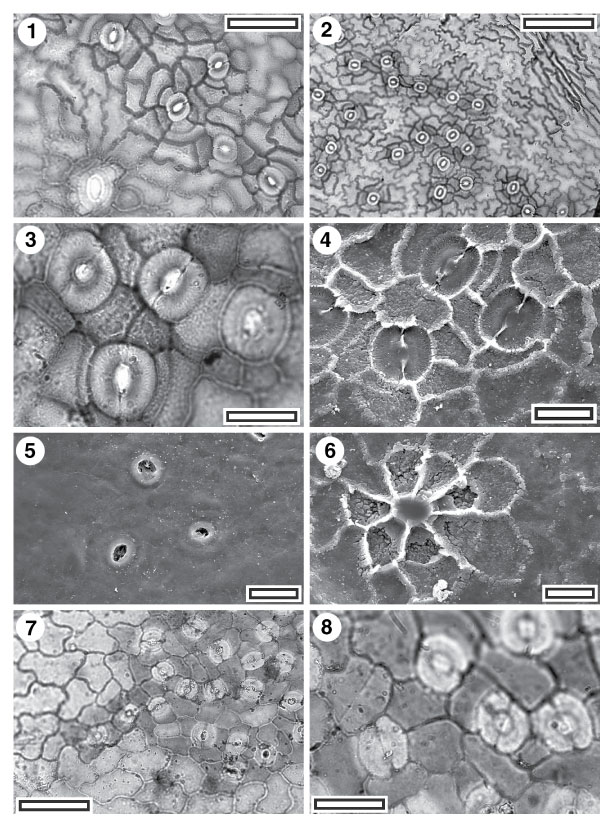
Figure 54. 1-8. Cuticle with stomatal complexes in islands: CUT-Z-EDC and CUT-Z-EIC. 1. CUT-Z-EDC, TLM view showing stomatal complexes. Note the giant stomatal complex at lower left (SL1908, scale-bar = 50 Ám); 2. CUT-Z-EDC, TLM view showing stomatal complexes (SL1908, scale-bar = 100 Ám); 3. CUT-Z-EDC, TLM view showing stomatal complexes. The lower complex has a brachyparacytic subsidiary cell arrangement while the others are less ordered and can be termed cyclocytic (SL1908, scale-bar = 20 Ám); 4. CUT-Z-EDC, SEM view of inner cuticular surface showing stomatal complexes. Note networking and granular texture (S-1048, scale-bar = 20 Ám); 5. CUT-Z-EDC, SEM view of outer cuticular surface showing three stomatal complexes with slight raised outer stomatal ledges and an otherwise smooth surface (S-1048, scale-bar = 20 Ám); 6. CUT-Z-EDC, SEM view of inner cuticular surface showing a trichome attachment scar (S-1048, scale-bar = 20 Ám); 7. CUT-Z-EIC, TLM view showing stomatal complexes. Note common networking (SL2438, scale-bar = 50 Ám); 8. CUT-Z-EIC, TLM view showing stomatal complexes (SL2438, scale-bar = 20 Ám).
|
