|
|
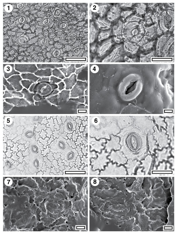
Figure 56. 1-8. Cuticle with unusual shaped outer stomatal ledges: CUT-Z-AJD and CUT-Z-FFG. 1. CUT-Z-AJD, TLM view showing stomatal complexes (SB1379, scale-bar = 50 Ám); 2. CUT-Z-AJD, TLM detail of single stomatal complex (SB1379, scale-bar = 20 Ám). Note key-hole shape to the stomatal pore; 3. CUT-Z-AJD, SEM view of inner cuticular surface showing a single stomatal complex (S-1200, scale-bar = 10 Ám); 4. CUT-Z-AJD, SEM view of outer cuticular surface showing a single stomatal complex with prominent outer stomatal ledges (S-1200, scale-bar = 10 Ám); 5. CUT-Z-FFG, TLM view showing stomatal complexes (SL2229, scale-bar = 50 Ám); 6. CUT-Z-FFG, TLM detail of single stomatal complex (SL2229, scale-bar = 20 Ám); 7. CUT-Z-FFG, SEM view of inner cuticular surface showing (near centre) a reasonably well preserved stomatal complex and (left) a poorly preserved one (S-1396, scale-bar = 10 Ám); 8. CUT-Z-FFG, SEM view of inner cuticular surface showing a single stomatal complex (S-1396, scale-bar = 10 Ám).
|
