|
|
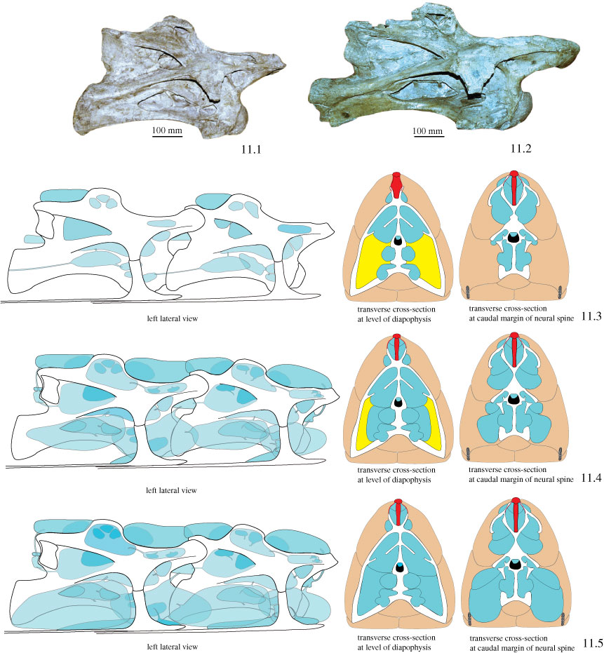
Figure 11. Reconstruction hypotheses for pneumatic diverticula in sauropod vertebrae, demonstrated at 4th and 5th cervical vertebra of Brachiosaurus brancai, all in right lateral view. Photographs of right lateral view of 11.1. 4th cervical vertebra (MB.R. 2180.25), and 11.2. 5th cervical vertebra (MB.R.2180.26). Schematic drawing of 4th and 5th cervical vertebra in lateral view and in transverse cross-sections, 11.3. Minimum expansion of pneumatic diverticula based on osteological evidence, reconstructed pneumatic ducts are hypothetical, based on the assumption that the diverticula are linked to each other and grow in the neck from the neck base in headward direction, 11.4. Intermediate expansion of pneumatic diverticula, based on ostelogical evidence and in analogy with extant birds, pneumatic ducts are again hypothetical, 11.5. Maximum expansion of pneumatic diverticula, which cannot be reconstructed by osteological evidence. Blue = pneumatic diverticula; orange = axial muscles; red = dorsal neck ligaments; yellow = connective tissue. With exception of 11.1 and 11.2 not to scale.
|