|
|
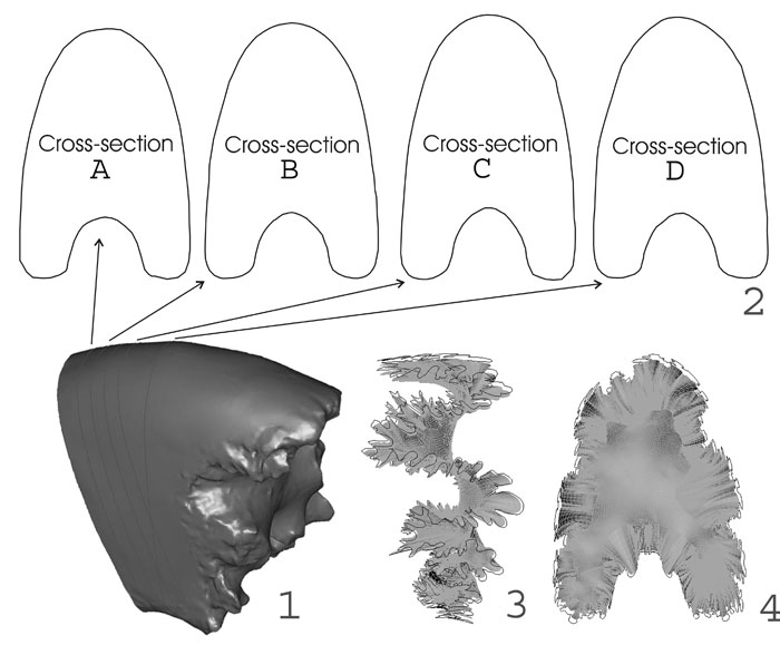
FIGURE 2. (1) Computer model of scanned fragment shown in Figure 1. (2) Cross-sections taken from model using Rhinoceros. Model of septum with superimposed suture line, (3) Lateral view, (4) Frontal view.
|
|
|
FIGURE 2. (1) Computer model of scanned fragment shown in Figure 1. (2) Cross-sections taken from model using Rhinoceros. Model of septum with superimposed suture line, (3) Lateral view, (4) Frontal view.
|