![]()
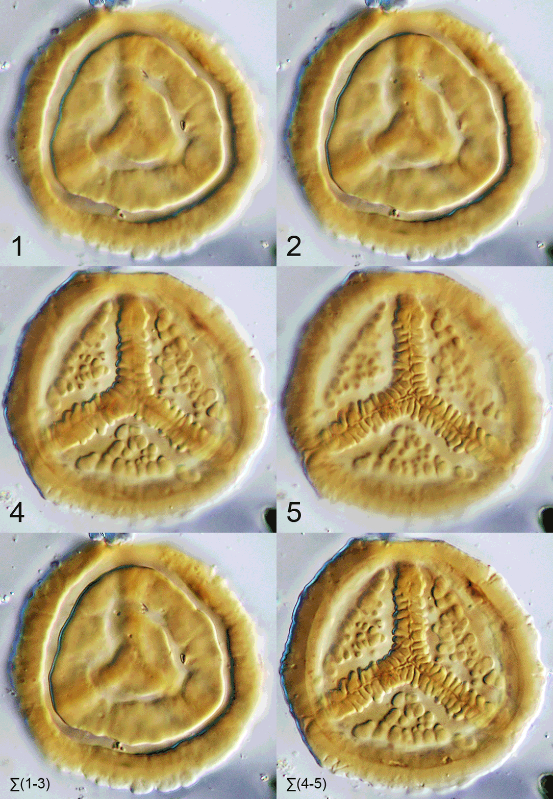
FIGURE 13. Example of reconstruction for Taurocusporites segmentatus (46 Ám, undyed, Utrillas Formation), with different morphological features on proximal and distal view. The final reconstructed images are the result of "Do Weighted Average" for the complete reconstruction, and of "Do Stack" for the reconstructions using only a subset of the original optical sections.
