![]()
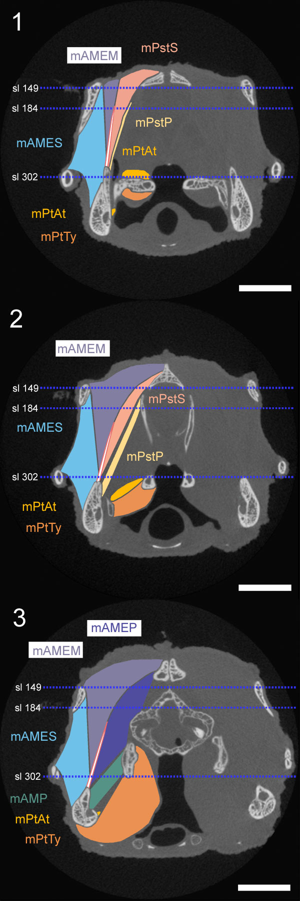
FIGURE 17. Coronal CT slices (YPM 9194) with schematic representations of the muscles and basal aponeurosis added. 17.1 Slice235, near the tallest point of the coronoid bone. 17.2 Slice 261 through the anterior edges of the epipterygoids. 17.3 Slice (309) through the posterior portion of the postorbital bone. Locations of the horizontal slices (see below) are shown in blue. Scale equals 10 mm.