![]()
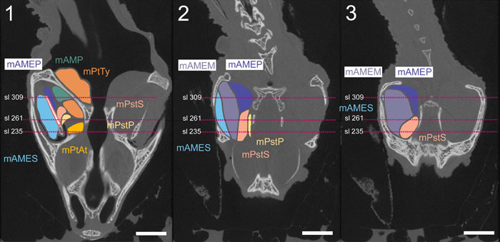
FIGURE 19. Horizontal CT slices with schematic representations of the muscles and basal aponeurosis added. 19.1 Slice 302 through the ventral part of the adductor chamber. 19.2 Slice 184 through the dorsal part of the adductor chamber. Locations of the coronal slices are shown in red. 19.2 Slice 302 through the dorsal part of the adductor chamber. Scale equals 10 mm.