![]()
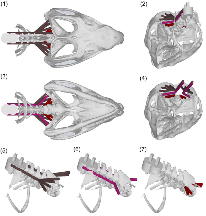
FIGURE 17. mSSpCa (brown [5]), mLCaL (violet_red [6]) and mLCaM (dark red [7]). (1) Dorsal view; (2) posteroventrolateral view; (3)ventral view; (4) posteroventrolateral view without the neck; (5) anterodorsolateral view of the mSSpCa without the skull; (6) anterodorsolateral view of the mLCaL without the skull; (7) anterodorsolateral view of the mLCaM without the skull. Where present the fascia is at 80% transparency.