![]()
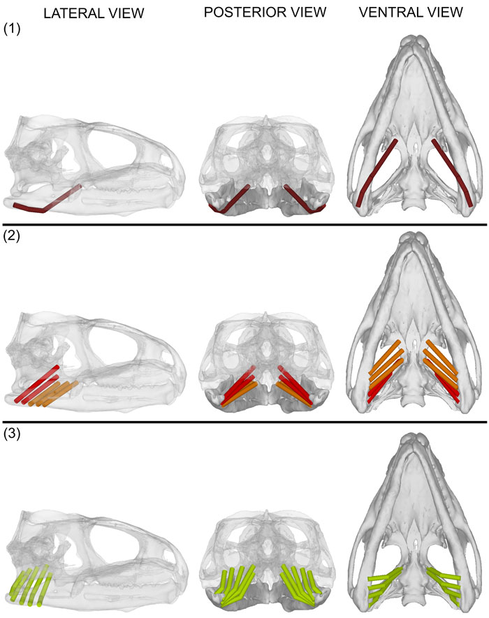
FIGURE 7. mPtTyp. Lateral, posterior and ventral views of (1) ventrolateral (brown red) portion of the mPtTyp; (2) lateral fibres of the middle portion (orange) and dorsal portion (red) of the mPtTyp; (3) medial fibres of the middle portion of the mPtTyp (green yellow). The transparency of all bone in the lateral view and the skull in the posterior view is at 80%. No fascia is present in this figure.