![]()
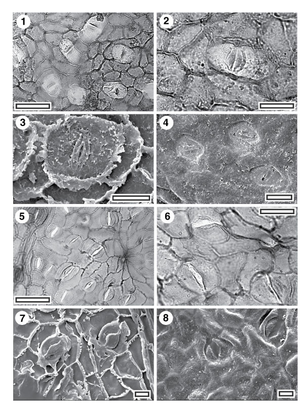
FIGURE 10. Lauraceae; (1) CUT-L-FCG, TLM view of stomatal complexes. Note cuticle over stomatal complexes is much thinner than over epidermal cells (SL2757, scale bar = 50 µm); (2) CUT-L-FCG, TLM detail of stomatal complex (SL2757, scale bar = 20 µm); (3) Inner SEM view of single stomatal complex. Note 'double' cuticular flanges (S-1427, scale bar = 10 µm); (4) Outer SEM view of three stomatal complexes (S-1427, scale bar = 20 µm); (5) CUT-L-FDG, TLM of stomatal complexes and (centre right) a trichome base (SL2772, scale bar = 50 µm); (6) CUT-L-FDG, TLM detail of three stomatal complexes and a trichome base (SL2772, scale bar = 20 µm); (7) Inner SEM view of stomatal complexes (S-1429, scale bar = 10 µm); (8) Outer SEM view of two stomatal complexes (S-1429, scale bar = 10 µm).