![]()
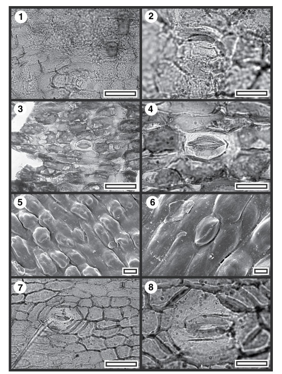
FIGURE 7. Kakahu podocarp sp. B and monocots; (1) Kakahu podocarp sp. B, TLM view showing two trichome bases (upper right) and a stomatal complex (lower centre) (SL2756, scale bar = 50 µm); (2) Kakahu podocarp sp. B, TLM detail of stomatal complex (SL2756, scale bar = 20 µm); (3) CUT-Mo-FDD, TLM view of stomatal complex and surrounding epidermal cells. Note the epidermal cells are broadly raised as papillae (SL2723, scale bar = 50 µm); (4) CUT-Mo-FDD, TLM detail of stomatal complex (SL2723, scale bar = 20 µm); (5) Outer SEM view (S-1428, scale bar = 20 µm); (6) Outer SEM view (S-1428, scale bar = 10 µm); (7) CUT-Mo-GED, TLM view of stomatal complex (SL5515; scale bar = 50 µm); (8) CUT-Mo-GED, TLM detail of stomatal complex (SL5515; scale bar = 20 µm).