![]()
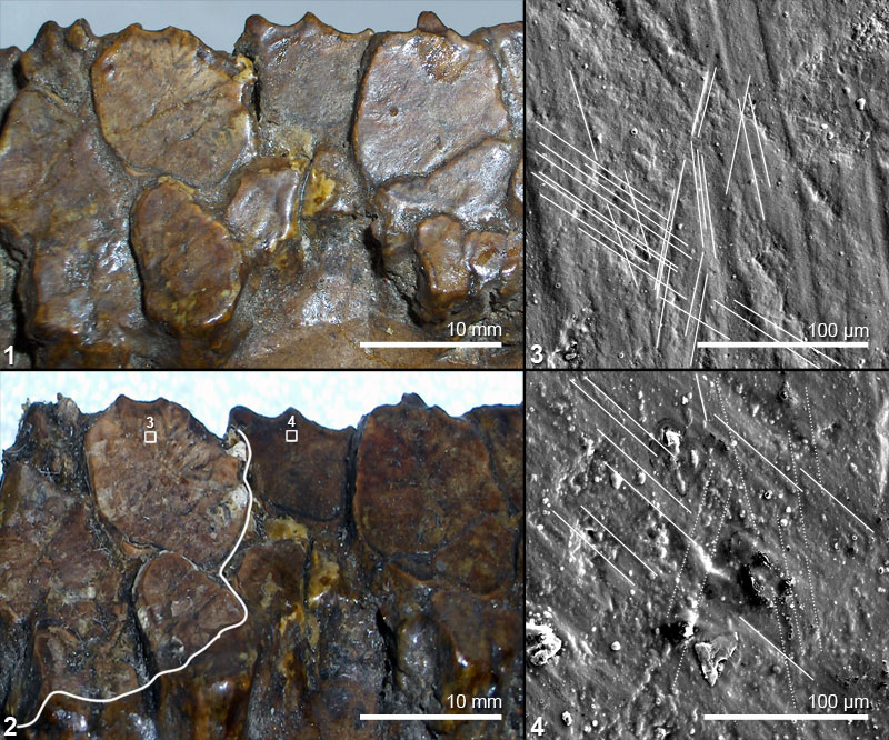
FIGURE 3. Occlusal surface of left dentary teeth of Corythosaurus casuarius (AMNH 3971), illustrating the results of cleaning with solvent gel. 3.1 Photograph of mesial fragment of left dentary prior to cleaning. 3.2 Post cleaning, shellac removed from teeth on the left by application of solvent gel; boxes show areas illustrated in 3.3 and 3.4. Broad white line drawn on to highlight the sharp boundary of the area from which shellac has been removed. 3.3 SEM micrograph of cast of central portion of a cleaned tooth (area in box 3 of 3.2); multiple microwear orientations (part highlighted with solid white lines) are visible with no remnant of varnish. 3.4 SEM micrograph of cast of central portion of an uncleaned tooth (area in box 4 of 3.2); microwear (part highlighted with solid white lines) is discernable but is largely obscured by varnish; in particular the near vertical microwear (part highlighted with dashed white lines), which is visible in 3.3, is barely noticeable here.