![]()
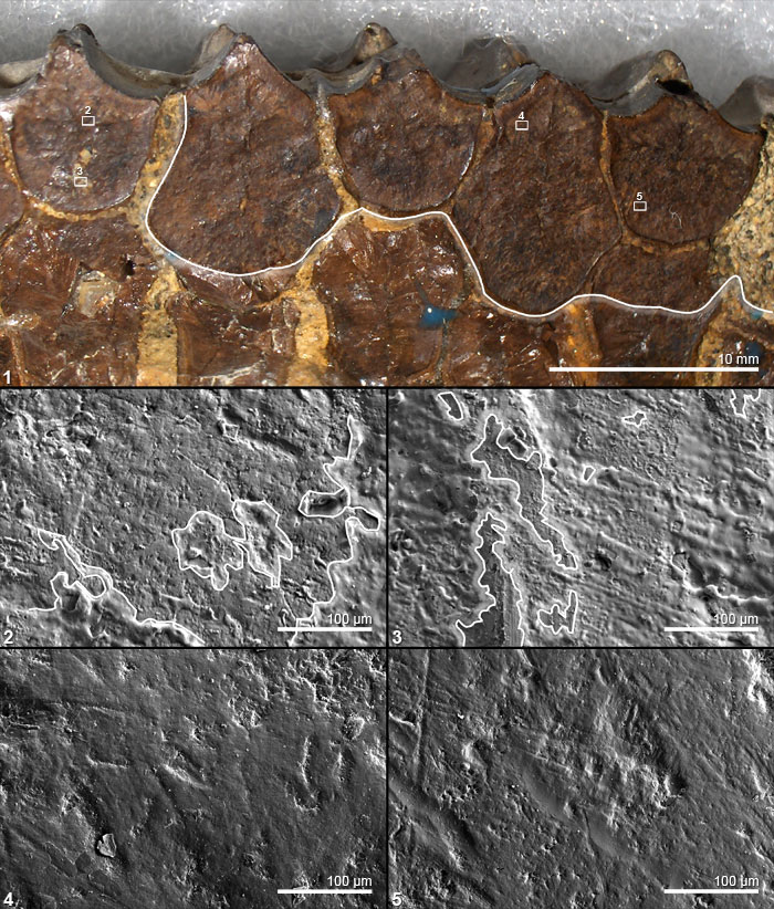
FIGURE 4. Occlusal surface of left dentary teeth of Edmontosaurus (SM 22102), illustrating the results of cleaning with solvent gel. 4.1 Photograph of distal section of a left dentary. White line drawn on to highlight the sharp boundary of the area from which shellac has been removed. The teeth on the top row and to the right have been cleaned by application of solvent gel. The teeth on the left and along the bottom row were not cleaned and remain coated in shellac. Boxes show areas of tooth illustrated in 4.2 to 4.5. 4.2 SEM micrograph of cast of site on uncleaned tooth (area in box 2 of 4.1); microwear is obscured by shellac. White lines drawn around areas of shellac, with shading in the shellac. 4.3 SEM micrograph of cast of site on uncleaned tooth (area in box 3 of 4.1); microwear is obscured by shellac. White lines drawn around areas of shellac, with shading in the shellac. 4.4 SEM micrograph of cast of site on cleaned tooth (area in box 4 of 4.1); showing microwear with no remnant of shellac. 4.5 SEM micrograph of cast of site on cleaned tooth (area in box 5 of 4.1); showing microwear with no remnant of shellac. (Note: the broad shallow grooves are typical of tool marks left by a vibro-tool during preparation of a fossil.)