![]()
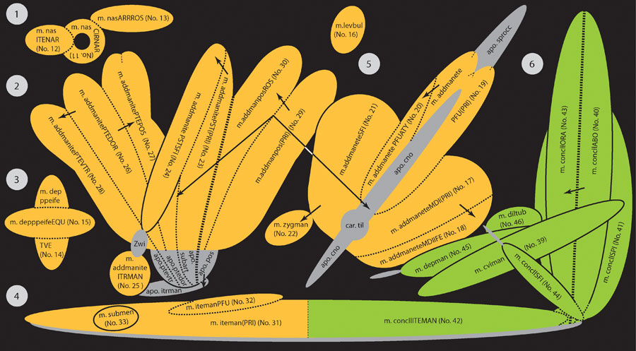
FIGURE 16. Scheme for the fluid pattern and plastic arrangement of n. trigeminus (V, orange) and n. facialis (VII, green) innervated musculature in turtles, including all potential muscular units as defined in the presented study; tendinuous structures are shown in grey. The scheme shows the maximum of partitions, which are observeable in turtles. There is no case, in which all muscular units are present. However, the arrows indicate potential phylogentic and ontogenetic origins of particular units. The thin dotted lines indicate different degrees of muscle separations, forming labelled muscle portions or non-labelled muscle heads. 1) Nasal musculature ((No. 11-13), 2) m. adductor mandibulae internus (No. 23-28) and posterior (No. 29-30), 3) lower eyelid muscle (No. 14-15), 4) intermandibular musculature: n. trigeminus (V) and n. facialis (VII) innervated muscular structures are fused (No. 31-33/42), 5) m. adductor mandibular externus (No. 17-21) and m. zygomaticomandibularis (No. 22), 6) n. facialis (VII) innervated musculature (No. 39-46). For abbreviations, see Appendix 1, Appendix 2, and Appendix 4.