![]()
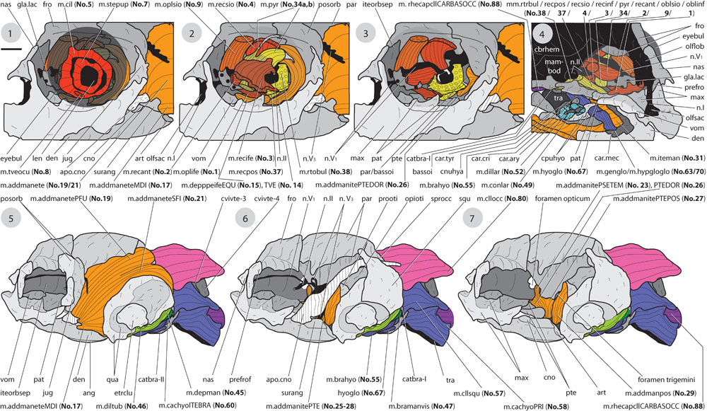
FIGURE 22. Hatchling specimen of Emydura subglobosa (collection of Prof. Dr. Wolfgang Maier / Tübingen). Different layers of cranial musculature mostly shown for the left body side. Drawings based on 3d reconstructions; superficial fibre courses based on manual dissections. 1-3) frontolateral view to the eye region, prefrontal removed, 1) internal eye muscles, 2) external eye muscles, 3) external eye muscles, deeper muscles, 4) frontomedial view to the eye-, hyoid-, and lower jaw region, 5-7) lateral view to the head/neck region, different layers of jaw adductor musculature (eye muscles and some neck muscles removed), 5) portions of m. adductor mandibulae externus (No. 17, 19, 21), 6) coronar aponeurosis (external adductor removed), 7) mm. adductor mandibulae internus et posterior (No. 23, 25-28, 29). Bar scale (under "1"): for 1-4) ca. 0.25 mm, for 5-7) ca. 0.5 mm. Continued in Figure 23.