

Article Search
Volume 27.1
January–April 2024
Full table of contents
ISSN: 1094-8074, web version;
1935-3952, print version
Recent Research Articles
See all articles in 27.1 January-April 2024
See all articles in 26.3 September-December 2023
See all articles in 26.2 May-August 2023
See all articles in 26.1 January-April 2023
Xijun Ni Division of Paleontology
American Museum of Natural History
Central Park West at 79th Street
New York, New York 10024
USA
and Key Laboratory of Evolutionary Systematics of Vertebrates
Institute of Vertebrate Paleontology and Paleonanthropology
Xi Zhi Men Wai Street 142
Beijing, 100044
China
Xijun Ni obtained his Ph.D. in ecology at the Beijing Normal University. He got postdoctoral training and rushed in paleontology in the Institute of Vertebrate Paleontology and Paleoanthropology (IVPP) and the American Museum of Natural History (AMNH). He was recruited in the Hundred-Talent Program of the Chinese Academy of Sciences, and appointed as a research professor in the IVPP. He is also a research associate in paleontology at the AMNH and the Carnegie Museum of Natural History. His research interests lie in four areas: searching for primate, tree shrew, flying lemur, rodent, and other mammalian fossils; applying high resolution CT imaging and 3-dimensional digital reconstruction techniques in evolutionary and functional morphology studies; reconstructing phylogenetic relationships of primates, tree shrews, flying lemurs, rodents and other major groups of mammals based on large combined data matrices; and paleoecology and biochronology inference based on studying mammalian faunas.
![]()
John J. Flynn Division of Paleontology and Richard Gilder Graduate School
American Museum of Natural History
Central Park West at 79th Street
New York, New York 10024
USA
John Flynn is the Frick Curator of Fossil Mammals & Dean of the Richard Gilder Graduate School at the American Museum of Natural History. Author of more than 120 scientific publications, Flynn's research focuses on the phylogeny and evolution of mammals and Mesozoic vertebrates, geological dating, plate tectonics, and biogeography. He also is curator for the Museum’s “Extreme Mammals” traveling exhibition, and has contributed articles to Scientific American, Natural History, and National Geographic, provided scientific expertise for several popular science books, and been featured in numerous television and radio shows, newspapers and magazines. Dr. Flynn has led more than 50 paleontological expeditions to Chile, Perú, Colombia, Madagascar, Angola, India, and the Rocky Mountains, supported by the U.S. National Science Foundation, the National Geographic Society, NASA, and other organizations. In 2001 Flynn received a Guggenheim Fellowship for a year of research, writing and expeditions in South America and was elected a Fellow of the American Association for the Advancement of Science in 2009. He is a member of the External Advisory Board for Yale's Peabody Museum, and has served the Society of Vertebrate Paleontology (SVP) as President (1999-2001) and member of the Board/Executive Committee (1993-2002), as received the Joseph T. Gregory Award (2007) and the Alfred Sherwood Romer Prize from the SVP. With a specialty in mammalian paleontology and paleomagnetism, Flynn has spent his career searching for important new fossil mammal localities, as well as developing better ways to read the age of rocks and fossils, leading to more accurate geological time scales. He has contributed to numerous public education projects (university, museum, web, and popular science), is actively pursuing research on mammalian evolution (particularly the anatomy, DNA and evolution of Carnivora and extinct relatives), and has current field programs focusing on the Andes Mountains of Chile, Amazon Basin of Perú, and Mesozoic deposits of Madagascar and India.
![]()
André R. Wyss Department of Earth Science
University of California- Santa Barbara
Santa Barbara, California 93106
USA
André Wyss is a Professor and Vice-Chair of the Department of Earth Science at the University of California- Santa Barbara, and a Research Associate in Paleontology at the American Museum of Natural History. Averse to such missives as this biography, only the editorial threat of being deemed "LAME" spurred his cooperation. Ensconced in his first floor, west-facing office, Wyss is enjoying his slow ride on a sliver of crust formerly belonging to North America. His abiding research interests are, 1) elucidating the history of mammals using fossils, and 2) applying the chronologic information offered by these data to a variety of geologic problems. His field activities are generally aimed at geographic regions and stratigraphic intervals having received little previous paleontological attention, including the Andes of central Chile and the early Mesozoic of western Madagascar.
FIGURE 1. Histogram of the grayscale distribution of the entire CT data set of the holotype skull of C. carrascoensis. Red line, original data set; green line, endocast of the bony labyrinth; blue line, skull with endocast and unprepared matrix removed.
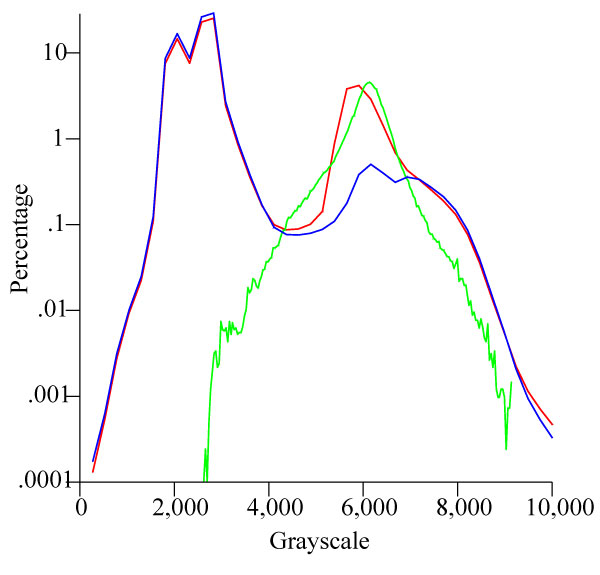
FIGURE 2. An example of the CT image segmentation process. The Half Maximum Height method (Baxter and Sorenson, 1981; Spoor et al., 1993) is used to assist in identifying the bone-matrix interface. 1, original CT images across the right ear region in coronal plane; 2, endocasts of the bony labyrinth and cranial cavity are segmented; 3, endocast of the bony labyrinth. 4, 5, and 6 indicate the grayscale distribution of 1, 2, and 3, respectively.
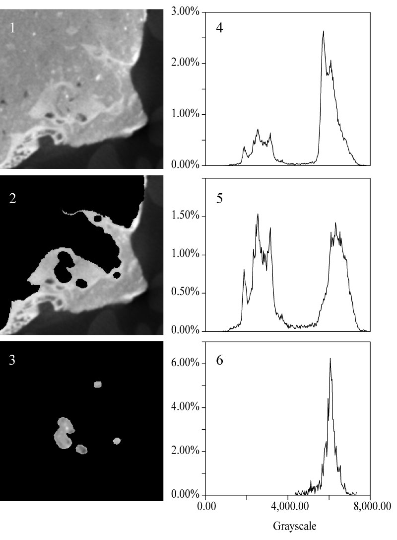
FIGURE 3. Three-dimensional virtual reconstruction of the bony labyrinth and the skull of C. carrascoensis. To show the position of the bony labyrinths, the rest of the skull is set to transparent. 1, dorsal view, un-restored; 2, dorsal view, restored with 7.7° offset.
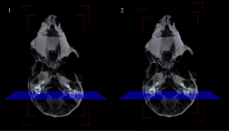
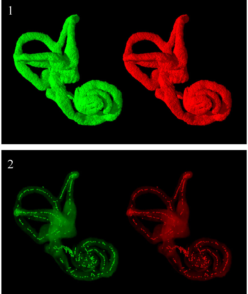
FIGURE 4. 1, Stereoscopic anterolateral view of the three-dimensional virtual reconstructions of the right bony labyrinth of C. carrascoensis; 2, stereoscopic view of the skeletonized right bony labyrinth. The bony labyrinth was set to be transparent. The white dots, generated via the skeletonization process, represent the central path of the bony labyrinth.
Imaging the inner ear in fossil mammals: High-resolution CT scanning and 3-D virtual reconstructions
Plain Language Abstract
The organs of hearing and balance in mammals are housed within an intricate cavity at the base of the skull, named, intriguingly, the “bony labyrinth.” Because it is lodged so deeply within the skull, it has been nearly impossible to study the bony labyrinth in fossils—since it is shrouded in bone and often encased in sediment matrix. Despite several technical challenges, high-resolution x-ray Computed Tomography (CT) scanning provides a powerful new tool for investigating this delicate and otherwise inaccessible structure. Challenges include addressing the complexly interwoven density patterns spanning the interface between the actual fossil bones and their encasing sediment, which frequently obscure the boundary between the two in CT images. Methods for reliably determining these boundaries and accurately segmenting the original CT images, both essential for producing anatomically accurate three-dimensional virtual reconstructions, have been lacking until now. In this study we introduce a protocol accomplishing both of these objective. Cyclically measuring the “Half Maximum Height grayscale thresholds”, a method emphasizing local gray-tone contrast differences rather than the range across the whole specimen, to better distinguish different structures between the fossil and rock or between the fossil and air, with constant interaction between operator and imaging software, reliably discriminates the bony labyrinth from the remainder of the skull as well as from surrounding sediment. To demonstrate the efficacy of this method, we CT scanned the skull of Chilecebus carrascoensis, a 20 million-year-old New World monkey fossil preserved in intractable, volcanically derived sediment, reconstructing its bony labyrinth successfully. We anticipate, therefore, that the protocol described here will be broadly applicable to a wide variety of fossils across a spectrum of sediment types and preservation styles, finally unveiling this long hidden region or other complex internal bony structures for detailed scientific analyses.
Resumen en Español
Obtención de imágenes del oído interno de los mamíferos: tomografía computarizada de alta resolución y reconstrucciones virtuales tridimensionales.
El laberinto óseo de los mamíferos es una delicada y compleja cavidad ubicada en la porción petrosa del hueso temporal que alberga los órganos de la audición y el equilibrio del oído interno. Dado que esta región se localiza en una zona poco accesible del cráneo, existen pocos estudios morfológicos del laberinto óseo en fósiles, en los que frecuentemente se encuentra completamente envuelto por los huesos que lo rodean y la matriz sedimentaria. El desarrollo reciente de la tomografía axial computarizada (TAC) de alta resolución proporciona una nueva y poderosa herramienta para la investigación de esas minúsculas y, a menudo, inaccesibles estructuras. En este artículo presentamos un protocolo para la obtención de imágenes virtuales tridimensionales (3D) del laberinto óseo a partir de tomografías computarizadas de alta resolución. A modo de ejemplo, hemos escaneado el cráneo de Chilecebus carrascoensis, un primate platirrino primitivo, mediante un tomógrafo axial de alta resolución de la Universidad Estatal de Pensilvania y reconstruido el molde interno del laberinto óseo a partir de los datos obtenidos. La segmentación de las imágenes TAC originales es un paso fundamental en la producción de reconstrucciones virtuales 3D precisas. Nuestros intentos de aislar el molde interno del laberinto óseo por medios automatizados fracasaron debido a la densidad similar que presentan el relleno sedimentario de las cavidades de los senos y del hueso esponjoso y el propio laberinto óseo. Fue necesario el uso valores umbrales medio-máximos para medir dinámicamente la diferencia de los contrastes de densidad en la interfase fósil/matriz y el solapamiento de las tonalidades de grises del fósil y la matriz. Resulta útil el uso de múltiples valores umbrales en el procesamiento de datos TAC de fósiles que son, intrínsecamente, heterogéneos en las propiedades y densidades de sus materiales. La interacción iterativa entre el operador y el ordenador ofrece el único medio disponible en la actualidad para discriminar de manera fiable el molde interno del laberinto óseo del resto del ejemplar.
Palabras clave: laberinto óseo; TAC de alta resolución; segmentación; reconstrucción virtual tridimensional (3D); Chilecebus; primate platirrino.
Traducción: Miguel Company
Résumé en Français
Représentation de l'oreille interne chez les mammifères fossiles: Balayage CT scan haute-résolution et reconstruction numérique virtuelle 3D
Le labyrinthe osseux des mammifères, une cavité délicate et complexe à l'intérieur du pétrosal, supporte les organes de l'audition et de l'équilibre de l'oreille interne. Du fait que cette région est située profondément dans le crâne – et souvent totalement enveloppée par d'autres os et le sédiment – peu d'études morphologiques du labyrinthe osseux on été réalisées dans le fossile. Les développements récents de la tomographie à rayon X assistée par ordinateur (Computed Tomography, CT) fournissent un outil puissant pour étudier des structures si petites et souvent inaccessibles. Nous présentons ici un protocole pour obtenir une représentation virtuelle tridimensionnelle (3D) du labyrinthe osseux à partir d'images de balayages CT haute-résolution.
Comme sujet d'étude, nous avons scanné un crâne de primate platyrrhinien basal, Chilecebus carrascoensis, utilisant le scanner tomographique haute-résolution de l'Université de l'État de Pennsylvanie, aboutissant à la reconstruction du moule interne du labyrinthe osseux à partir de données obtenues. La segmentation des images originales du balayage tomographique est un point crucial dans la production d'une reconstruction virtuelle 3D précise. Nous n'avons pas réussi à isoler le moule interne du labyrinthe osseux par des moyens automatisés, dû à la similarité de densité de la matrice de remplissage dans les sinus et les cavités spongieuses de l'os, et le labyrinthe osseux lui-même. Les différentes densités de contraste au travers de l'interface fossile/matrice et le recoupement des nuages de gris du fossile et de la matrice, ont nécessité des seuils à moitié du 'Half Maximum Height' pour être mesuré de manière dynamique. De multiples seuils sont avantageux pour traiter les données tomographiques du fossile dont les propriétés et les densités des matériaux sont par nature hétérogènes. Les interactions répétées entre l'operateur et l'ordinateur sont pour le moment le seul moyen disponible permettant une discrimination fiable entre le moule interne du labyrinthe osseux et le reste du spécimen.
Mots clés : Labyrinthe osseux, Tomographie haute-résolution à rayon X assistée par ordinateur ; segmentation ; reconstruction virtuelle tridimensionnelle (3D) ; Chilecebus; primate platyrrhinien
Translator: Olivier Maridet
Deutsche Zusammenfassung
Abbildung des Innenohrs von fossilen Säugetieren: hochaufgelöste Computertomographie und 3D virtuelle Rekonstruktionen
Das knöcherne Labyrinth der Säugetiere ist ein zierlicher und komplexer Hohlraum innerhalb des Petrosums der beherbergt die Gehörorgane und das Gleichgewichtsorgan im Innenohr beherbergt. Da sich diese Region normalerweise tief im Inneren des Schädels befindet, gibt es nur wenige morphologische Untersuchungen zum knöchernen Labyrinth bei Fossilien, da es häufig komplett von Knochen und Sediment umgeben ist. Die neueste Entwicklung der hochaufgelösten Röntgen-Computertomographie (CT) bietet ein leistungsfähiges Instrument für solche kleinen, häufig schwer zugänglichen Strukturen. Wir stellen hier ein Protokoll vor, wie eine dreidimensionale (3D) virtuelle Veranschaulichung des knöchernen Labyrinths aus hochauflösenden CT-Aufnahmen gewonnen werden kann. Als Fallstudie scannten wir den Schädel des basalen Platyrrhinen Chilecebus carrascoensis. Wir benutzten die hochauflösende CT-Anlage der Pennsylvania State University und rekonstruierten den Innenausguss des knöchernen Labyrinths aus den gewonnenen Daten. Das Aufteilen der Original CT-Aufnahmen ist ein entscheidender Schritt für die Herstellung einer präzisen 3D virtuellen Rekonstruktion. Es ist uns nicht gelunge,n den knöchernen Labyrinth-Innenausguss durch automatisierte Weise zu isolieren, aufgrund der ähnlichen Dichten zwischen der Matrix, den Sinussen und den spongiösen Knochenholräumen des Stückes und dem knöchernen Labyrinth selbst.
Unterschiedliche Dichtekontraste entlang der Fossil/Matrix Grenze und die überlappenden Grauskalen des Fossils und der Matrix erforderten Half Maximum Height Schwellenwerte, die dynamisch gemessen werden mussten. Multiple Schwellenwerte sind für die Verarbeitung von CT-Daten von Fossilien vorteilhaft die inhärent heterogen sind, was Materialeigenschaften und Dichte betrifft. Die iterative Interaktion zwischen Anwender und Computer bietet das derzeit einzig verfügbare Mittel für die zuverlässige Unterscheidung des knöchernen Labyrinth-Innenausgusses vom Rest des Stückes.
SCHLÜSSELWÖRTER: knöchernes Labyrinth; hochauflösendes Röntgen- CT; Segmentation; dreidimensionale (3-D) virtuelle Rekonstruktion; Chilecebus; platyrrhiner Primat
Translators: Eva Gebauer and Anke Konietzka
Arabic
Translator: Ashraf M.T. Elewa
Polski Abstrakt
Obrazowanie ucha wewnętrznego u ssaków kopalnych: wysokorozdzielczośćowa tomografia komputerowa i trójwymiarowe rekonstrukcje wirtualne
Błędnik kostny ssaków, delikatne i złożone wgłębienie w kości skalistej, mieści narządy słuchu i równowagi w uchu wewnętrznym. Ponieważ obszar ten zazwyczaj znajduje się głęboko w czaszce, i w wypadku skamieniałości często jest całkowicie otoczony przez kości i osad, przeprowadzono niewiele badań morfologicznych błędnika kostnego u materiału kopalnego. Niedawny rozwój skenowania za pośrednictwem wysokorozdzielczośćowej tomografii komputerowej (CT) stanowi potężne narzędzie do badania drobnych i często niedostępnych struktur. Poniżej przedstawiamy protokół do wytwarzania trójwymiarowych (3-D) wizualizacji wirtualnych błędnika kostnego z wysokorozdzielczościowych obrazów CT. Dla przykładu użycia zeskanowaliśmy za pośrednictwem wysokorozdzielczościowego CT z Pennsylvania State University czaszkę bazalnej małpy szerokonosej (Platyrrhini) z gatunku Chilecebus carrascoensis i z odzyskanych danych zrekonstruowaliśmy budowę wewnętrzną błędnika kostnego. W ramach odtworzenia dokładnych trójwymiarowych wizualizacji wirtualnych, istotnym krokiem jest segmentacja pierwotnych obrazów z CT. Ze względu na podobnej gęstości matrycę wypełniającą zatoki i jamę kości gąbczastej badanej próbki, jak zarówno właściwego błędnika kostnego, nie udało nam się automatycznie odizolować obraz budowy wewnętrznej błędnika kostnego. Odmienna gęstość kontrastuje w ramach całego interfejsu materiału kopalnego/matrycy i nakładających się szarości skamieniałości i matrycy, i wymaga, by wartości progowe pół-maksymalnej wysokości mierzono dynamicznie. Wielokrotne wartości progowe są korzystne dla przetwarzania danych z CT dotyczących skamieniałości, które są nieodłącznie heterogeniczne pod względem właściwości oraz gęstości materiału. Iteracyjne interakcje między użytkownikiem i komputerem oferują jedyny dostępny obecnie sposób na wiarygodną dyskryminację budowy wewnętrznej błędnika kostnego od reszty próbki.
Słowa kluczowe: Błędnik kostny; wysokorozdzielczośćowa tomografia komputerowa; segmentacja; trójwymiarowe rekonstrukcje wirtualne; Chilecebus; małpa szerokonosa
Translators: Dawid Mazurek, Robert Bronowicz, and Daniel Madzia
-
-
-
Review: The Princeton Field Guide to Mesozoic Sea Reptiles
 The Princeton Field Guide to Mesozoic Sea Reptiles
Article number: 26.1.1R
April 2023

 Poster Winners 2024