

Article Search
Volume 27.1
January–April 2024
Full table of contents
ISSN: 1094-8074, web version;
1935-3952, print version
Recent Research Articles
See all articles in 27.1 January-April 2024
See all articles in 26.3 September-December 2023
See all articles in 26.2 May-August 2023
See all articles in 26.1 January-April 2023
FIGURE 3. Variation in enamel thickness in the teeth of Revueltosaurus callenderi 1-9, premaxillary tooth (P-33799--longitudinal section); 10-14, maxillary tooth (P-33798--transverse section). 1, overview of premaxillary tooth indicating approximate place where measurements and micrographs shown in this figure and Figure 4 were taken; 2-9, close-up views showing enamel thickness variation, with enamel-dentine junction (EDJ) oriented relative to overview in (1); and 10, overview of maxillary tooth section, indicating approximate place where measurements and micrographs shown in this figure and Figure 5 were taken; 11-14, close-up views showing enamel thickness variation, with enamel-dentine junction (EDJ) oriented relative to overview in (10). Scale bars = 100 µm except for 1 (5 mm), 2 (10 µm) and 10 (2 mm). Numbers in image are linked to further enlargements. Author note: Figure 3.10 is part of overview.
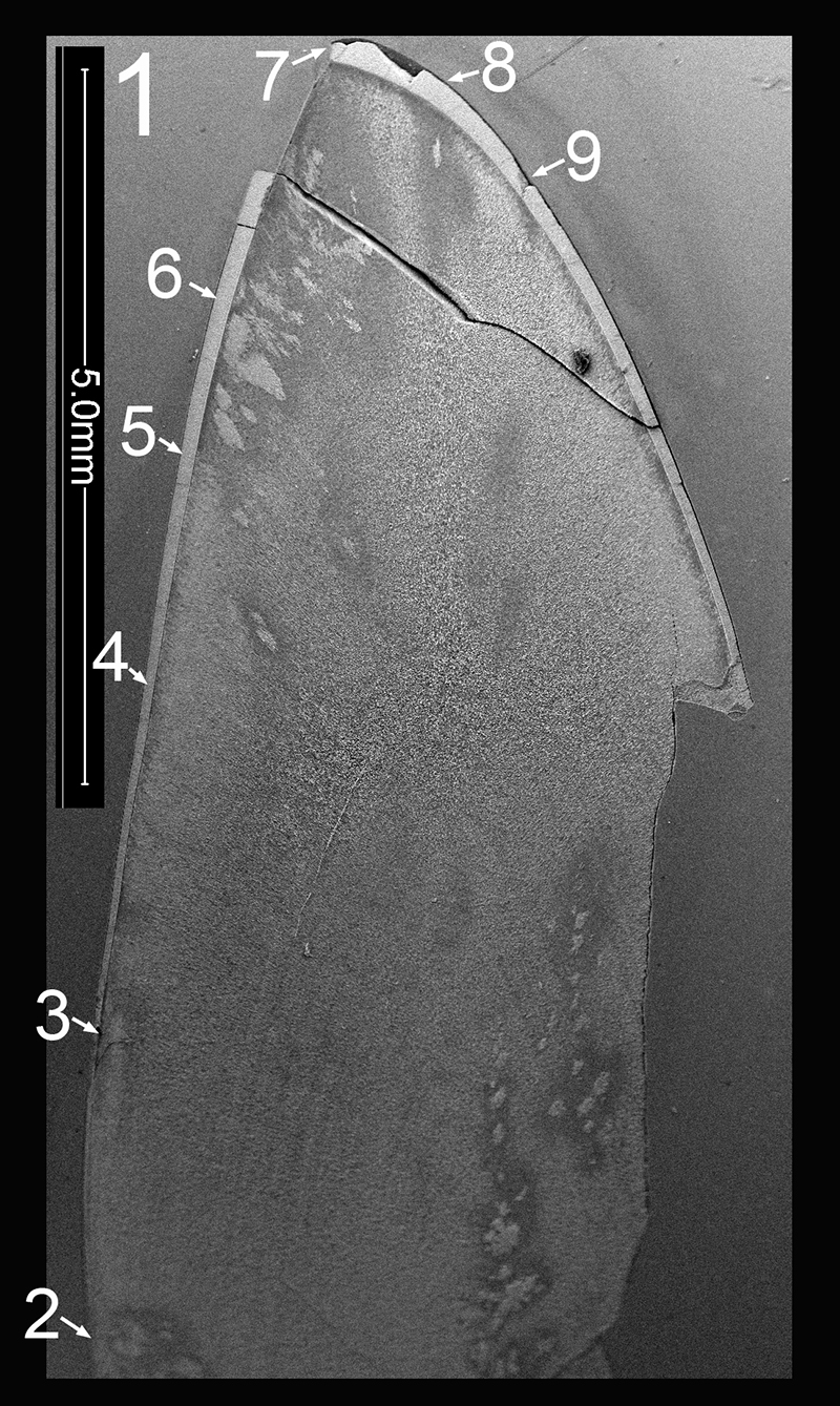
FIGURE 3.2. Original micrograph showing tooth enamel microstructure of premaxillary tooth of Revueltosaurus callenderi (P-33799) in longitudinal section near the base of the tooth; EDJ oriented to bottom of picture.
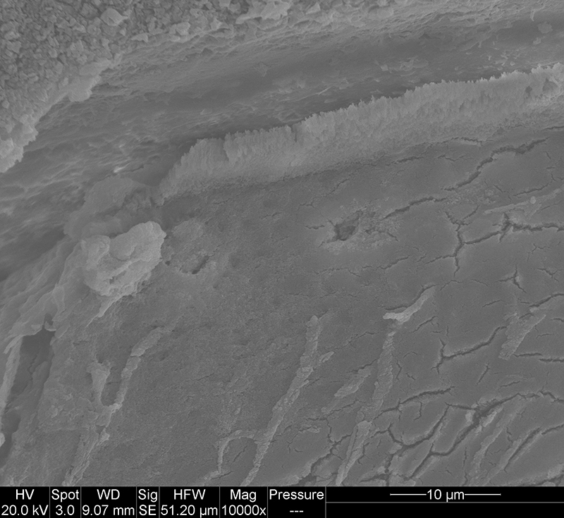
FIGURE 3.3. Original micrograph showing tooth enamel microstructure of premaxillary tooth of Revueltosaurus callenderi (P-33799) in longitudinal section near the base of the tooth; EDJ oriented to bottom of picture.
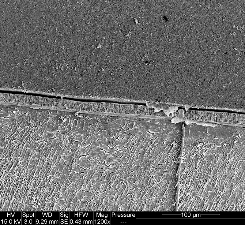
FIGURE 3.4. Original micrograph showing tooth enamel microstructure of premaxillary tooth of Revueltosaurus callenderi (P-33799) in longitudinal section near the middle of the tooth; EDJ oriented to bottom of picture.
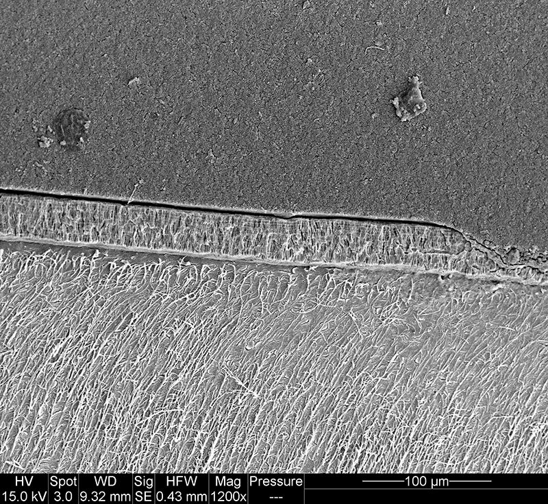
FIGURE 3.5. Original micrograph showing tooth enamel microstructure of premaxillary tooth of Revueltosaurus callenderi (P-33799) in longitudinal section near the middle of the tooth; EDJ oriented to bottom of picture.
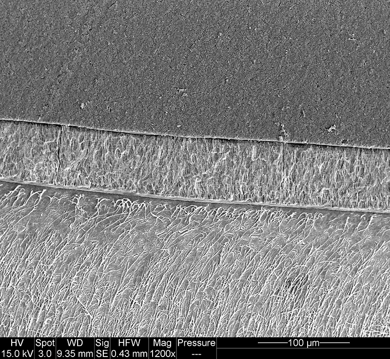
FIGURE 3.6. Original micrograph showing tooth enamel microstructure of premaxillary tooth of Revueltosaurus callenderi (P-33799) in longitudinal section high on the crown; EDJ oriented to bottom of picture.
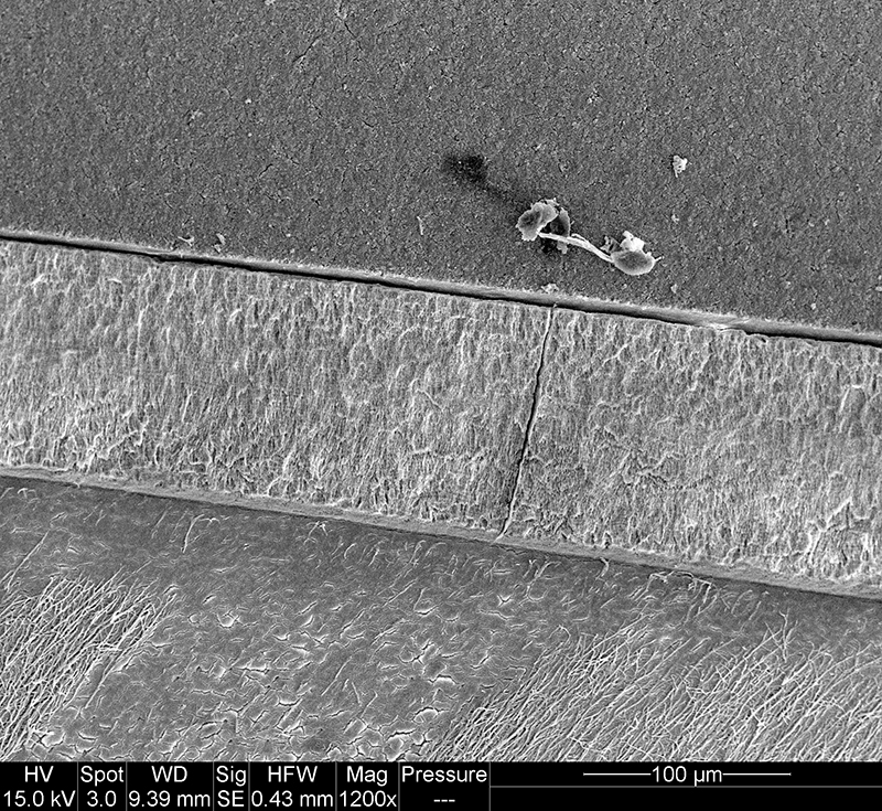
FIGURE 3.7. Original micrograph showing tooth enamel microstructure of premaxillary tooth of Revueltosaurus callenderi (P-33799) in longitudinal section at the tip of the crown; EDJ oriented to left of picture.
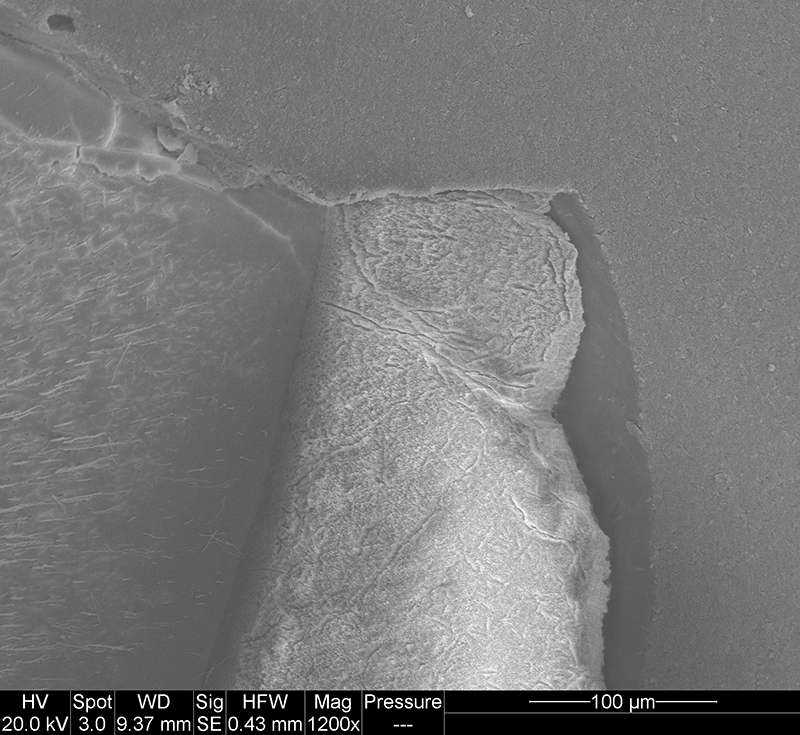
FIGURE 3.8. Original micrograph showing tooth enamel microstructure of premaxillary tooth of Revueltosaurus callenderi (P-33799) in longitudinal section near the tip of the crown; EDJ oriented to bottom of picture.
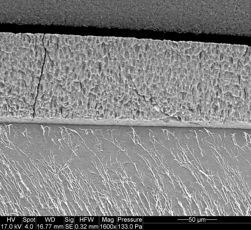
FIGURE 3.9. Original micrograph showing tooth enamel microstructure of premaxillary tooth of Revueltosaurus callenderi (P-33799) in longitudinal section near the base of the tooth; EDJ oriented to upper left of picture.
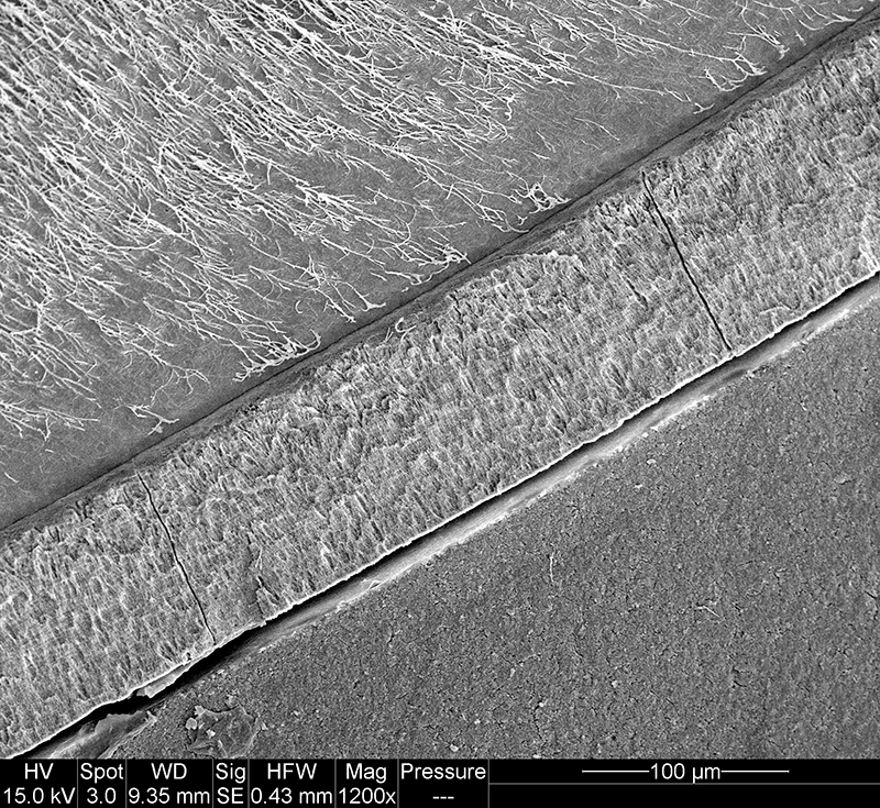
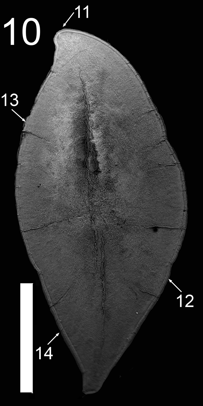
FIGURE 3.11. Original micrograph showing tooth enamel microstructure of maxillary tooth of Revueltosaurus callenderi (P-33798) in transverse section across one denticle.
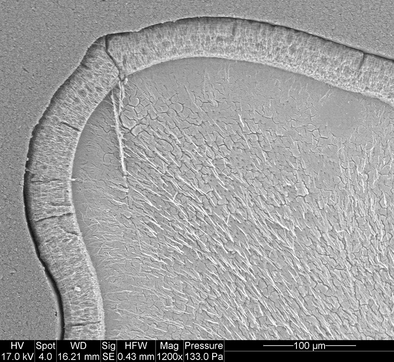
FIGURE 3.12. Original micrograph showing tooth enamel microstructure of maxillary tooth of Revueltosaurus callenderi (P-33798) in transverse section on labial side; EDJ to upper right.
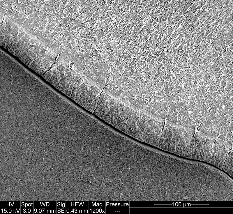
FIGURE 3.13. Original micrograph showing tooth enamel microstructure of maxillary tooth of Revueltosaurus callenderi (P-33798) in transverse section on lingual side; EDJ to bottom.
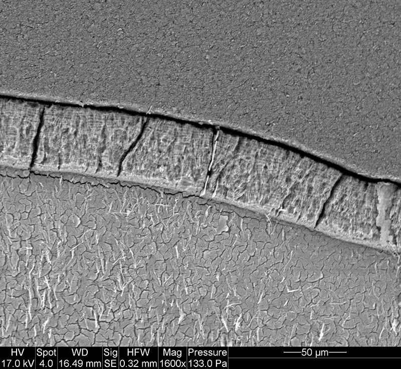
FIGURE 3.14. Original micrograph showing tooth enamel microstructure of maxillary tooth of Revueltosaurus callenderi (P-33798) in transverse section on labial side; EDJ to bottom.
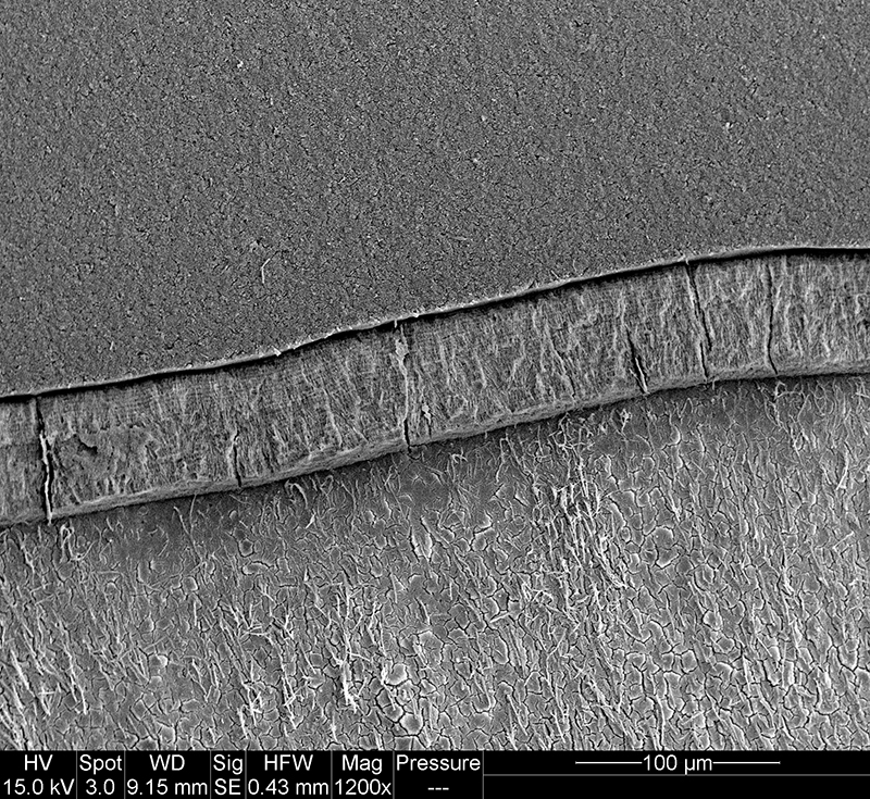
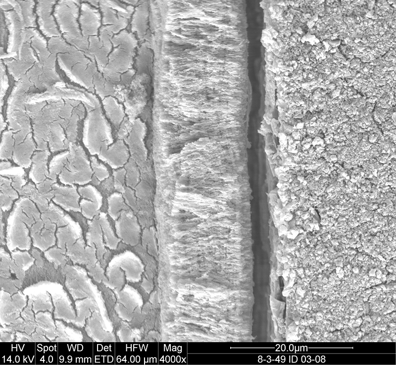
FIGURE 7. Scanning electron microscope images of (UCMP 165211), Krzyzanowskisaurus hunti premaxillary tooth enamel microstructure in transverse section. 1, overview of tooth indicating approximate place where measurements and micrographs shown in this figure were taken; 2-7, close-up views showing enamel thickness variation, with EDJ oriented relative to overview in (1) and OES away from the same overview. Scale bars = 20 µm except 1 (2 mm). Numbers in image are linked to further enlargements.
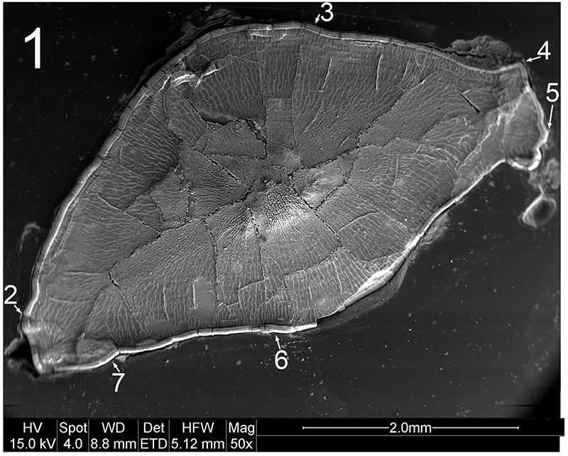
FIGURE 7.2. Original micrograph showing tooth enamel microstructure of (UCMP 165211), Krzyzanowskisaurus hunti in transverse section on the labial side; EDJ oriented to right of picture.
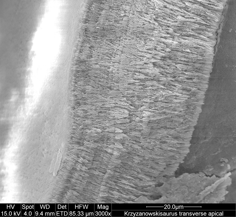
FIGURE 7.3. Original micrograph showing tooth enamel microstructure of (UCMP 165211), Krzyzanowskisaurus hunti in transverse section on the labial side; EDJ oriented to left of picture.
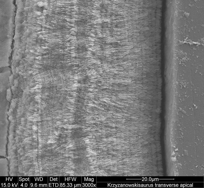
FIGURE 7.4. Original micrograph showing tooth enamel microstructure of (UCMP 165211), Krzyzanowskisaurus hunti in transverse section across a denticle; EDJ oriented to lower left of picture.
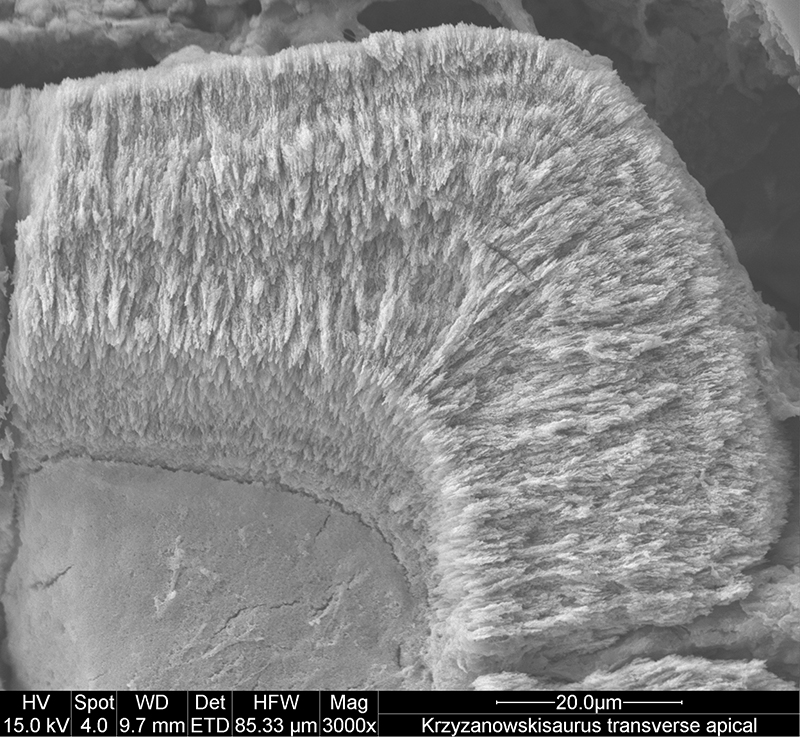
FIGURE 7.5. Original micrograph showing tooth enamel microstructure of (UCMP 165211), Krzyzanowskisaurus hunti in transverse section across a denticle; EDJ oriented to left of picture.
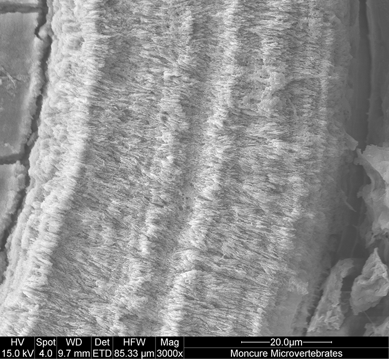
FIGURE 7.6. Original micrograph showing tooth enamel microstructure of (UCMP 165211), Krzyzanowskisaurus hunti in transverse section on the lingual side; EDJ oriented to left of picture.
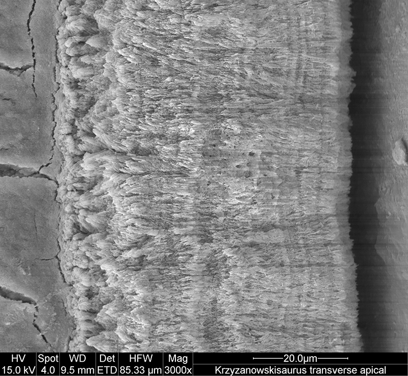
FIGURE 7.7. Original micrograph showing tooth enamel microstructure of (UCMP 165211), Krzyzanowskisaurus hunti in transverse section on the lingual side; EDJ oriented to left of picture.
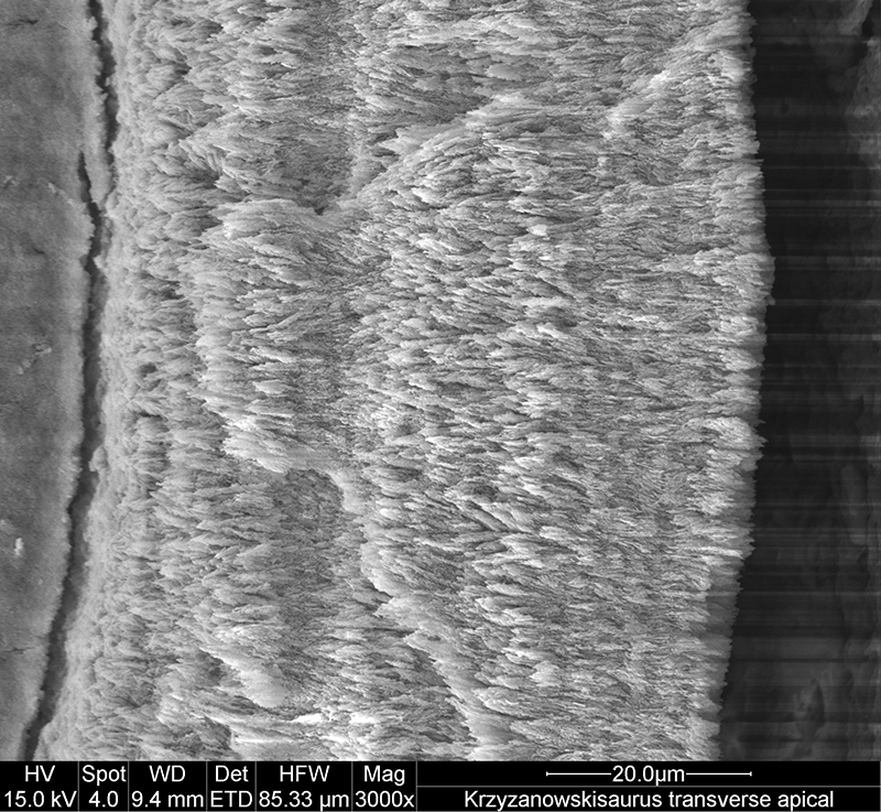
FIGURE 6. Scanning electron microscope images of (UCMP 165213), Krzyzanowskisaurus hunti tooth enamel microstructure in longitudinal section. 1, overview of tooth indicating approximate place where measurements and micrographs shown in this figure were taken; 2-4, close-up views showing enamel thickness variation, with EDJ oriented relative to overview in (1) and OES away from the same overview. Scale bars = 2 mm (1), 50 µm (2-3), 100 µm (4). Numbers in image are linked to further enlargements.
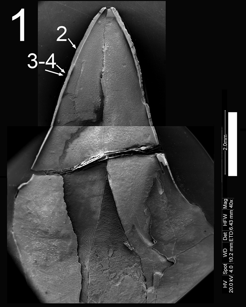
FIGURE 6.2. Original micrograph showing tooth enamel microstructure of (UCMP 165213), Krzyzanowskisaurus hunti in longitudinal section high on the crown; EDJ oriented to bottom of picture.
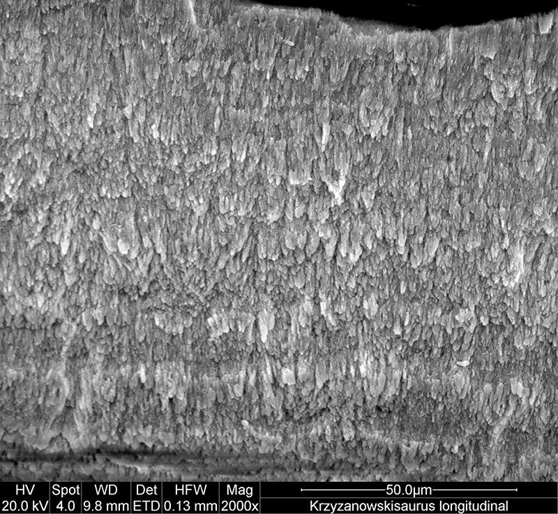
FIGURE 6.3. Original micrograph showing tooth enamel microstructure of (UCMP 165213), Krzyzanowskisaurus hunti in longitudinal section high on the crown; EDJ oriented to bottom of picture.
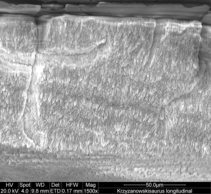
FIGURE 6.4. Original micrograph showing tooth enamel microstructure of (UCMP 165213), Krzyzanowskisaurus hunti in longitudinal section high on the crown; EDJ oriented to bottom of picture.
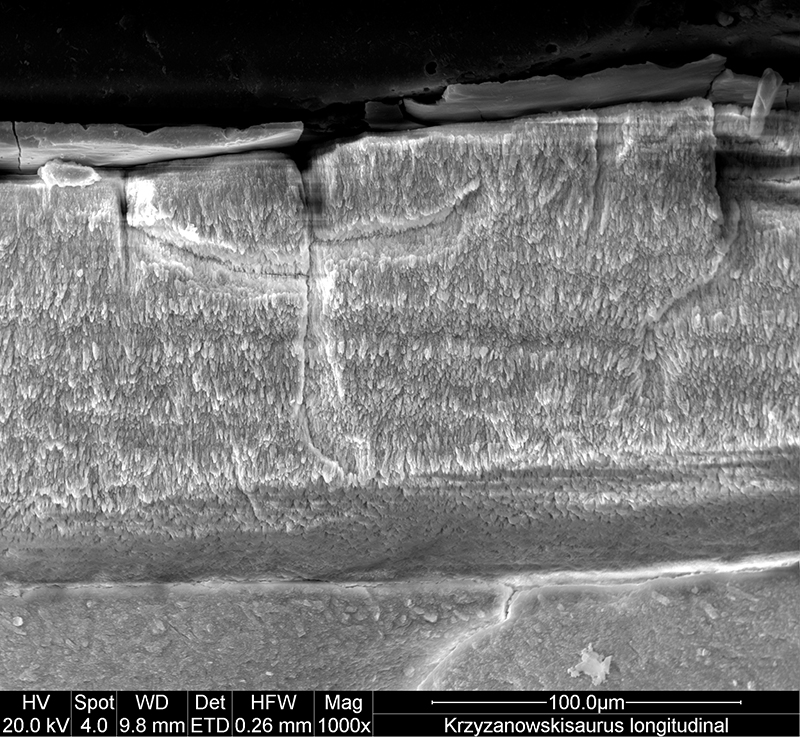
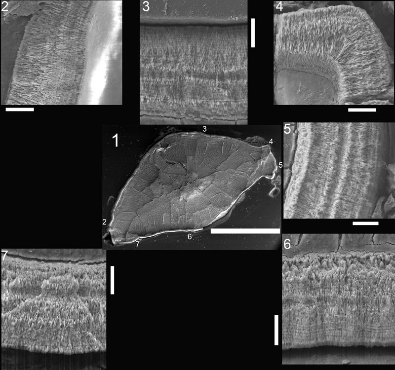
FIGURE 5. Scanning electron microscope images of NMMNH P-33798, Revueltosaurus callenderi maxillary tooth enamel microstructure in transverse section. 1, overview of tooth indicating approximate place where measurements and micrographs shown in this figure were taken; 2-8, close-up views showing enamel thickness variation, with EDJ oriented relative to overview in (1) and OES away from the same overview. Scale bars = 20 µm except 1 (2 mm), 3 (50 µm). Numbers in image are linked to further enlargements.
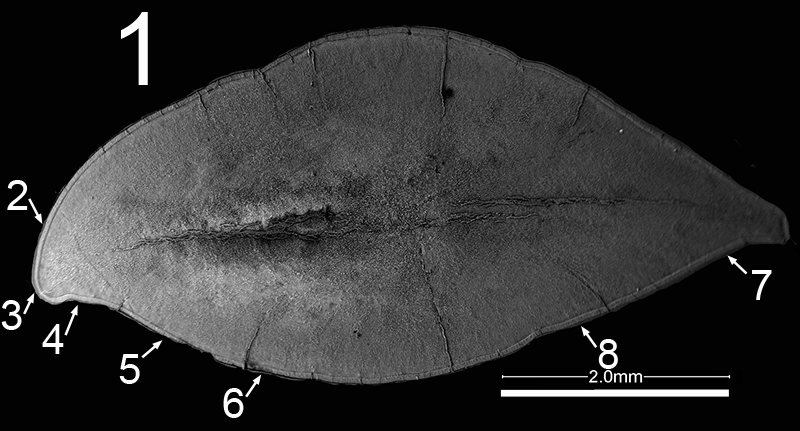
FIGURE 5.2. Original micrograph showing tooth enamel microstructure of maxillary tooth of Revueltosaurus callenderi (P-33798) in transverse section on labial side; EDJ to left.
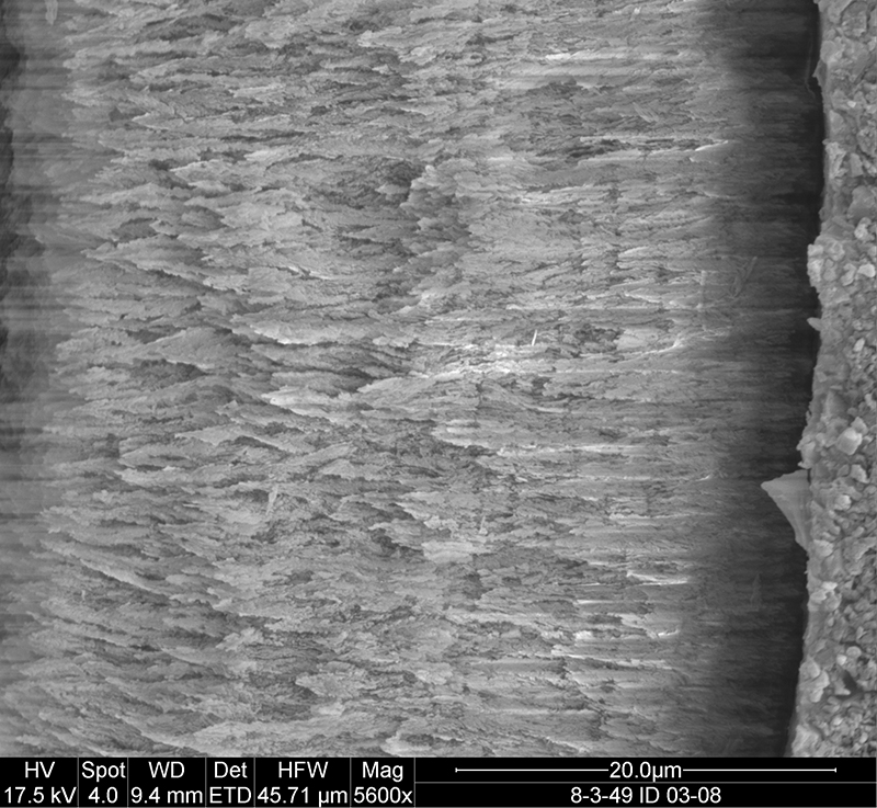
FIGURE 5.3. Original micrograph showing tooth enamel microstructure of maxillary tooth of Revueltosaurus callenderi (P-33798) in transverse section across one denticle; EDJ to right.
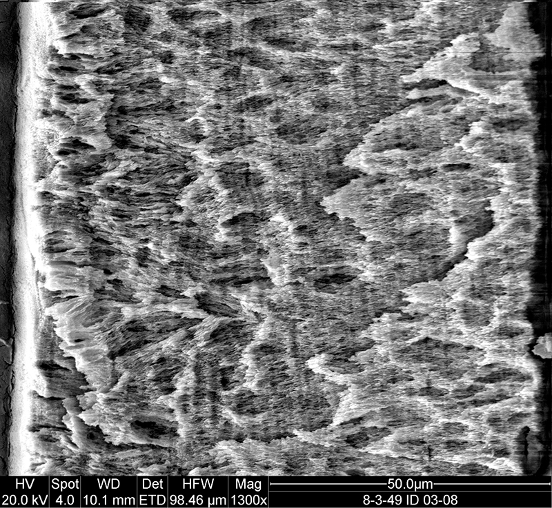
FIGURE 5.4. Original micrograph showing tooth enamel microstructure of maxillary tooth of Revueltosaurus callenderi (P-33798) in transverse section on lingual side; EDJ to left.
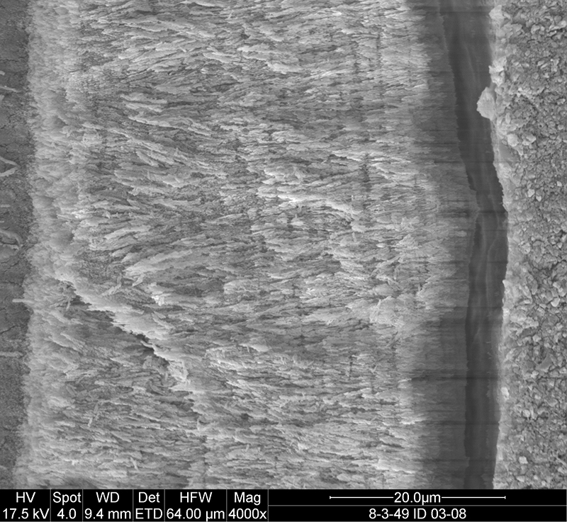
FIGURE 5.5. Original micrograph showing tooth enamel microstructure of maxillary tooth of Revueltosaurus callenderi (P-33798) in transverse section on lingual side; EDJ to left.
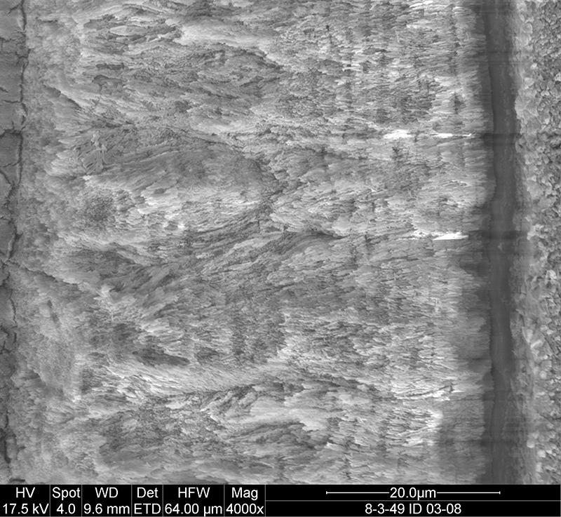
FIGURE 5.6. Original micrograph showing tooth enamel microstructure of maxillary tooth of Revueltosaurus callenderi (P-33798) in transverse section on lingual side; EDJ to left.
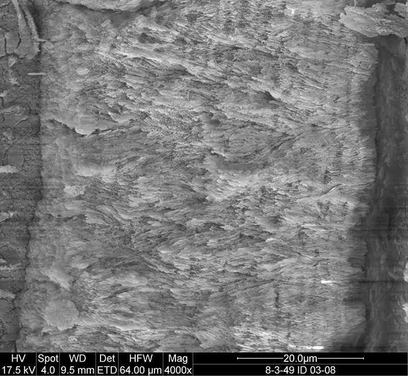
FIGURE 5.7. Original micrograph showing tooth enamel microstructure of maxillary tooth of Revueltosaurus callenderi (P-33798) in transverse section on lingual side; EDJ to left.
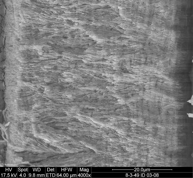
FIGURE 5.8. Original micrograph showing tooth enamel microstructure of maxillary tooth of Revueltosaurus callenderi (P-33798) in transverse section on lingual side; EDJ to left.
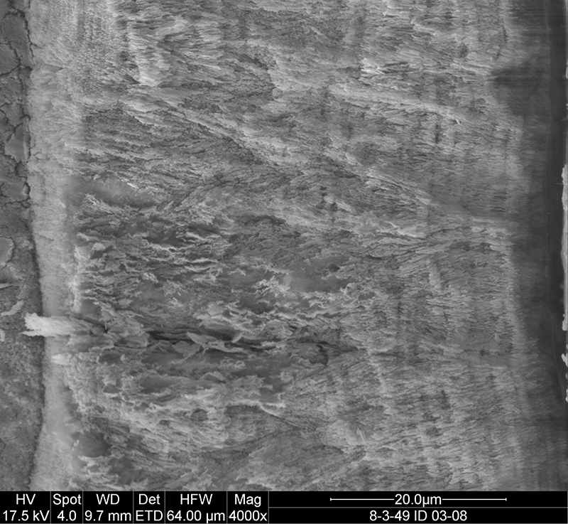
FIGURE 4. Scanning electron microscope images of NMMNH P-33799, Revueltosaurus callenderi premaxillary tooth showing variation in enamel microstructure in longitudinal section. 1, overview of tooth indicating approximate place where measurements and micrographs shown in this figure were taken; 2-8, close-up views showing enamel thickness variation, with enamel-dentine junction (EDJ) oriented relative to overview in (1) and outer enamel surface (OES) away from the same overview. White scale bars = 20 µm except for 1 (5 mm) and 4 (50 µm). Numbers in image are linked to further enlargements.
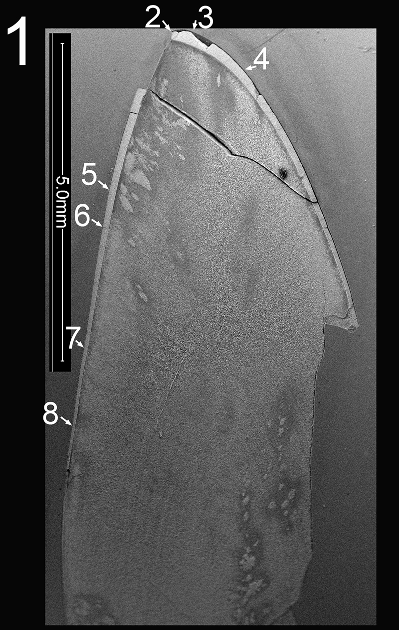
FIGURE 4.2. Original micrograph showing tooth enamel microstructure of premaxillary tooth of Revueltosaurus callenderi (P-33799) in longitudinal section at the tip of the crown; EDJ oriented to left of picture.
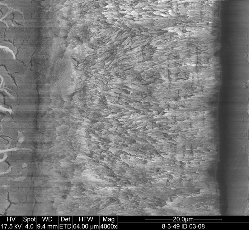
FIGURE 4.3. Original micrograph showing tooth enamel microstructure of premaxillary tooth of Revueltosaurus callenderi (P-33799) in longitudinal section near the tip of the crown; EDJ oriented to left of picture.
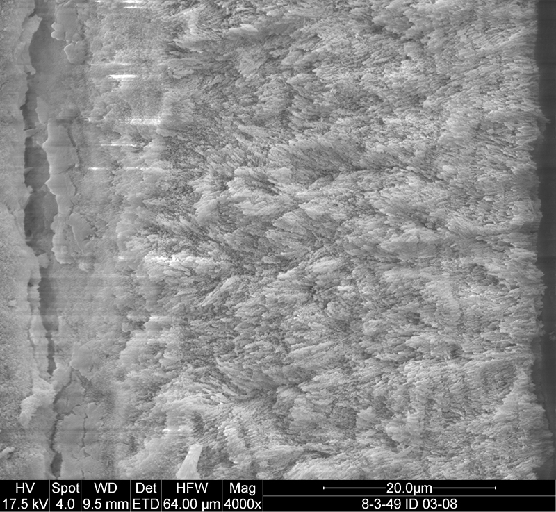
FIGURE 4.4. Original micrograph showing tooth enamel microstructure of premaxillary tooth of Revueltosaurus callenderi (P-33799) in longitudinal section high on the crown; EDJ oriented to left of picture.
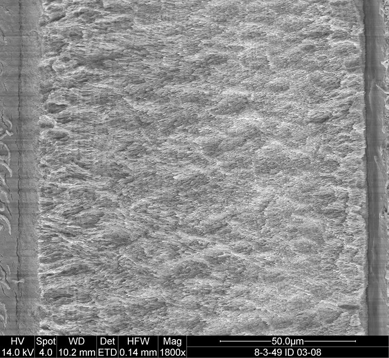
FIGURE 4.5. Original micrograph showing tooth enamel microstructure of premaxillary tooth of Revueltosaurus callenderi (P-33799) in longitudinal section high on the crown; EDJ oriented to left of picture.
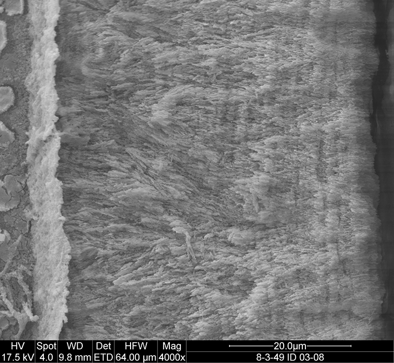
FIGURE 4.6. Original micrograph showing tooth enamel microstructure of premaxillary tooth of Revueltosaurus callenderi (P-33799) in longitudinal section high on the crown; EDJ oriented to right of picture.
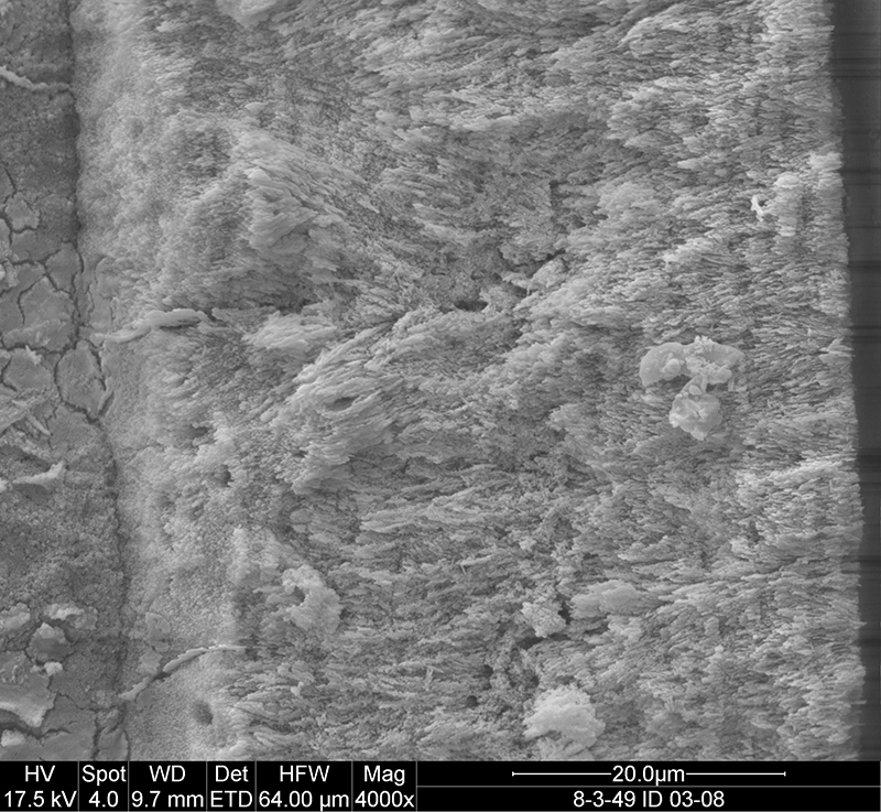
FIGURE 4.7. Original micrograph showing tooth enamel microstructure of premaxillary tooth of Revueltosaurus callenderi (P-33799) in longitudinal section low on the tooth; EDJ oriented to left of picture.
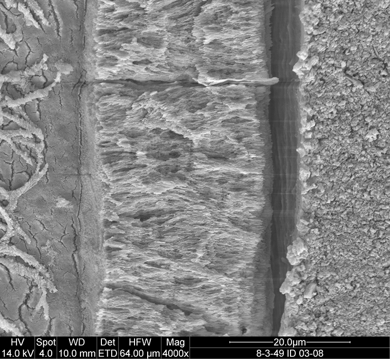
FIGURE 4.8. Original micrograph showing tooth enamel microstructure of premaxillary tooth of Revueltosaurus callenderi (P-33799) in longitudinal section near the middle of the tooth; EDJ oriented to left of picture.
Andrew B. Heckert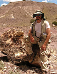 Department of Geology
Appalachian State University
ASU Box 32067
Boone, North Carolina 28608-2067
U.S.A.
website
Andrew Heckert earned a B.S. in Geology summa cum laude from Denison University in 1993 before earning an M.S. (1997) and Ph.D. (2001) from the Department of Earth & Planetary Sciences at the University of New Mexico. Subsequent to this he worked as the Geoscience Collections Manager at the New Mexico Museum of Natural History (2002-2005) before taking a post at Appalachian State University, where he is an associate professor in geology and director of the McKinney Geology Teaching Museum.
Heckert's research interests revolve around Late Triassic stratigraphic, biostratigraphic, and paleontologic issues, focusing primarily on microvertebrates, but he enjoys ranging up and down the section, and has collected vertebrates ranging in age from Devonian to Pleistocene, conducting field work across the American West and now in his new home state of North Carolina.
Photo by Alan Erickson.
![]()
Jessica A. Miller-Camp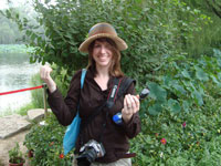 Department of Geology
Appalachian State University
ASU Box 32067
Boone, North Carolina 28608-2067, U.S.A.
currently: Department of Geoscience
121 Trowbridge Hall
University of Iowa
Iowa City, Iowa, 52242
U.S.A.
Jessica Miller-Camp earned a B.S. in Geology from Appalachian State University in 2007 where she studied the tooth enamel microstructure of several archosaurs. Since then she earned an M.S. in Geoscience at the University of Iowa (2010) for a geometric morphometrics study of the dicynodont Lystrosaurus. She is currently enrolled as a Ph.D. student at the University of Iowa undertaking a dissertation on the phylogenetics, taxonomy, biogeography, and morphology of Alligatorinae.
TABLE 1. Comparison of tooth enamel thickness and enamel microstructural features in different archosauromorphs compiled from the literature.
Tooth Enamel Microstructure
| Higher Taxon | Taxon | Sampler | minimum thickness |
maximum thickness |
Parallel | Columnar | BUL | LIG | Wavy | Tubules | Comments |
|
Archosauromorpha |
Trilophosaurus |
Sander (1999) |
20 |
20 |
|
|
x |
x |
|
|
|
|
Crurotarsi |
Phytosauridae (Dockum) |
Sander (1999) |
20 |
20 |
x |
|
|
x |
|
|
I and III |
|
|
Phytosauridae (Dockum) |
Sander (1999) |
|
150 |
x |
x |
x |
x |
|
|
II |
|
|
Phytosauridae (Hallau) |
Sander (1999) |
|
60 |
|
x |
|
|
|
|
|
|
|
Rauisuchidae |
Sander (1999) |
60 |
100 |
x |
x |
x |
x |
|
|
95% columnar; outer is parallel; rare LIG |
|
Mesosuchia |
Machimosaurus hugi |
Sander (1999) |
|
350 |
x |
x |
x |
|
|
|
LIG rare |
|
Crocodylia: Alligatoridae |
Allognathosuchus sp. |
Sander (1999) |
|
300 |
x |
x |
|
x |
|
|
up to 50/50 columnar/parallel; sometimes 95/5; LIG only in parallel |
|
|
Alligator mississippiensis |
Sander (1999) |
|
1000 |
x |
x |
x |
x |
|
|
Mostly columnar; LIG in columnar enamel |
|
Crocodylia: Crocodylidae |
Deinosuchus riograndensis |
Sander (1999) |
<150 |
525 |
|
x |
x |
|
|
|
40% columnar; 55% microunit; only specimen w/described microunits |
|
|
Asiatosuchus |
Sander (1999) |
|
500 |
|
x |
x |
|
|
|
compound unit enamel/weakly columnar |
|
|
Pristichampsa |
Sander (1999) |
20 |
50 |
x |
x |
|
x |
|
|
LIG best in the carinae |
|
|
Eusuchia indet |
Sander (1999) |
50 |
125 |
x |
x |
|
x |
|
|
Columnar yields to parallel |
|
Dinosauria:Saurischia: Theropoda |
|||||||||||
|
|
Basal Theropoda indet. |
Hwang (2009) |
|
|
x |
x |
x |
|
|
x |
100 µm average thickness; Kayenta Fm "carnosaur" in UCMP collections |
|
D:S:T:Ceratosauria |
Coelophysis bauri |
Hwang (2005, 2011) |
10 |
10 |
x |
|
|
x |
|
|
C. bauri of Hwang (2005) a distinct taxon (Hwang, 2011, p. 192) |
|
|
Ceratosaurus nasicornis |
Hwang (2011) |
20 |
70 |
x |
x |
x |
x |
|
x |
120 µm average thickness; varies wildly |
|
|
Majungasaurus sp. |
Hwang (2011) |
<50 |
>90 |
x |
x |
x |
x |
|
x |
|
|
D:S:T:Allosauroidea |
Allosaurus fragilis |
Hwang (2011) |
20 |
>85 |
x |
x |
x |
x |
|
|
|
|
|
cf. Allosaurus |
Sander (1999) |
10 |
15 |
x |
|
|
|
|
|
Not even LIG |
|
|
Charcharodontosaurus |
Buffetaut et al. (1986) |
|
|
|
x |
|
|
|
|
called "prisms" in published description |
|
|
Spinosaurus |
Buffetaut et al. (1986) |
|
|
|
x |
|
|
|
|
called "prisms" in published description |
|
|
"Carnosaur" |
Buffetaut et al. (1986) |
|
|
x |
? |
x |
|
|
|
|
|
D:S:T: Coelurosauria |
|
|
|
|
|
x |
|
|
|
|
|
|
D:S:T:C: Tyrannosauridae |
Tyrannosauridae indet. (TX) |
Sander (1999) |
150 |
200 |
x |
x |
|
|
|
|
Thickest at base of carina; parallel only at the outer edge, great columnar |
|
|
Tyrannosauridae indet. (MT) |
Sander (1999) |
|
120 |
x |
x |
x |
|
|
|
|
|
|
Tyrannosauridae indet. |
Hwang (2005, 2009) |
|
|
x |
x |
x |
|
|
x |
115 µm average thickness |
|
|
Tyrannosauridae indet. (="Nanotyrannus") |
Hwang (2009) |
|
|
x |
x |
|
|
|
x |
60µm average thickness; juvenile tyrannosaurid assigned to "Nanotyrannus" in UWGM collections |
|
|
Daspletosaurus torosus |
Hwang (2011) |
|
|
|
x |
|
x |
|
x |
80 µm average thickness |
|
|
cf. Gorgosaurus sp. |
Hwang (2009) |
|
|
x |
x |
x |
|
|
x |
80 µm average thickness |
|
|
Gorgosaurus libratus |
Hwang (2011) |
|
|
x |
x |
x |
x |
|
x |
|
|
|
Albertosaurus sarcophagus |
Hwang (2005) |
60 |
180 |
x |
x |
|
|
|
|
|
|
|
Albertosaurus sp. |
Stokosa (2005) |
100 |
120 |
x |
x |
|
|
|
x |
|
|
|
?Albertosaurus gen. A. indet. |
Stokosa (2005) |
40 |
55 |
x |
x |
|
x |
|
|
SDSM 12737; tip of tooth |
|
|
?Albertosaurus gen. A. indet. |
Stokosa (2005) |
180 |
200 |
x |
x |
|
x |
|
|
SDSM 15143 |
|
|
?Albertosaurus gen. A. indet. |
Stokosa (2005) |
95 |
100 |
x |
x |
|
x |
|
|
SDSM 64351 |
|
|
Tyrannosaurus sp. |
Hwang (2011) |
|
|
|
x |
|
|
|
x |
200 µm average thickness |
|
|
cf. Tyrannosaurus rex |
Stokosa (2005) |
45 |
50 |
x |
x |
|
x |
|
x |
SDSM 15135; Poorly developed columnar |
|
|
cf. Tyrannosaurus rex |
Stokosa (2005) |
60 |
75 |
|
|
|
|
|
|
SDSM 64287 |
|
|
cf. Tyrannosaurus rex |
Stokosa (2005) |
80 |
90 |
|
|
|
|
|
|
SDSM 15115 |
|
|
Tarbosaurus |
Dauphin et al. (1989) |
|
|
|
x |
|
|
|
|
intrepreted from pl. 2, fig. 4 |
|
|
Tarbosaurus |
Hwang (2005) |
|
300 |
x |
x |
|
|
|
|
|
|
D:S:T:C:incertae sedis |
Richardoestesia cf. R. gilmorei |
Stokosa (2005) |
10 |
10 |
x |
|
|
|
|
|
|
|
|
Richardoestesia gilmorei |
Hwang (2011) |
~10 |
~10 |
x |
|
x |
x |
|
x |
10 µm average thickness |
|
|
Richardoestesia isosceles |
Hwang (2011) |
~13 |
~13 |
x |
|
x |
|
|
x |
13 µm average thickness |
|
|
Richardoestesia sp. |
Stokosa (2005) |
10 |
15 |
x |
|
|
|
|
|
|
|
D:S:T:C:Maniraptora |
Troodontid indet. A |
Hwang (2005) |
|
60 |
x |
|
|
|
|
|
|
|
|
Troodontid indet. B |
Hwang (2005) |
|
|
x |
x |
|
|
|
|
|
|
|
Troodontid n. gen. et. Sp. |
Hwang (2005) |
|
30 |
x |
|
|
x |
|
|
|
|
|
Troodon sp. |
Sander (1999) |
|
20 |
x |
x |
x |
|
|
|
Columnar units small (2µm) and weak, seem to arise from BUL (Sander, p. 65) |
|
|
Troodon sp. |
Hwang (2009, 2011) |
|
|
x |
|
x |
|
|
|
20µm average thickness; parallel crystallites |
|
|
Troodon sp. cf. T. formosus |
Stokosa (2005) |
10 |
15 |
x |
|
|
|
|
|
|
|
|
Paronychodon cf. P. lacustris |
Stokosa (2005) |
0 |
15 |
x |
|
|
|
|
|
|
|
|
Paronychodon lacustris |
Sander (1999) |
|
20 |
x |
|
|
x |
|
|
LIG are few and weak |
|
|
Paronychodon (Troodontid) |
Hwang (2005) |
|
|
x |
|
|
x |
|
|
|
|
|
Byronosaurus jaffei |
Hwang (2005) |
|
13 |
x |
|
|
x |
|
|
|
|
|
Paronychodon (Dromaeosaurid) |
Hwang (2005) |
|
|
|
|
|
|
|
|
|
|
|
Velociraptor |
Dauphin et al. (1989) |
|
|
x |
|
|
|
|
|
interpreted from pl. 1, figs. 4-5 |
|
|
Velociraptor mongoliensis |
Hwang (2005) |
|
24 |
x |
|
x |
|
|
|
|
|
|
Bambiraptor feinbergi |
Hwang (2005) |
|
|
x |
|
|
x? |
|
|
No LIG in Hwang (2005), but faint in Hwang (writ. Comm.) |
|
|
Dromaeosauridae indet. |
Hwang (2005) |
|
55 |
x |
|
x |
|
|
|
|
|
|
Deinonychus antirrhopus |
Hwang (2011) |
17 |
30+ |
x |
|
x |
x |
|
x |
BUL poorly developed; LIG faint |
|
|
Saurornitholestes sp. |
Hwang (2011) |
7 |
20+ |
x |
|
x |
|
|
x |
BUL half of enamel thickness |
|
|
Dromaemosaurus sp. |
Hwang (2011) |
25 |
35 |
x |
|
x |
x |
|
x |
BUL half of enamel thickness, but not well-developed |
|
|
Dromaeosaurus sp. cf. D. albertensis |
Stokosa (2005) |
40 |
45 |
x |
x |
|
x |
|
|
Columnar at EDJ, divergent parallel more at OES |
|
D:S:T:Avialae |
Indeterminate Avialan A |
Hwang (2011) |
|
|
x |
|
x |
|
|
x |
20 µm average thickness |
|
|
Indeterminate Avialan B |
Hwang (2011) |
|
|
x |
|
x |
|
|
x |
16 µm average thickness |
|
Sauropodomorpha |
|||||||||||
|
|
Plateosaurus engelhardti |
Sander (1999) |
10 |
40 |
x |
? |
|
x |
|
|
Columnar is very poorly developed; probably more divergent parallel |
|
|
cf. Diplodocus |
Sander (1999) |
|
150 |
|
|
x |
|
|
|
Pseudo-wavy (Sander, p. 68) |
|
|
Diplodocus longus |
Hwang (2011) |
440 |
490 |
|
x |
x |
x |
|
x |
465 µm average thickness |
|
|
Camarasaurus sp. |
Hwang (2011) |
700 |
1000+ |
x |
x |
x |
x |
|
|
850 µm average thickness |
|
|
Titanosauridae indet. |
Hwang (2011) |
|
|
x |
x |
x |
x |
|
x |
170 µm average thickness |
|
Dinosauria:Ornithischia |
|||||||||||
|
D:O:Stegosauria |
Stegosaurus sp. |
Hwang (2011) |
|
|
x |
|
x |
x |
|
|
30-40 µm average thickness |
|
D:O:Ankylosauria |
Ankylosauria indet. |
Sander (1999) |
|
60 |
|
x |
x |
|
|
|
|
|
|
Ankylosaurus magniventris |
Hwang (2005) |
|
60 |
x |
x |
x |
x |
|
|
|
|
|
Edmontonia rugosidens |
Hwang (2005) |
|
100+ |
x |
x |
x |
x |
|
|
|
|
|
Sauropelta edwardsi |
Hwang (2005) |
|
105 |
x |
x |
x |
x |
|
|
|
|
|
Euplocephalus |
Hwang (2009, 2011) |
35 |
65 |
x |
x |
x |
x |
|
x |
55 µm average thickness; originally identified as a pachycephalosaurid |
|
Dinosauria:Ornithischia:Euornithopoda |
|||||||||||
|
D:O:E: Hypsilophodontidae |
Thescelosaurus sp. |
Sander (1999) |
14 |
140 |
x |
|
x |
|
|
|
BUL thin |
|
|
Thescelosaurus sp. |
Hwang (2011) |
20 |
90 |
x |
x |
x |
|
|
x |
Hwang (2011) suspects Sander's (1999) specimen is not Thescelosaurus |
|
D:O:E:Dryomorpha |
Dryosaurus altus |
Hwang (2011) |
|
|
|
|
x |
x |
x |
x |
55-65 µm average thickness |
|
|
Camptosaurus dispar |
Hwang (2011) |
|
|
|
|
x |
|
x |
x |
|
|
D:O:E: Iguanodontidae |
Iguanodon sp. |
Sander (1999) |
100 |
150 |
|
|
x |
|
x |
|
Inner and outer wavy enamel |
|
|
Tenontosaurus tilleti |
Hwang (2005) |
|
100+ |
x |
x |
x |
|
|
x |
|
|
D:O:E: Hadrosauridae |
Hadrosauridae indet. |
Sander (1999) |
160 |
210 |
|
|
x |
|
x |
|
|
|
|
Hadrosaurinae indet. |
Hwang (2009) |
|
|
|
x |
|
|
|
x |
|
|
|
Anatosaurus sp. |
Sander (1999) |
100 |
100+ |
|
|
x |
|
x |
x |
BUL inverted |
|
|
Saurolophus sp. |
Hwang (2011) |
|
165+ |
|
|
x |
|
x |
|
115 µm average thickness |
|
|
Gilmoreosaurus mongoliensis |
Hwang (2005) |
|
|
|
|
x |
|
x |
|
|
|
|
Bactrosaurus johnsoni |
Hwang (2005) |
|
|
|
|
x |
|
x |
|
|
|
|
Kritosaurus navajovius |
Hwang (2005) |
|
|
|
|
x |
|
x |
|
|
|
|
Hypacrosaurus altispinus |
Hwang (2005) |
|
|
|
|
x |
|
x |
|
|
|
|
Corythosaurus casuaris |
Hwang (2005) |
|
|
|
|
x |
|
x |
|
|
|
|
Prosaurolophus maximus |
Hwang (2011) |
|
~200 |
|
|
x |
|
x |
|
135 µm average thickness |
|
Dinosauria:Ornithischia: Ceratopsia |
|||||||||||
|
|
Neoceratopsia indet. |
Hwang (2009) |
|
|
|
x |
|
|
|
|
Average 285 µm; originally assigned to Thescelosaurus sp. |
|
|
Psittacosaurus sp. |
Hwang (2005) |
|
|
|
x |
x |
|
|
? |
enamel voids |
|
|
Protoceratops |
Dauphin et al. (1988) |
|
|
x |
x |
x |
|
|
|
interpreted from pl. 1, figs. 7-8 (maxilla) and 12 (premaxilla) |
|
|
Leptoceratops gracilis |
Hwang (2005) |
|
420 |
|
x |
x |
|
|
x |
|
|
|
Protoceratops sp. |
Hwang (2005) |
|
120+ |
|
x |
x |
|
|
x |
|
|
D:O:C:Ceratopsidae |
Ceratopsidae indet. (Can) |
Sander (1999) |
150 |
|
|
x |
x |
x |
|
x |
|
|
|
Ceratopsidae indet. (WY) |
Sander (1999) |
|
|
|
|
|
|
|
|
|
|
|
Triceratops sp. |
Hwang (2005) |
|
325 |
|
x |
x |
|
|
x |
|
|
|
Centrosaurus apertus |
Hwang (2011) |
|
|
|
x |
x |
x |
|
x |
170-270 µm average thickness depending on position |
|
|
Pachyrhinosaurus canadensis |
Hwang (2011) |
|
|
|
x |
x |
x |
|
x |
170-270 µm average thickness depending on position |
|
Dinosauria:Ornithischia:Pachycephalosauridae |
|||||||||||
|
|
Pachycephalosauridae indet. A |
Hwang (2005) |
|
20 |
x |
|
|
|
|
|
anterior tooth, diverging parallel |
|
|
Pachycephalosauridae indet. B |
Hwang (2005) |
|
40 |
x |
x |
x |
|
|
|
posterior tooth, incipient columnar |
|
|
Pachycephalosauridae indet. C |
Hwang (2005) |
|
50 |
x |
x |
x |
|
|
|
posterior tooth, incipient columnar |
TABLE 2. Measurements of enamel thickness in the teeth of Revueltosaurus and Krzyzanowskisaurus sampled here.
|
Revueltosaurus callenderi enamel measurements from longitudinal section of NMMNH P-33798 |
|||
|
|
|
|
|
|
Figure |
Section |
Enamel thickness (µm) |
Measured at: |
|
3.2 |
Longitudinal |
4.87 |
end of [full width of] enamel (thinnest enamel) |
|
3.3 |
Longitudinal |
20.42 |
middle of frame |
|
3.4 |
Longitudinal |
36.23 |
middle of frame |
|
3.5 |
Longitudinal |
68.48 |
middle of frame |
|
3.6 |
Longitudinal |
115.56 |
top of frame (thickest enamel) |
|
3g |
Longitudinal |
114.88 |
left of frame (thickest enamel) |
|
3h |
Longitudinal |
152.13 |
bottom of frame (thickest enamel) |
|
3i |
Longitudinal |
103.34 |
middle of frame |
|
4.2 |
Longitudinal |
46.88 |
middle of frame |
|
4.3 |
Longitudinal |
50.08 |
middle of frame |
|
4.4 |
Longitudinal |
52.43 |
middle of frame |
|
4.5 |
Longitudinal |
53.93 |
middle of frame |
|
4.6 |
Longitudinal |
15.47 |
middle of frame |
|
4.7 |
Longitudinal |
31.36 |
middle of frame |
|
4.8 |
Longitudinal |
127.6 |
middle of frame |
|
"Tooth has total crown height (TCH) of 13.1 mm, total crown length (TCL) 8.1 mm" |
|||
|
|
|
|
|
|
Revueltosaurus callenderi enamel measurements from transverse section of NMMNH P-33797 |
|||
|
Figure |
Section |
Enamel thickness (µm) |
Measured at: |
|
3.11 |
Transverse |
51.5 |
on denticle |
|
3.12 |
Transverse |
53.41 |
middle of frame |
|
3.13 |
Transverse |
55.15 |
middle of frame |
|
3.14 |
Transverse |
59.66 |
middle of frame |
|
5.1 |
Transverse |
42.8 |
left of frame (EDJ visible) |
|
5.3 |
Transverse |
92.47 |
middle of frame |
|
5.4 |
Transverse |
50.77 |
middle of frame |
|
5.5 |
Transverse |
54.48 |
middle of frame |
|
5.6 |
Transverse |
53.32 |
middle of frame |
|
5.7 |
Transverse |
62.79 |
middle of frame |
|
5.8 |
Transverse |
60.04 |
middle of frame |
|
TCH: 6 mm+ (broken) |
|
TCL: ~6 mm |
|
|
|
|
|
|
|
Krzyzanowskisaurus hunti enamel measurements from longitudinal section of UCMP 165213 |
|||
|
Figure |
Section |
Enamel thickness (µm) |
Measured at: |
|
6.2 |
Longitudinal |
112.8 |
middle of frame |
|
6.3 |
Longitudinal |
138.89 |
left of frame (EDJ visible) |
|
6.4 |
Longitudinal |
151.25 |
right of frame (unbroken outer edge of enamel) |
|
TCH: ~6 mm (incomplete) |
|
TCL: 6 mm |
|
|
|
|
|
|
|
Krzyzanowskisaurus hunti enamel measurements from transverse sectionof UCMP 165211 |
|||
|
|
|
|
|
|
Figure |
Section |
Enamel thickness (µm) |
Measured at: |
|
7.1 |
Transverse |
55.38 |
middle of bend |
|
7.2 |
Transverse |
64.34 |
middle of frame |
|
7.3 |
Transverse |
50.49 |
middle of frame |
|
7.4 |
Transverse |
65.54 |
middle of frame |
|
7.5 |
Transverse |
63.38 |
middle of frame |
|
7.6 |
Transverse |
43.47 |
middle of frame |
|
7.7 |
Transverse |
61.82 |
middle of frame |
|
TCH: ~10.4 mm |
TCL: ~7.4 mm |
|
|
FIGURE 1. Location map and stratigraphic section showing the geographic and stratigraphic distribution of Revueltosaurus and Krzyzanowskisaurus. This includes the localites of the Revueltosaurus (NMMNH locality 1) and Krzyzanowskisaurus (UCMP locality 7307) teeth described here. Tooth illustrations after Heckert (2002, 2005). LVF = land vertebrate faunachron (following Lucas et al., 2007); L-1171 = type location of Krzyzanowskisaurus hunti; PFV = locality yielding abundant Revueltosaurus fossils in the Petrified Forest National Park.
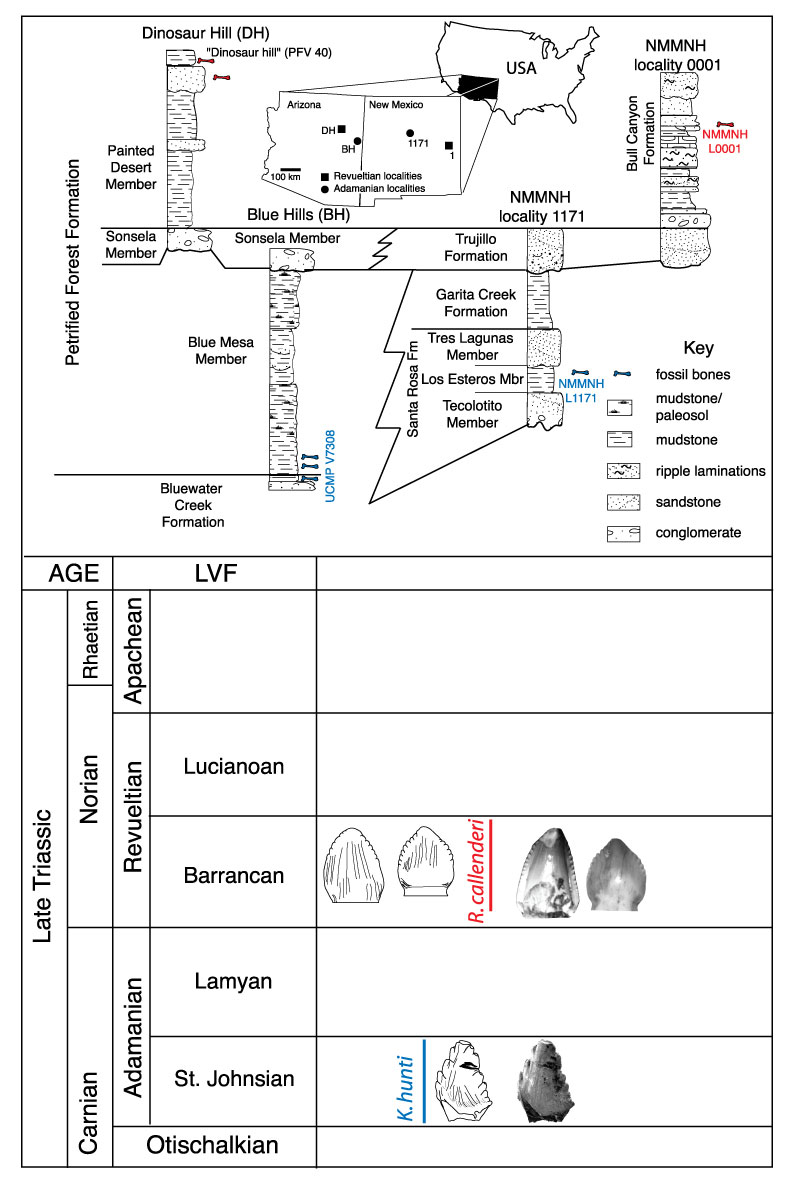
FIGURE 2. Generalized archosaurian phylogeny showing the relationships of taxa sampled by Sander (1999) and Hwang (2005) (in black) as well as Revueltosaurus (red) and Krzyzanowskisaurus (blue). Dashed lines demonstrate the two hypothesized positions of Krzyzanowskisaurus, either as a crurotarsan closely allied to Revueltosaurus (e.g., Parker et al., 2005; Irmis et al., 2007) or a basal ornithischian (Heckert, 2002, 2005).
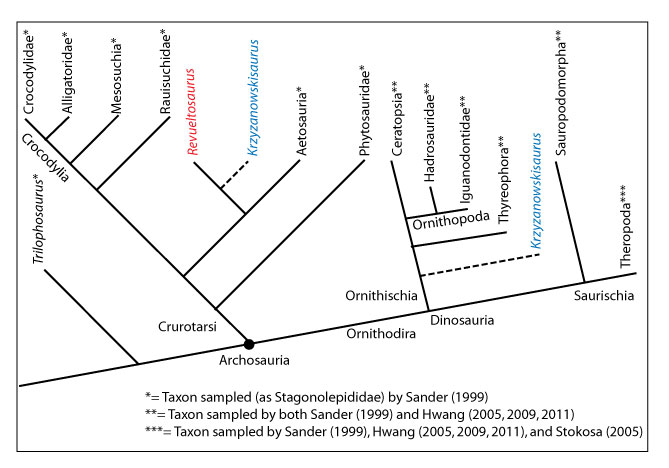
FIGURE 3. Variation in enamel thickness in the teeth of Revueltosaurus callenderi. 1-9, premaxillary tooth (P-33799); 10-14, maxillary tooth (P-33798). 1, overview of premaxillary tooth indicating approximate place where measurements and micrographs shown in this figure and Figure 4 were taken; 2-9, close-up views showing enamel thickness variation, with enamel-dentine junction (EDJ) oriented relative to overview in (1); and 10, overview of maxillary tooth section, indicating approximate place where measurements and micrographs shown in this figure and Figure 5 were taken; 11-14, close-up views showing enamel thickness variation, with enamel-dentine junction (EDJ) oriented relative to overview in (10). Scale bars equal 100 µm except for 1 (5 mm), 2 (10 µm), and 10 (2 mm). Click to see image close-ups.
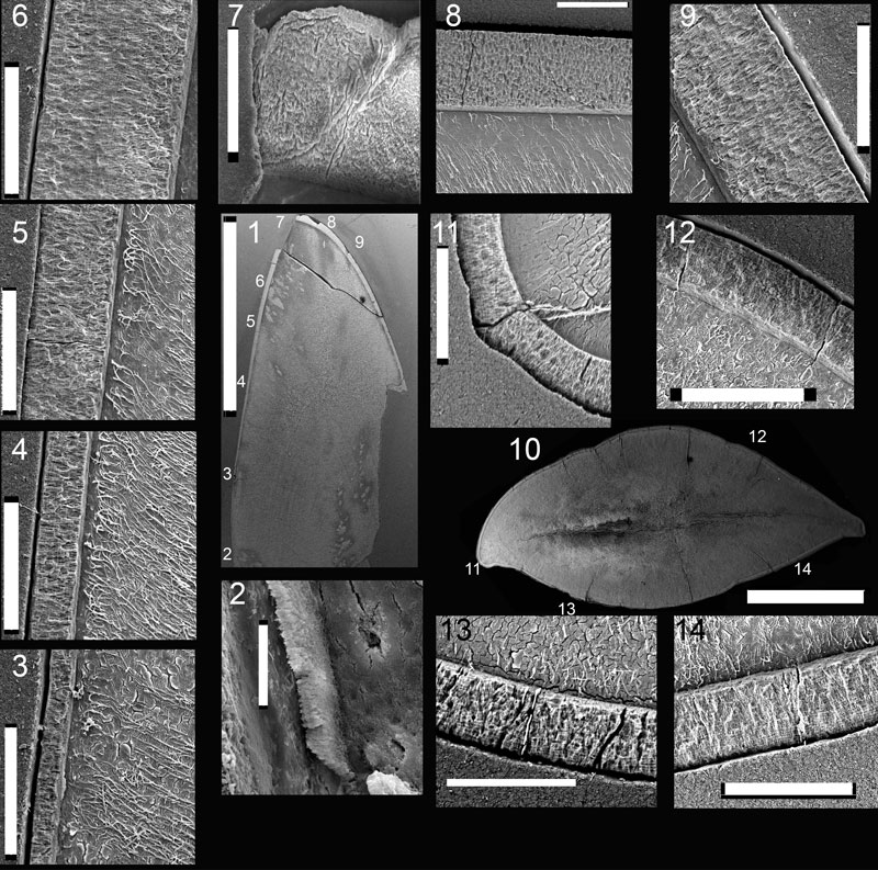
FIGURE 4. Scanning electron microscope images of NMMNH P-33799, Revueltosaurus callenderi premaxillary tooth showing variation in enamel microstructure in longitudinal section. 1, overview of tooth indicating approximate place where measurements and micrographs shown in this figure were taken; 2-8, close-up views showing enamel thickness variation, with enamel-dentine junction (EDJ) to the left and the outer enamel surface (OES) to the right. White scale bars = 20 µm except for 1 (5 mm) and 4 (50 µm). Click to see image close-ups.
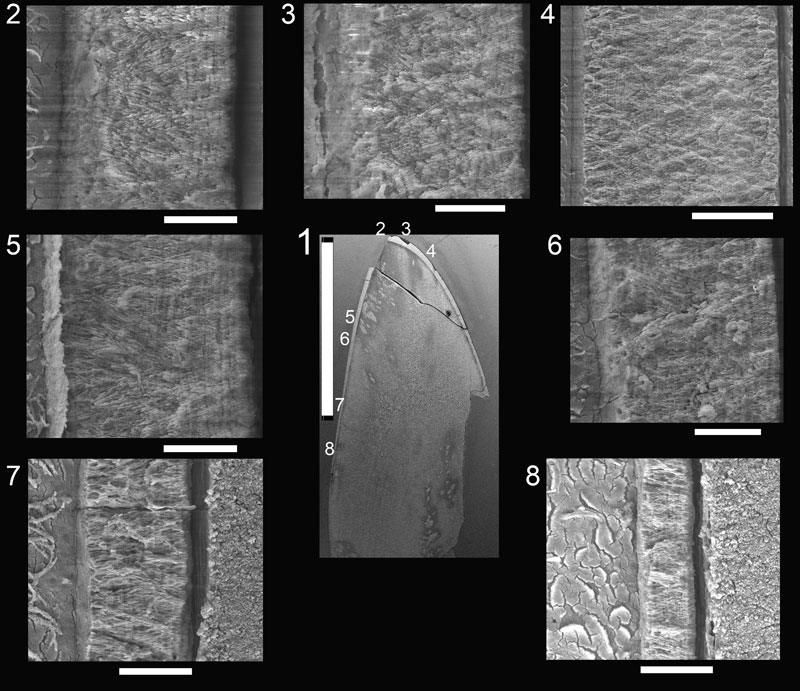
FIGURE 5. Scanning electron microscope images of NMMNH P-33798, Revueltosaurus callenderi maxillary tooth enamel microstructure in transverse section. 1, overview of tooth indicating approximate place where measurements and micrographs shown in this figure were taken; 2-8, close-up views showing enamel thickness variation, with EDJ oriented relative to overview in (1) and OES away from the same overview. Scale bars equal 20 µm except 1 (2 mm), 3 (50 µm). Click to see image close-ups.

FIGURE 6. Scanning electron microscope images of UCMP 165213, Krzyzanowskisaurus hunti tooth enamel microstructure in longitudinal section. 1, overview of tooth indicating approximate place where measurements and micrographs shown in this figure were taken; 2-4, close-up views showing enamel thickness variation, with EDJ oriented relative to overview in (1) and OES away from the same overview. Scale bars equal 2 mm (1), 50 µm (2-3), and 100 µm (4). Click to see image close-ups.
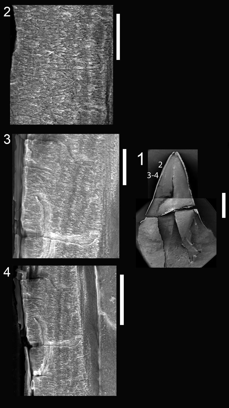
FIGURE 7. Scanning electron microscope images of UCMP 165211, Krzyzanowskisaurus hunti premaxillary tooth enamel microstructure in transverse section. 1, overview of tooth indicating approximate place where measurements and micrographs shown in this figure were taken; 2-7, close-up views showing enamel thickness variation, with EDJ oriented relative to overview in (1) and OES away from the same overview. Scale bar equals 20 µm except 1 (2 mm). Click to see image close-ups.
Tooth enamel microstructure of Revueltosaurus and Krzyzanowskisaurus (Reptilia:Archosauria) from the Upper Triassic Chinle Group, USA: Implications for function, growth, and phylogeny
Andrew B. Heckert and Jessica A. Miller-Camp
Plain Language Abstract
This is a study of microscopic tooth structures in two extinct Late Triassic (~225-210 million years old) reptiles. Revueltosaurus is thought to be more closely related to crocodiles and Krzyzanowskisaurus to dinosaurs and therefore birds. Using a scanning electron microscope (SEM) reveals extremely fine details of the enamel, or hardest and outermost, portion of the tooth. Similarities in the anatomical details of tooth enamel may mean that taxa are closely related, but could also simply imply that they used their teeth in similar ways and therefore may have had similar diets. Particularly interesting features of the enamel of both Revueltosaurus and Krzyzanowskisaurus are that the enamel is relatively thick, especially given the relatively small size of their teeth, and that they possess discontinuities (lines of incremental growth—similar in concept to tree rings) that may indicate that, while small, they maintained individual teeth in their dentition for long periods of time, a trait that is somewhat unusual among reptiles. The enamel of Krzyzanowskisaurus and Revueltosaurus is similar enough that they are probably closely related, but also displays evolutionary convergence with some dinosaurs.
Resumen en Español
Microestructura del esmalte dental de Revueltosaurus y Krzyzanowskisaurus (Reptilia, Archosauria) del Grupo Chinle (Triásico superior, EE.UU.): implicaciones para el análisis funcional, del crecimiento y de la filogenia
La microestructura del esmalte dental puede aportar importante información filogenética, ontogenética y funcional en los amniotas. En este trabajo se describe por primera vez la microestructura del esmalte dental de dos taxones del Triásico superior, el crurotarso Revueltosaurus callenderi Hunt y el generalmente aceptado como ornitisquio Krzyzanowskisaurus hunti (Heckert), que algunos autores consideran estrechamente emparentados. Para contrastar las hipótesis de que el grosor del esmalte depende de la función y/o de la filogenia, hemos analizado el esmalte de cada una de las especies a varias escalas, midiendo el grosor del esmalte y examinando las características microestructurales del esmalte tanto longitudinal como transversalmente, utilizando técnicas previamente establecidas para facilitar las comparaciones. Ambos taxones poseen esmaltes gruesos (hasta ~150μm) en relación con su tamaño (<20 mm de altura de la corona). El grosor del esmalte de R. callenderi varía entre ~5 y 152 μm en una sección longitudinal de un diente premaxilar y entre ~42 y 92 μm en una sección transversal de un diente maxilar/dentario. El espesor del esmalte de K. hunti varía entre ~18 y 155 μm en sección longitudinal y entre ~29 y 75 μm en sección transversal. Ambas especies muestran capas de la unidad basal bien desarrolladas y una microestructura columnar poco desarrollada. También son patentes en ambos taxones las líneas de crecimiento incremental, a lo largo de las cuales el esmalte columnar pasa gradualmente a esmalte paralelo. La microestructura de su esmalte es, por tanto, muy similar a la de varios taxones ornitisquios, especialmente los anquilosaurios, con los que muestran fuertes convergencias y también es comparable a la de los rauisuquios y tiranosáuridos. La combinación relativamente única de características microestructurales en el schmelzmuster de R. callenderi y K. hunti apoya la hipótesis de su parentesco cercano, aunque no excluye irrefutablemente la posibilidad de una posición taxonómica diferente para K. hunti, por lo que mantenemos una designación genérica separada.
PALABRAS CLAVE: arcosaurio; esmalte dental; microestructura; Triásico; Revueltosaurus; crurotarso
Traducción: Miguel Company
Résumé en Français
Microstructure de l'émail dentaire de Revueltosaurus et Krzyzanowskisaurus (Reptilia:Archosauria) du Trias supérieur du Groupe Chinle, USA : implications pour le fonctionnement, la croissance et la phylogénie.
La microstructure de l'émail des dents peut comporter des informations phyllogénétiques, ontogénétiques et fonctionnelles significatives chez les amniotes. Nous présentons ici les premières descriptions de la microstructure de l'émail dentaire de deux taxa du Trias supérieur, le crurotarsien Revueltosaurus callenderi Hunt et le supposé ornithischien Krzyzanowskisaurus hunti (Heckert), que certains considèrent comme de proches parents. Pour tester l'hypothèse que l'épaisseur de l'émail dépend du fonctionnement et/ou de la phylogénie, nous avons analysé l'émail de chacun des deux taxa à différentes échelles, mesurant l'épaisseur de l'émail et examinant les caractères microstructuraux au travers de sections transversales et longitudinales, utilisant des techniques établies pour faciliter les comparaisons. Les deux taxa possèdent un émail épais (jusqu'à ~150 µm) pour leur taille (< 20 mm de haute de couronne). L'émail chez R. callenderi varie de ~5 à 152 µm sur une section longitudinal d'une dent prémaxillaire, et de ~42 à 92 µm dans une section transverse de dent maxillaire/mandibulaire. L'épaisseur de l'émail de K. hunti était de ~18-155 µm longitudinalement et ~29-75 µm transversalement. Les deux avaient aussi les couches de l'unité de base (basal unit layers, BUL) bien développée, et des microstructures colonnaires faiblement développées. Des lignes de croissances bien développées (lines of incremental growth, LIG) sont présentes chez les deux taxa, au travers desquelles l'émail colonnaire évolue en émail cristallite parallèle. La microstructure de leur émail est donc grossièrement similaire à celles de plusieurs taxa ornithischiens, spécialement les ankylosaures, avec lesquels ils sont fortement convergents, et ressemble bien également aux rauisuchidés et tyrannosauridés. L'association relativement unique de caractéristiques microstructurale du schmelzmuster de R. callenderi et K. hunti supporte l'hypothèse qu'ils sont de proches parents, mais n'exclue pas définitivement une attribution taxinomique différent pour K. hunti, de fait nous le gardons sous une désignation générique séparée.
Mots clés : archosaure; émail dentaire; microstructure; Trias; Revueltosaurus; crurotarsien
Translator: Olivier Maridet
Deutsche Zusammenfassung
Zahnschmelz-Mikrostruktur von Revueltosaurus und Krzyzanowskisaurus (Reptilia:Archosauria) aus der obertriassischen Chinle Gruppe, USA: Auswirkungen auf Funktion, Wachstum und Phylogenie
Die Zahnschmelz-Mikrostruktur kann bei Amnioten maßgebliche Informationen zur Phylogenie, Ontogenese und Funktion liefern. Wir bieten die erste Beschreibung von Zahnschmelz-Mikrostruktur bei zwei spättriassischen Taxa, die einige als eng verwandt ansehen: der crurotarsale Revueltosaurus callenderi Hunt und der vermeintliche Ornitischier Krzyzanowskisaurus hunti (Heckert). Um die Hypothese zu testen, dass die Dicke des Zahnschmelzes der Funktion und/oder Phylogenie entspricht, haben wir den Schmelz der beiden in unterschiedlichen Maßstäben analysiert. Dabei haben wir die Zahnschmelzdicke gemessen, und mikrostrukturelle Eigenschaften sowohl über die Längs - als auch Querschnittsdicke untersucht. Um die Vergleiche zu vereinfache, benutzten wir vorher ermittelte Techniken.
Beide Taxa haben für ihre Größe (< 20 mm Kronenhöhe) einen dicken Schmelz (bis zu ~150 µm). Der Schmelz bei R. callenderi reichte von ~5-152 µm über einen Prämaxlliar-Zahn im Längsschnitt und ~42-92 µm bei einem Zahn im Maxillare/Dentale im Querschnitt. Der Schmelz von K. hunti hatte eine Dicke von ~18-155 µm im Längsschnitt und ~29-75 µm im Querschnitt. Beide hatten gut entwickelte Basal Unit Layers (BUL) und eine schwach entwickelte kolumnare Mikrostruktur. Die Schrittweise-Wachstumslinien (LIG) durch die der kolumnare Schmelz in den parallelen kristallinen Schmelz übergeht, sind bei beiden Taxa gut entwickelt. Ihr Schmelz ist daher grob dem einiger Ornitischier ähnlich, besonders Ankylosauriern, mit denen sie eine starke Konvergenz aufweisen und ebenso mit dem von Rauisuchiden und Tyrannosauriden. Die relativ einzigartige Kombination der mikrostrukturellen Charakteristika im Schmelzmuster von R. callenderi und K. hunti unterstützt die Hypothese einer engen Verwandtschaftsbeziehung. Damit ist jedoch eine unterschiedliche taxonomische Zuweisung von K. hunti nicht endgültig ausgeschossen, so dass wir die separate Gattungsbezeichnung beibehalten.
SCHLÜSSELWÖRTER: Archosaurier; Zahnschmelz; Mikrostruktur; Trias; Revueltosaurus; crurotarsal
Translators: Eva Gebauer
Arabic
Translator: Ashraf M.T. Elewa
-
-
-
Review: The Princeton Field Guide to Mesozoic Sea Reptiles
 The Princeton Field Guide to Mesozoic Sea Reptiles
Article number: 26.1.1R
April 2023

 Poster Winners 2024