

Article Search
Volume 27.1
January–April 2024
Full table of contents
ISSN: 1094-8074, web version;
1935-3952, print version
Recent Research Articles
See all articles in 27.1 January-April 2024
See all articles in 26.3 September-December 2023
See all articles in 26.2 May-August 2023
See all articles in 26.1 January-April 2023
FIGURE 1. General view of Cambrian echinoderm types of preservation. 1.1. Stereom microstructure of a Cambrian echinoderm preserved as iron-oxide replacement (after Clausen and Smith, 2008). 1.2. Stereom microstructure of a Cambrian echinoderm preserved as silica coating (after Clausen and Smith, 2008). 1.3. Stereom of a recent echinoderm. 1.4. Specimen of Protocicnctus mansillaensis preserved in recrystallized calcite, as specimens presented in this study (after Rahman and Zamora, 2009). 1.5. Latex cast of the edrioasteroid Aragocystites belli preserving superficial traces of stereom on plates around central mouth (after Zamora, 2013). 1.6. Same specimen preserved as a natural mould. 1.7. Isolated echinoderm element obtained after acid etching (image courtesy of Olaf Elicki).
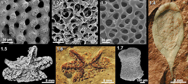
FIGURE 2. Microstructural organization and basic geochemical characteristics of the Middle Cambrian echinoderms: 2.1, Transverse section of the integument plate of Protocinctus in SEM; 2.2, transverse section of the integument plate of Protocinctus in SEM back-scattered electron (BSE) images; 2.3, geochemical analyses of the integument plate of Protocinctus (o) and the sediment (s); 2.4, CL-activated UV-VIS spectrum of the intense orange-red luminescent integument plate of Protocinctus showing Mn2+ emission maximum at ~620 nm; 2.5, Transverse section of the thecal plate of Stromatocystites in SEM; 2.6, transverse section of the thecal plate of Stromatocystites in BSE images; 2.7, geochemical analyses of the ossicle of thecal plate of Stromatocystites and the sediment; 2.8, CL-activated UVVIS spectrum of the intense orange-red luminescent thecal plate of Stromatocystites showing Mn2+ emission maximum at ~610 nm; 2.9, Axial section of the thecal plate of Dibrachicystidae in SEM; 2.10, transverse section of the thecal plate of Dibrachicystidae in BSE images; 2.11, geochemical analyses of the ossicle of thecal plate of Dibrachicystidae and the sediment; 2.12, CL-activated UV-VIS spectrum of the intense orange-red luminescent thecal plate of Dibrachicystidae showing Mn2+ emission maximum at ~610 nm. Scale bar equals 100µm.

FIGURE 3. Cathodoluminescence photomicrographs of the Middle Cambrian echinoderms showing orange-red luminescent stereom and pore system filled with non-luminescent likely ferroan calcite: 3.1.-3.3, 3.5, Integument plates of Protocinctus; 3.4, 3.6, marginal plates of Protocinctus; 3.7, ambulacral plate of Stromatocystites; 3.8, thecal plate of Stromatocystites; 3.9, ambulacral plate of Stromatocystites; 3.10, 3.11, thecal plate of Dibrachicystidae; 3.12, columnal? plate of Dibrachicystidae. Scale bar equals 100µm.
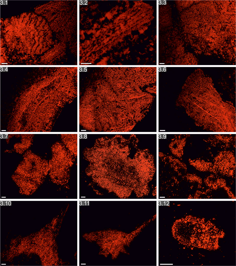
TABLE 1. List of some Cambrian echinoderm examples with preserved stereom microstructure.
|
Cambrian taxa and ossicle type |
Preservation style |
Stereom type |
Locality and age |
Reference |
|
Theca of Cymbionites craticula (Eocrinoidea) |
Silica |
Labyrinthic, galleried |
Australia |
Smith (1982) |
|
Pelmatozoan columnals (Blastozoa) |
Iron oxides |
Galleried |
Bornholm |
Berg-Madsen (1986) |
|
Thecal and spine-like elements of unknown echinoderms |
Phosphates |
Labyrinthic, galleried |
Newfoundland |
Smith (1989) |
|
Stylocone and distal ossicles of Ceratocystis? (Stylophora) |
Iron oxides |
Labyrinthic, galleried |
Morocco |
Clausen and Smith (2005) |
|
Pelmatozoan columnals (Blastozoa) |
Iron oxides and silica |
Labyrinthic, fascicular, galleried |
Morocco |
Clausen and Smith (2008) |
|
Ambulacral plates of edrioasteroids and other plates of unknown echinoderms |
Phosphates and glauconites |
Labyrinthic, rectilinear, galleried |
Siberia |
Kouchinsky et al. (2011) |
|
Pelmatozoan columnals and ambulacral plates of Edrioasteroidea |
Phosphates |
Labyrinthic, fascicular, galleried |
Greenland |
Clausen and Peel (2012) |
|
Marginal and integument plates of Protocinctus (Cincta) |
Calcite |
Labyrinthic, fascicular, galleried, microperforate, imperforate |
Spain |
Present paper |
|
Thecal and ambulacral plates of Stromatocystites (Edrioasteroidea) |
Calcite |
Labyrinthic |
Morocco |
Present paper |
|
Thecal plates of Dibrachicystidae (Basal Rhombifera) |
Calcite |
Labyrinthic, galleried, microperforate, imperforate |
Spain |
Present paper |
APPENDIX
FIGURE A1. Photomicrographs of Protocinctus under cathodoluminescence (a), optical (b) and polarizing microscopy (c), respectively: 1.1-1.7, transversal to slightly oblique sections of various integument plates. Scale bar equals 100µm.
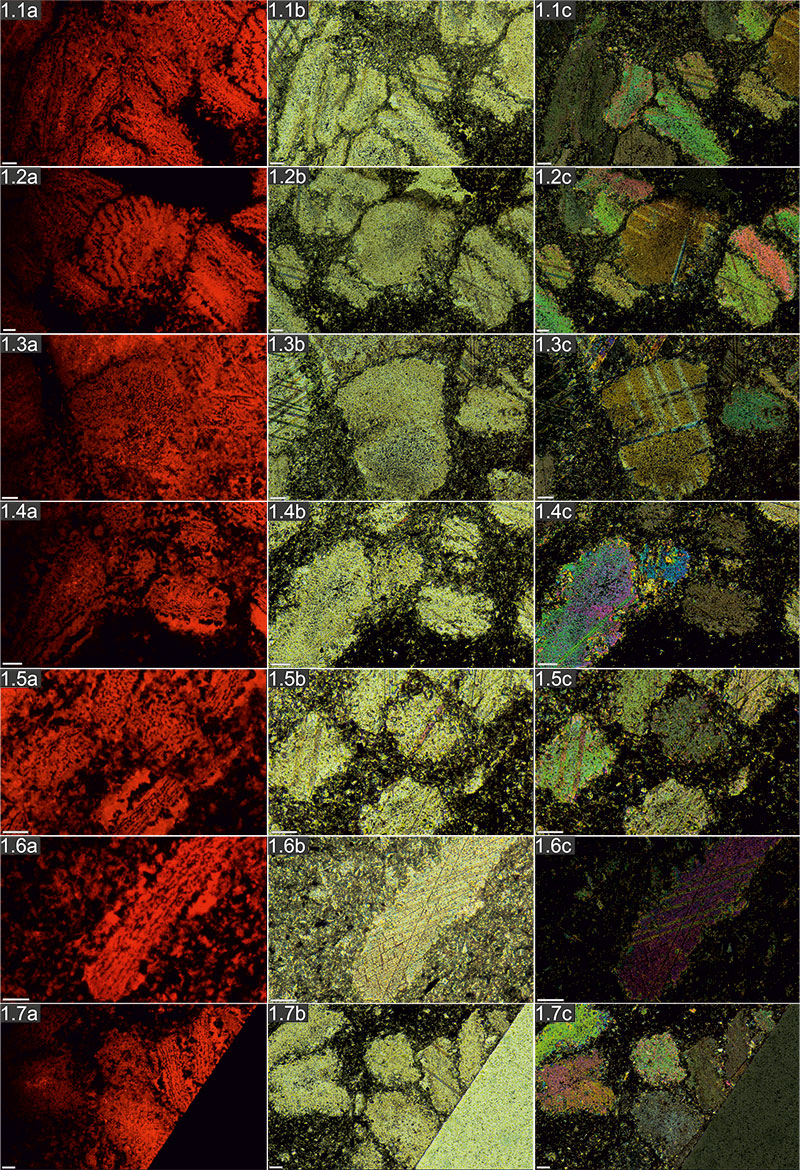
FIGURE A2. Photomicrographs of Protocinctus under cathodoluminescence (a), optical (b) and polarizing microscopy (c), respectively: 2.1-2.2, Axial to slightly oblique sections of various integument plates; 2.3, transversal section of marginal plate; 2.4, transversal to slightly oblique sections of various integument plates; 2.5, Axial to oblique section of marginal plate; 2.6, transversal section of marginal plate; 2.7, Axial to slightly oblique section of thecal? plate. Scale bar equals 100µm.
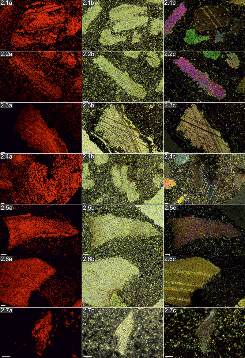
FIGURE A3. Photomicrographs of Protocinctus under cathodoluminescence (a), optical (b) and polarizing microscopy (c), respectively: 3.1-3.3, 3.5m, Axial to slightly oblique sections of various integument plates; 3.4, 3.6, Axial sections of marginal plates. Scale bar equals 100µm.
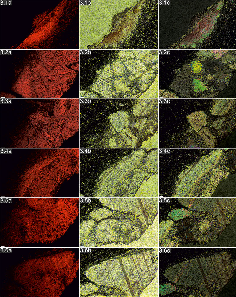
FIGURE A4. Photomicrographs of Stromatocystites under cathodoluminescence (a), optical (b) and polarizing microscopy (c), respectively: 4.1, 4.2, 4.4, 4.6, transversal to slightly oblique sections of various ambulacral plates; 4.3, 4.5, 4.7, transversal to slightly oblique sections of thecal plates. Scale bar equals 100µm.
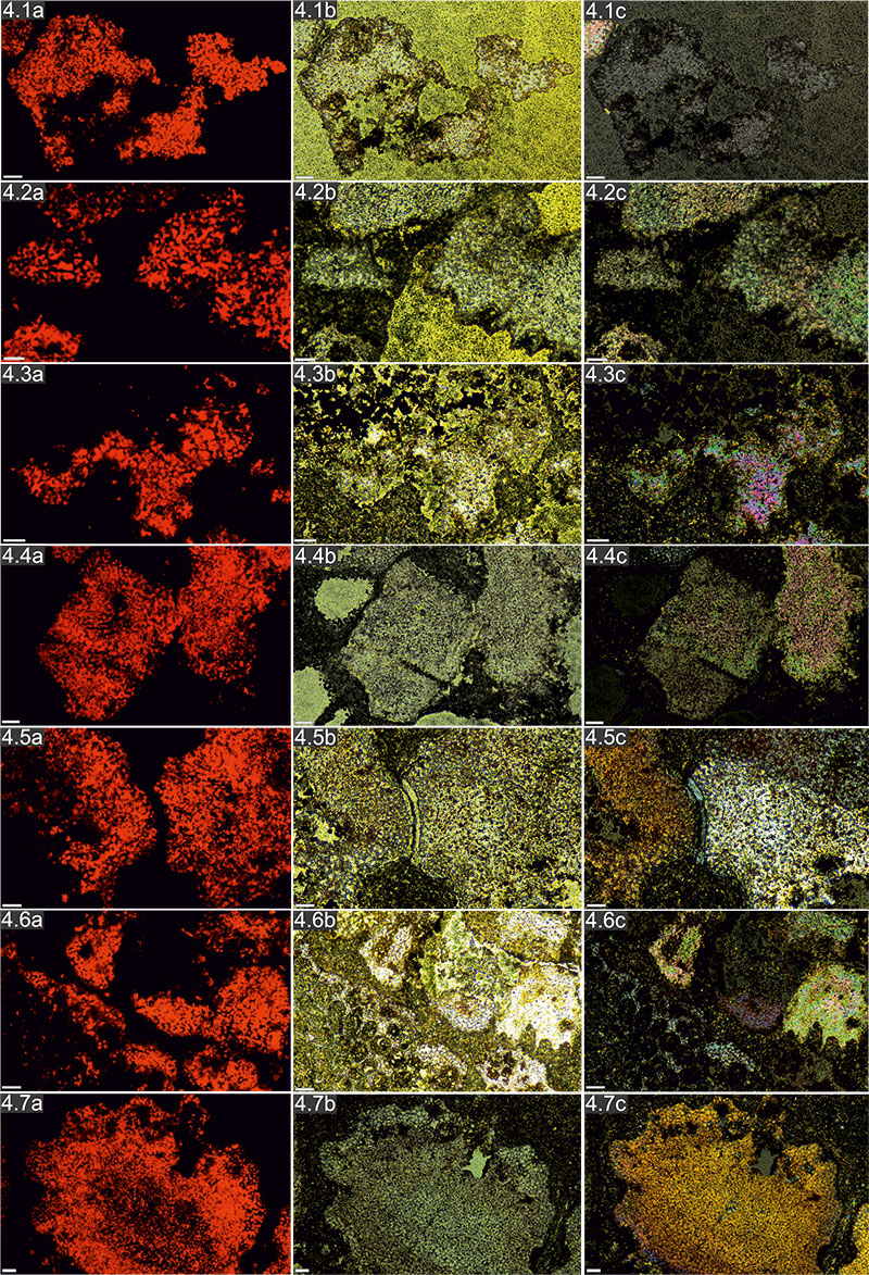
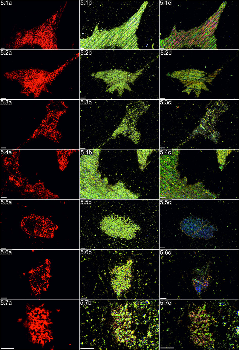
FIGURE A5. Photomicrographs of Dibrachicystidae under cathodoluminescence (a), optical (b) and polarizing microscopy (c), respectively: 5.1-5.4, longitudinal to slightly oblique sections of thecal plates; 5.5-5.7, Slightly oblique sections of columnal? plates. Scale bar equals 100µm.
 Przemysław Gorzelak
Department of Biogeology
Institute of Paleobiology
Polish Academy of Sciences
Twarda Str. 51/55
PL 00-818 Warsaw
Poland
pgorzelak@twarda.pan.pl
Przemysław Gorzelak is a paleontologist who got his Ph.D. at the Institute of Paleobiology, Polish Academy of Sciences (Warsaw) in 2012. He is interested in the morphology and paleoecology of fossil echinoderms, with special value given to the Mesozoic crinoids. His research interests include also predator-prey interactions and their effects on morphology, ecology, and evolution. He is currently involved in projects on biomineralization and diagenesis of the stereom in extant and fossil echinoderms.

 Samuel Zamora
Department of Paleobiology
National Museum of Natural History
Smithsonian Institution
Washington DC, 20013–7012
USA
samuel@unizar.es
Samuel Zamora is a post-doctoral researcher at the Smithsonian Institution (Washington DC) funded by the Springer fund. He is a specialist on the early evolutionary history of echinoderms. He completed his Ph.D. in 2009 at the University of Zaragoza (Spain) base on Cambrian echinoderm faunas from North Spain and he spent later two years post-Doc at the Natural History Museum (London). He has published extensively on the morphology and a phylogenetic relationship of a broad range of Cambrian and Ordovician echinoderms and is currently researching the origins of the echinoderm body plan and the extrinsic and intrinsic factors that drove the early diversification of echinoderms.
Stereom microstructures of Cambrian echinoderms revealed by cathodoluminescence (CL)
Plain Language Abstract
Echinoderms possess a skeleton with a characteristic meshlike microstructure called stereom. Stereom is considered the major echinoderm character and is recognized already in some Cambrian echinoderms. However, data on the skeletal microstructures of early echinoderms are still sparse and come only from isolated plates of limited taxonomic value in which the primary calcite has been replaced by other minerals.
Here, we applied for the first time cathodoluminescence (CL) in distinguishing stereom microstructures of the diagenetically altered calcitic skeletons of some Cambrian echinoderms (in particular Protocinctus, Stromatocystites and Dibrachicystidae). CL not only provides insights into the diagenesis of their skeletons but also reveals primary microstructural details that are not visible under optical or scanning electron microscopes. Different stereom types comparable to those observed in extant echinoderms have been recognized for the first time in these Cambrian taxa. Our results show that the stereom microstructures widely occur in various Cambrian echinoderms, which suggest that they likely evolved the same genetically controlled biomineralization mechanisms as those observed in modern echinoderms.
These results underline that the CL technique can be a powerful tool in the detection of the microstructural organization in even the most recrystallized echinoderm specimens. Given the close association between the skeletal microstructure and the investing soft tissues, the method presented here opens new possibilites for revealing skeletal growth and soft tissue paleoanatomy of fossil echinoderms.
Resumen en Español
Microestructuras estereómicas en equinodermos cámbricos proporcionadas por catodoluminiscencia (CL)
Los equinodermos poseen un esqueleto con una microestructura única que se conoce con el nombre de estereoma y que está controlada por una familia de genes específica. El estereoma es por tanto considerado como la principal sinapomorfía del grupo y es reconocible en los primeros equinodermos cámbricos. Sin embargo los datos de esta microestructura en los equinodermos más antiguos son todavía insuficientes y provienen de osículos aislados cuyo valor taxonómico es muy limitado y que aparecen conservados por reemplazamiento de la calcita por fosfato, sílice u óxidos de hierro.
En este trabajo, aplicamos la catodoluminiscencia (CL) para estudiar la microestructura estereómica en algunos equinodermos Cámbricos conservados en calcita muy alterada diagenéticamente (en concreto los taxones Protocinctus, Stromatocystites y Dibrachicystidae). La CL no sólo aporta datos sobre la diagénesis del esqueleto de los equinodermos sino también revela detalles microestructurales primarios que no eran observable con luz trasmitida o SEM. Se reconocen diferentes tipos de estereoma en estos taxones cámbricos (parecidos al laberíntico, fasciculado, fenestrado, microperforado e imperforado) y que son comparables a los que aparecen en equinodermos modernos. Estos resultados demuestran que estas estructuras estereómicas aparecen en diferentes clados de equinodermos cámbricos sugiriendo que su formación está controlada por los mismos mecanismos genéticos que están presentes en las especies actuales.
Estos resultados sugieren que la CL puede ser una técnica muy interesante para detectar las microestructuras en equinodermos que han sufrido una recristalización importante. Dada la relación que existe en los equinodermos modernos entre la microestructura y los tejidos blandos asociados a ésta, el método presentado puede contribuir de manera importante a comprender la paleoanatomía de grupos extintos de equinodermos.
Palabras clave: esqueleto; biomineralización; Cámbrico; equinodermos; estereoma; catodoluminiscencia
Traducción: los autores
Résumé en Français
Microstructures stéréome d'échinodermes du Cambrien révélées par cathodoluminescence (CL)
Les échinodermes possèdent un squelette avec une microstructure unique et distinctive ressemblant à un filet, appelé stéréome qui est soutenu par une famille de gènes spécifiques . Stéréome est donc considéré comme la principale synapomorphie des échinodermes et est déjà reconnu dans certains clades d'échinodermes du Cambrien. Toutefois, les données sur les microstructures du squelette des premiers échinodermes sont encore rares et proviennent uniquement d'osselets isolés de valeur taxonomique limitée dans laquelle le carbonate de calcium primaire a été remplacé ou finement recouvert par des phosphates , des silices ou des oxydes de fer .
Ici, nous avons appliqué de la cathodoluminescence (CL) pour révéler les microstructures stéréomes des squelettes calcitiques diagénétiquement modifiés de certains échinodermes du Cambrien (en particulier Protocinctus, Stromatocystites et Dibrachicystidae). CL non seulement donne un aperçu de la diagenèse de leurs squelettes, mais aussi révèle des détails de la microstructure primaires qui ne sont pas visibles par la lumière transmise ou SEM . Plusieurs types de stéréome différents comparables à ceux observés chez les échinodermes existants ont été reconnu pour la première fois dans ces taxons du Cambrien. Nos résultats montrent que les microstructures de stéréome se produisent dans la plupart des différents clades d'échinodermes du Cambrien, ce qui suggère qu'ils ont probablement évolué les mêmes mécanismes de biominéralisation génétiquement contrôlées que ceux observées chez les échinodermes modernes.
Ces résultats soulignent que la technique CL peut être un outil puissant pour la détection de l'organisation des microstructures, même pour des spécimens d'échinodermes fortement recristallisés. Compte tenu de l'étroite association entre la microstructure du squelette et leurs tissus mous, la méthode présentée ici ouvre de nouvelles possibilités pour révéler la croissance du squelette et la palaeoanatomie des tissus mous des échinodermes fossiles.
Mots-clés: squelette; biominéralisation; Cambrien; échinoderme ; stéréome; cathodoluminescence
Translator: Kenny J. Travouillon
Deutsche Zusammenfassung
Stereom-Mikrostrukturen kambrischer Echinodermen, aufgezeigt mit Kathodenluminiszens (CL)
Echinodermen besitzen ein Skelett mit einer einzigartigen und charakteristischen maschenartigen Mikrostruktur, genannt Stereom, die durch eine spezifische Genfamilie unterstützt wird. Stereom wird deshalb als die hauptsächliche Synapomorphie der Echinodermen angesehen und wurde bereits bei einigen kambrischen Echinoderm-Kladen erkannt. Allerdings sind Daten zur Skelettmikrostruktur früher Echinodermen immer noch selten und stammen nur von isolierten Stückchen von geringem taxonomischem Wert. Außerdem wurde bei ihnen das primäre Kalziumkarbonat ersetzt oder sie sind dünn mit Phosphaten, Silikaten oder Eisenoxiden überzogen.
Wir wendeten hier Kathodenluminiszens (CL) an, um Stereom-Mikrostrukturen von diagenetisch veränderten Kalzitskeletten einiger kambrischer Echindodermen (besonders Protocinctus, Stromatocystites and Dibrachicystidae) aufzuweisen. CL gibt nicht nur Einsichten in die Skelett-Diagenese, sondern zeigt auch primäre Mikrostrukturen-Details auf, die nicht unter Durchlicht oder SEM sichtbar sind. Unterschiedliche Stereom Typen (ähnlich labyrinthisch, fascicular, gallerid, mikroperforierte und unperforierte Mikrostrukturen) vergleichbar mit denen heutiger Echinodermen, wurden zuerst bei diesen kambrischen Taxa beobachtet. Unsere Ergebnisse zeigen, dass die Stereom-Mikrostrukturen umfassend bei verschiedenen kambrischen Echinodermen-Kladen auftreten, was darauf hinweist, dass sie wahrscheinlich dieselben genetisch kontrollierten Biomineralisationsmechanismen entwickelt haben wie diese, welche in modernen Echinodermen beobachtet werden. Diese Ergebnisse unterstreichen, dass die CL-Technik ein starkes Werkzeug sein kann, um mikrostrukturelle Organisationen selbst bei stark rekristallisierten Echinodermen zu entdecken. Im Hinblick auf den engen Zusammenhang zwischen der Skelettmikrostruktur und den anliegenden Weichteilen, eröffnet die hier vorgestellte Methode neue Möglichkeiten, um Skelettwachstum und Weichteilpaläoanatomie bei fossilen Echinodermen aufzuzeigen.
SCHLÜSSELWÖRTER: Skelett; Biomineralisation; Kambrium; Echinodermen; Stereom; Kathodenluminiszens
Translator: Eva Gebauer
Arabic
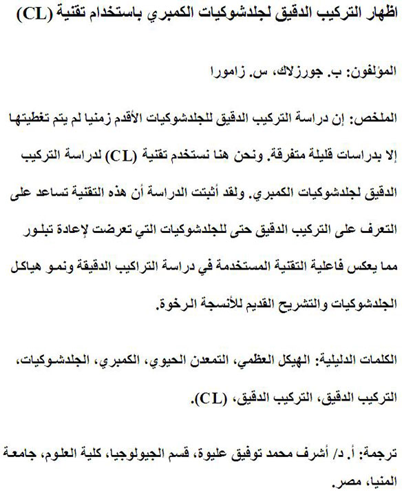
Translator: Ashraf M.T. Elewa
-
-
-
Review: The Princeton Field Guide to Mesozoic Sea Reptiles
 The Princeton Field Guide to Mesozoic Sea Reptiles
Article number: 26.1.1R
April 2023

 Poster Winners 2024