

Article Search
Volume 27.1
January–April 2024
Full table of contents
ISSN: 1094-8074, web version;
1935-3952, print version
Recent Research Articles
See all articles in 27.1 January-April 2024
See all articles in 26.3 September-December 2023
See all articles in 26.2 May-August 2023
See all articles in 26.1 January-April 2023
Figure 1. Example of how the selected Micro-CT scanned specimens were virtually sectioned at several heights parallel to a plane approximating the occlusal surface.
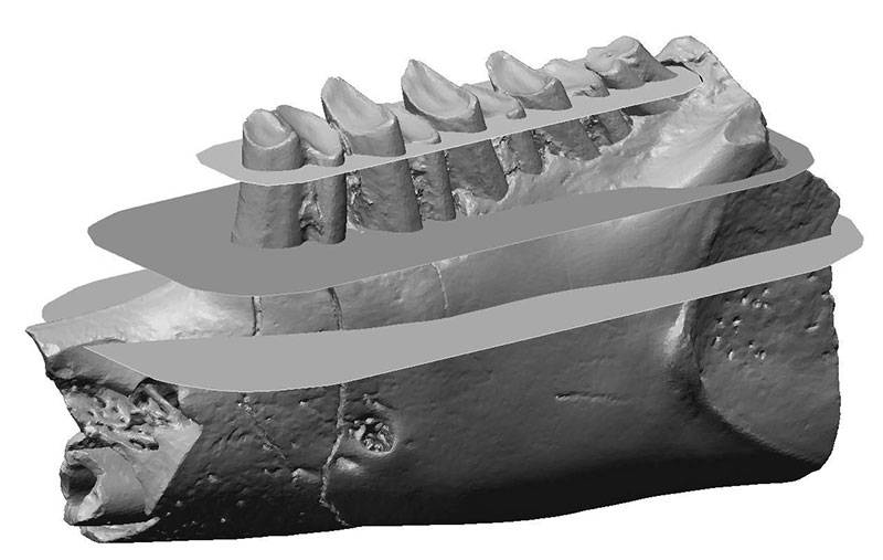
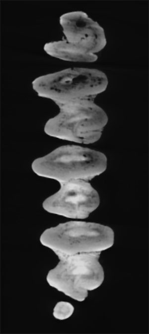
Figure 2. Virtual sections of the ontogenetic development of the p3 of "Amphilagus" antiquus. (1) Occlusal surface. The hypoconulid shallows up (2) and a central connection interrupts the hypoflexid forming a mesofossettid (3). Both hypoflexid and mesofossettid progressively disappear in further stages of wear (4, 5). White arrow: hypoflexid; grey arrow: hypoconulid; dotted white arrow: lingual flexid; dotted white arrow: mesofossettid.
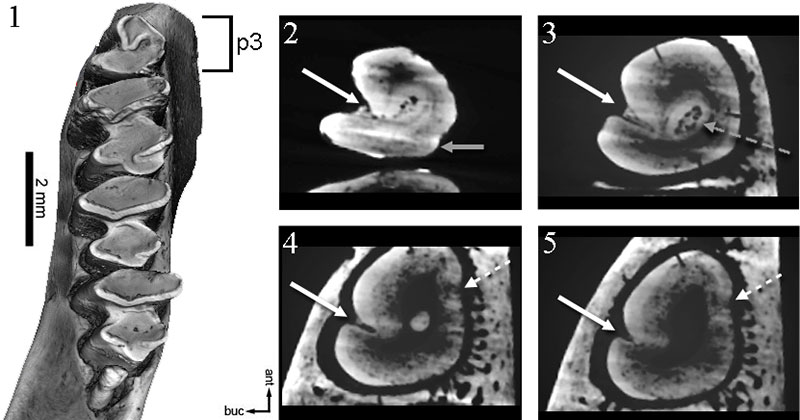
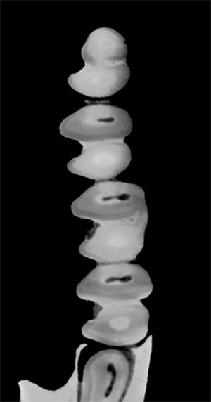
Figure 3. Virtual sections of the ontogenetic development of the p3 of Desmatolagus cf. youngi. (1) Occlusal surface. The hypoflexid (white arrow) progressively shallows up (2, 3).
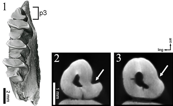
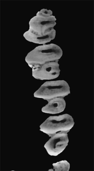
Figure 4. Virtual sections of the ontogenetic development of the p3 of Palaeolagus sp. (a) Occlusal surface. Beginning with an hourglass shape (1, 2), an additional lingual connection between trigonid and talonid forms a mesofossettid (3); the hypoflexid does not substantially shallow up (4). White arrow: mesoflexid; grey arrow: hypoflexid; dotted white arrow: mesofossettid.
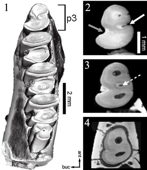
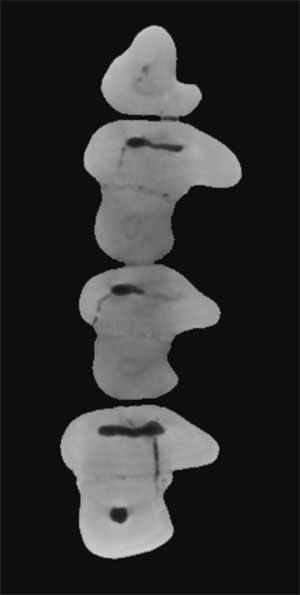
Figure 5. Left lower jaw of Piezodus branssatensis virtually sliced along the mesiodistal midline. In the younger specimen (1) we can observe dp3 and dp4 lying upon p3 and p4 (white arrow), and the lack of the anteroconid along the shaft (grey dotted line); the roots are not present. In the older specimen (2) we see the incipient roots (white dotted arrow).
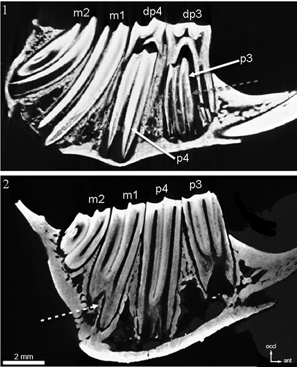
Figure 6. Virtual sections of the ontogenetic development of the p3 of Piezodus branssatensis. (1, 4) Occlusal surfaces. In the younger specimen the dp3 covers the p3 (1); the unworn p3 has the shape of an hourglass (2) which remains even at maximum stages of wear. The anteroconid (3) does not appear. In the older specimen of Piezodus the anteroconid is already present at the level of the occlusal surface (4), and eventually merges lingually with the trigonid (5) (white arrow); trigonid and talonid merge lingually (6) (dotted white arrow) and the hypoflexid (grey arrow) progressively shallows up (7).
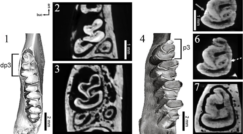
Figure 7. Virtual sections of the ontogenetic sequence of the trigonid-talonid connection in Palaeolagus sp. From unconnected trigonids and talonids on the occlusal surface (1) progressive wear produces connections (indicated by the white arrow) in m1 (2) then in m2 (3) and at last in p4 (4).
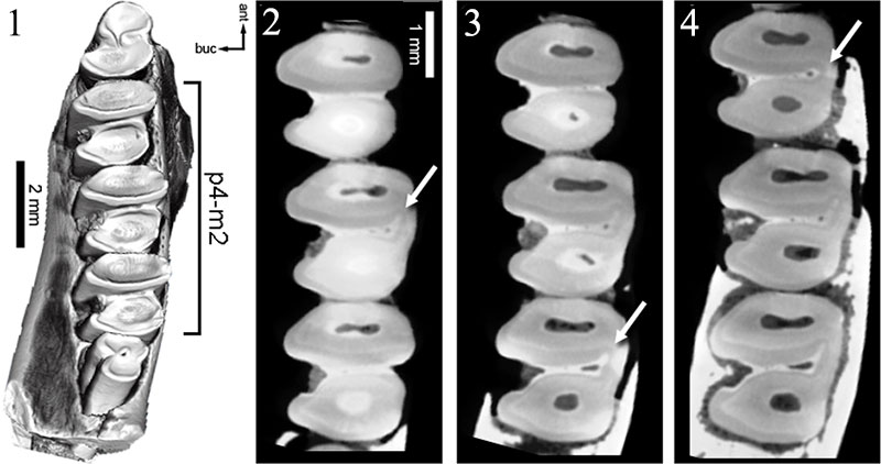
Figure 8. Virtual sections of the ontogenetic sequence of the trigonid-talonid connection in Desmatolagus cf. youngi. From unconnected trigonids and talonids on the occlusal surface (1) progressive wear produces connections (indicated by the white arrow) in m2 (2) then in m1 (3) and at last in p4 (4).
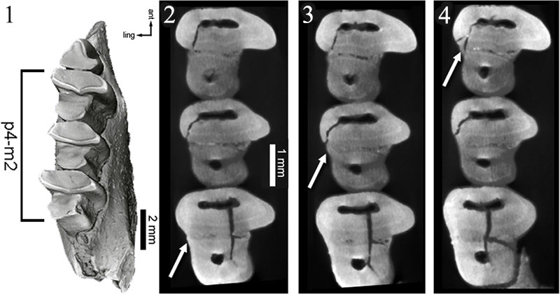
Figure 9. Virtual sections of the ontogenetic sequence of the trigonid-talonid connection in "Amphilagus" antiquus. From unconnected trigonids and talonids in the occlusal surface (1) progressive wear starts an incipient connection (indicated by the white arrow) in m2 (2) then in m1 (3) and at last in p4 (4).
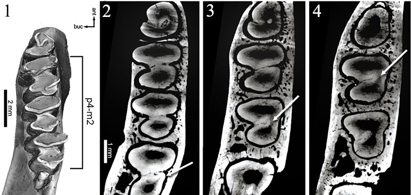
Figure 10. Virtual sections of the ontogenetic sequence of the trigonid-talonid connection in Piezodus branssatensis. In the younger specimen trigonid and talonid remain separated even with maximum wear. In the occlusal surface of the older specimen (1) wear starts an incipient connection (white arrow) between trigonids and talonids in m2 (2) then in m1 (3) and at last in p4 (4).
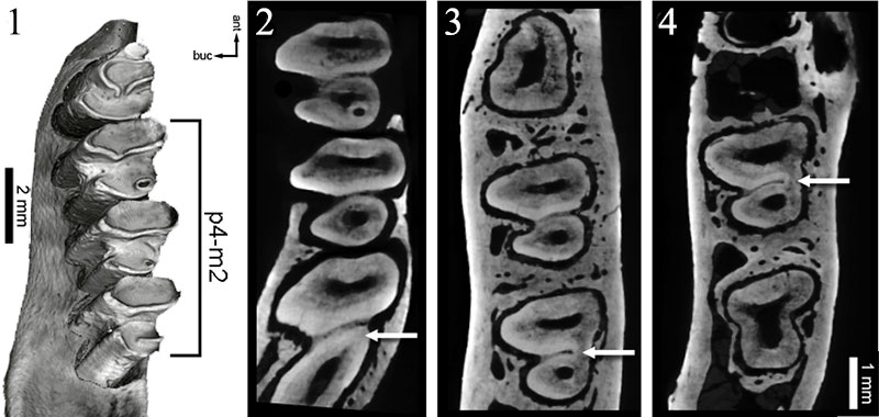
Figure 11. Virtual sections of the ontogenetic sequence of hypoconulid/fossettes development in "Amphilagus" antiquus. From lingually directed hypoconulids in the occlusal surface (1) progressive wear develops a fossette (white arrow) in m1 (2) then in m2 (3) and at last in p4 (4).
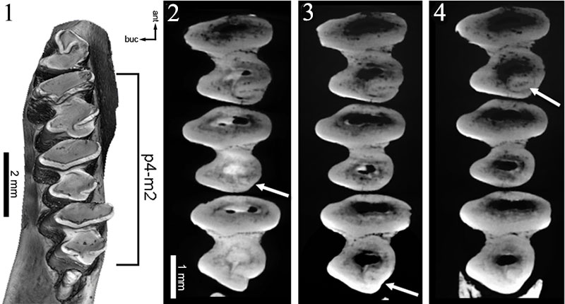
Figure 12. Virtual sections of the ontogenetic sequence of hypoconulid/fossettes development in Piezodus branssatensis. In the original occlusal surface only m1 and m2 are visible (1); the hypoconulid in m1 forms a fossette in very early stages of wear (2) that rapidly disappears (3); eventually a fossette is formed in p4 (4) and in m2 (5).
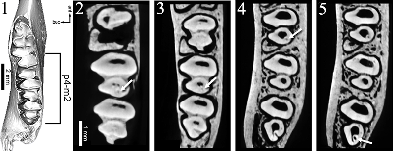
Table 1. Sequences of appearance/development of p3-m2 of lobe connections, and of appearance/disappearance of flexids/fossettes in the studied specimens.
|
"Amphilagus" antiquus |
Piezodus branssatensis |
Desmatolagus cf. youngi |
Palaeolagus sp. |
||
|
p3 |
development posterior flexids/fossettes |
deep hypoflexid --> hypoflexid+mesofossettid --> shallowing of hypoflexid + disappearance of mesoflexid |
hypoflexid + mesoflexid --> hypoflexid + mesofossettid--> shallowing of hypoflexid + disappearance of mesoflexid |
shallowing of hypoflexid |
hypoflexid + mesoflexid --> hypoflexid + mesofossettid --> hypoflexid alone |
|
p4-m2 |
trigonid-talonid connection |
m2-m1-p4 |
m2-m1-p4 |
m2-m1-p4 |
m1-m2-p4 |
|
hypoconulid --> fossette |
m1-m2-p4 |
m1-p4-m2 |
- |
- |
|
|
fossette disappearance |
m1-m2-p4 |
m1-m2-p4 |
- |
- |
ANIMATION 1. Virtual sectioning of the lower jaw of "Amphilagus" antiquus shows the ontogenetic development of lower cheek teeth. Click on image to run animation.
ANIMATION 2. Virtual sectioning of the lower jaw of Desmatolagus cf. youngi shows the ontogenetic development of lower cheek teeth. Click on image to run animation.
ANIMATION 3. Virtual sectioning of the lower jaw of Palaeolagus sp. shows the ontogenetic development of lower cheek teeth.Click on image to run animation.
ANIMATION 4. Virtual sectioning of the lower jaw of Piezodus branssatensis (senile specimen) shows the ontogenetic development of lower cheek teeth. Click on image to run animation.
 Chiara Angelone
Institut Català de Paleontologia
Universitat Autònoma de Barcelona
08193 Cerdanyola del Vallès
Barcelona, Spain
chiara.angelone@icp.cat
Research interests: Taxonomy of Eurasian Neogene-Quaternary fossil lagomorphs and applications in biochronology and palaeogeography.

 Julia A. Schultz
Steinmann-Institut für Geologie
Mineralogie und Paläontologie
Universität Bonn
Nussallee 3, 53115 Bonn
Germany
schultzj@uni-bonn.de
The research interest of Julia A. Schultz is functional morphology of the early mammalian dentition. Her PhD thesis was concerned with the reconstruction of mastication pattern of fossil mammal species using striation analysis, facet formation and wear pattern. Since 2012 she is in a post-doc position in the Steinmann-Institut (Universität Bonn) studying mammalian evolution during the Mesozoic focusing on 3D-reconstruction, virtual simulation and micro-computed tomography.
 Margarita A. Erbajeva
Geological Institute
Siberian Branch
Russian Academy of Sciences
Ulan-Ude
Russia 670047
erbajeva@gin.bscnet.ru
Research palaeontologist of the Cenozoic Laboratory of the Geological Institute, Siberian Branch of the Russian Academy of Sciences, Ulan-Ude. Specialist on the Pliocene-Recent small mammals of Siberia and Paleogene-Recent ochotonids of the world. She is the author and co-author of a number of papers on small mammals and ochotonids and of 8 monographs.
The main subject of interest is systematics, evolution and phylogeny of small mammals, moreover the paleonvironmental reconstruction, biostratigraphy and interregional correlation of the Late Cenozoic deposits and faunas.
Determining the ontogenetic variation of lower cheek teeth occlusal surface patterns in lagomorphs using Micro CT-technology - preliminary results and perspectives
Chiara Angelone, Julia A. Schultz, and Margarita A. Erbajeva
Plain Language Abstract
Lagomorph teeth morphology changes considerably during the lifetime of an animal. The reconstruction of tooth development during lifetime is very important (particularly in lagomorphs with rooted teeth), so as to not overestimate the number of species in an assemblage and avoid consequent taxonomic and phylogenetic repercussions.
We performed Micro CT-scans of lower cheek teeth of some selected lagomorph taxa, in order to obtain virtual slices at different heights of the teeth and to obtain reliable reconstructions of the teeth development during lifetime.
We expected that every taxon could be univocally individuated by its teeth development, and our results (though limited to a few specimens) seem to confirm such hypothesis. Actually the analyzed taxa taken into consideration in our preliminary work show different sequences of appearance/development of some structures of the occlusal surface. Our results also suggest that ontogenetic teeth development can give suggestions about the relationship among taxa.
RIASSUNTO
Una serie di scansioni microtomografiche è stata effettuata su alcune mandibole appartenenti a differenti taxa dell'ordine Lagomorpha, con l'intento di ricostruire il loro controverso sviluppo ontogenetico. Le analisi hanno riguardato lo sviluppo di p3, mentre su p4-m2 le osservazioni si sono concentrate sulla sequenza di apparizione/sparizione dei flessi e delle fossette e sulla sequenza di connessione trigonide/talonide.
Questa è la prima volta che l'analisi microtomografica viene applicata allo studio dei lagomorfi fossili ed apre scenari molto promettenti specialmente per quanto riguarda la tassonomia e la filogenesi.
Resumen en Español
Determinación de la variación ontogenética de los patrones de la superficie oclusal de la dentición yugal inferior para lagomorfos con el uso de tecnología de Micro CT
Se han realizado análisis de las mandíbulas inferiores de algunos taxones seleccionados de lagomorfos con el uso de digitalización de Micro-CT para reconstruir fidedignamente su controvertido desarrollo ontogenético. Los análisis se centraron en el desarrollo del p3, y en las secuencias de conexiones de los lóbulos y en la secuencia de aparición/desaparición de fléxidos/fosetas de los p4-m2.
Esta es la primera vez que este enfoque se aplica al estudio de lagomorfos, la cual abre perspectivas prometedoras, especialmente en lo que se refiere a la taxonomía y la filogenia de este complejo orden.
Palabras clave: Micro-CT; Lagomorpha; ontogenia; dentición inferior
Traducción: Enrique Peñalver
Résumé en Français
Déterminer la variation ontogénétique des motifs de surfaces occlusales de dents de joues inférieurs chez les lagomorphes utilisant la technologie Micro CT
Des scans Micro CT ont été effectués sur des mâchoires inférieures d'une sélection de taxons de lagomorphes pour reconstruire sans équivoque leur développement ontogénétique controversé. Les analyses ont été concentrés sur le développement de la p3, et sur les séquences des connexions de lobe et de la séquence d'apparition / disparition des flexids/fossettes de p4-m2.
C'est la première fois que cette approche a été appliquée chez les lagomorphes et ouvre des perspectives prometteuses en particulier pour la taxonomie et la phylogénie de cet ordre complexe.
Mots-clés: Micro-CT; Lagomorpha; ontogenèse; dents inférieures
Translator: Kenny J. Travouillon
Deutsche Zusammenfassung
Bestimmung der ontogenetischen Variation okklusaler Kauflächenmuster der unteren Backenzähne bei Lagomorphen mittels Mikro-Ct-Technologie
Es wurde Mikro-Ct-Scanning bei Unterkiefern von einigen ausgewählten lagomorphen Taxa durchgeführt, um deren kontroverse ontogenetische Entwicklung unzweideutig zu rekonstruieren. Die Analysen waren auf die Entwicklung des p3, auf die Sequenzen der Lobenverbindungen und auf die Aufeinanderfolge von Vorkommen/ nicht Vorkommen von Flexiden/Fossetten von p4-m2 konzentriert.
Hiermit wurde zum ersten Mal ein solcher Ansatz in Bezug auf Lagomorphe angewendet, der vielversprechende Perspektiven zur Taxonomie und Phylogenie dieser komplexen Ordung öffnet.
Schlüsselwörter: Mikro-CT; Lagomorpha; Ontogenie; untere Zähne
Translator: Eva Gebauer
Arabic

Translator: Ashraf M.T. Elewa
-
-
-
Review: The Princeton Field Guide to Mesozoic Sea Reptiles
 The Princeton Field Guide to Mesozoic Sea Reptiles
Article number: 26.1.1R
April 2023

 Poster Winners 2024