

Article Search
Volume 27.1
January–April 2024
Full table of contents
ISSN: 1094-8074, web version;
1935-3952, print version
Recent Research Articles
See all articles in 27.1 January-April 2024
See all articles in 26.3 September-December 2023
See all articles in 26.2 May-August 2023
See all articles in 26.1 January-April 2023
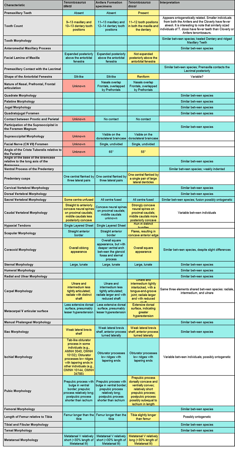
TABLE 1. Comparisons of non-coded characteristics between the two species of Tenontosaurus from their originally described ranges (T. tilletti from Montana, USA and T. dossi from Texas, USA) and specimens of T. tilletti from the Antlers Formation of Oklahoma. Short descriptors of each characteristic are listed in the first column, and explanations of those characteristics from Tenontosaurus specimens in each of the three known ranges are listed in the second through fourth columns. A short interpretation is included in the fifth column with additional information. Cells colored in blue represent commonalities between one or both of the known Tenontosaurus species with individuals from the Antlers Formation, cells colored in yellow represent differences, and cells colored in red represent unknowns. This table is based off of observations from Ostrom (1970), Forster (1990), and Winkler et al. (1997), as well as personal observations made by the author.
Appendix 1 - Taxa and Specimen Resource List
The following is a list of taxa analyzed and compared in this study, with references to descriptive papers used in the analysis of each.
Outgroup:
Lesothosaurus diagnosticus Galton, 1978–Thulborn (1970, 1972), Santa Luca (1984), Weishampel and Witmer (1990a), Sereno (1991a), Norman, (2004)
Basal Ornithopod Taxa:
Hypsilophodon foxii Huxley, 1869–Galton (1974a), Norman (2004)
Parksosaurus warreni Parks, 1926–Parks (1926), Sternberg (1940), Galton (1973)
Thescelosaurus neglectus Gilmore, 1913–Gilmore (1913), Gilmore (1915), Sternberg (1940), Galton (1974b, 1995, 1997), Norman et al. (2004)
Basal Iguanodontian Taxa:
Camptosaurus dispar (Marsh, 1879)–Marsh (1879), Gilmore (1909), Norman (2004)
Cumnoria prestwichii Hulke 1880–Hulke (1880), Galton and Powell (1980), Norman (2004)
Dryosaurus altus (Marsh, 1878)–Marsh (1878), Galton (1983), Norman (2004)
Eolambia caroljonesa Kirkland, 1998–Kirkland (1998), Head (2001), Norman (2004), McDonald et al. (2012)
Iguanodon bernissartensis Boulenger, 1881–Boulenger (1881), Norman (1980), Norman (2004)
Jinzhousaurus yangi Wang and Xu, 2001–Wang and Xu (2001), Barrett et al., (2009), Wang et al. (2011)
Mantellisaurus atherfieldensis (Hooley, 1925)–Hooley (1925), Norman (1986), Norman (2004)
Ouranosaurus nigeriensis Taquet, 1976–Taquet (1976), Norman (2004)
Tenontosaurus dossi Winkler et al., 1997–Winkler et al. (1997), Norman (2004)
Tenontosaurus tilletti Ostrom, 1970–Ostrom (1970), Forster (1990), OMNH 58340 (subadult, primary specimen), OMNH 2531 (juvenile), OMNH 16562 (subadult, skull transversely compressed, palpebral), OMNH 34191 (premaxilla)
Zalmoxes robustus (Nopcsa, 1900)–Nopcsa (1900), Weishampel et al. (2003), Norman (2004)
Hadrosauroid Taxa:
Corythosaurus casuarius Brown, 1914–Brown (1914, 1916), Ostrom (1961), Horner et al. (2004)
Edmontosaurus regalis Lambe 1917–Lambe (1917), Lull and Wright (1942), Norman (2004), Horner et al. (2004)
Probactrosaurus gobiensis Rozhdestvensky, 1966–Rozhdestvensky (1966), Norman (2002), Norman (2004)
Protohadros byrdi Head, 1998–Head (1998), Norman (2004)
Telmatosaurus transsylvanicus (Nopcsa, 1900)–Nopcsa (1900), Nopcsa (1903), Weishampel et al. (1993), Horner et al. (2004)
Appendix 2 - Character Description
The following is a list of character states used in this analysis. States were determined independently, but similarities with previously published analyses (Norman, 2004; McDonald et al., 2010) have been noted.
Facial Characters:
1. Presence of nasal contact with the maxilla and reason for exclusion of contact (Modified from Norman, 2004: Character 6):
1:0. Contact
1:1. No contact due to an anterior extension of the lachrymal
1:2. No contact due to a posterior extension of the premaxilla
2. Character of the occlusal margin of the premaxilla (Modified from McDonald et al., 2010: Character 33):
2:0. Smooth
2:1. Denticulate
2:2. Scooped, possibly to accommodate the denticles of the predentary
3. Premaxillary teeth (Modified from Norman, 2004: Character 3; McDonald et al., 2010: Character 29):
3:0. Present
3:1. Absent
4. Posterior extent of the nasal process of the premaxilla:
4:0. Does not reach antorbital fenestra
4:1. Reaches antorbital fenestra or, if no fenestra present, posterior border of dorsal process of maxilla
5. Beak position relative to tooth row (Modified from Norman, 2004: Character 2):
5:0. At level
5:1. Below
6. Width of combined frontals compared to length (Modified from Norman, 2004: Character 15):
6:0. About 2/3 length of frontal
6:1. About equal to length of frontal
6:2. Twice length of frontal
7. Transverse length of frontals' suture with parietal:
7:0. About equal with combined width of frontals
7:1. About two-thirds combined width of frontals
7:2. About half combined width of frontals
7:3. About one-third combined width of frontals
8. Frontal contribution to dorsal orbital rim (Modified from Norman, 2004: Character 16; McDonald et al., 2010: Character 68):
8:0. More than half
8:1. Less than half
8:2. Does not contribute
9. Triangular process of the parietal:
9:0. Present
9:1. Absent
10. Ratio of axial lengths of the frontal and the nasal:
10:0. Roughly equal in length
10:1. Nasal is one and one half times length of the frontal
10:2. Nasal is twice length of the frontal
10:3. Nasal is three times length of the frontal
10:4. Nasal is four times length of the frontal
11. Position of nasal contribution to the border of the external naris:
11:0. Posterodorsal border
11:1. Dorsal border
11:2. Does not contribute
12. Nature of the articulation of the nasal, frontal, and prefrontal:
12:0. Nasal overlaps frontal, overlapped by prefrontal
12:1. Nasal overlaps frontal
12:2. Nasal has transverse suture with frontal
12:3. Frontal overlaps nasal
13. Nature of the articulation of the premaxillae with the nasals:
13:0. Nasal processes wedge between nasals
13:1. Nasal overlapped by nasal process of premaxilla
13:2. Tip to tip
13:3. Nasals lie in beveled grooves in lateral surface of nasal processes of premaxilla
13:4. Nasals overlap nasal processes of premaxilla
13:5. Nasal processes split into dorsal and ventral branches, which overlap the nasals
14. Supratemporal fensestra imprints frontal:
14:0. Present
14:1. Absent
15. Lateral suture of the laterosphenoid with the:
15:0. Frontal and postorbital
15:1. Postorbital only
15:2. Frontal only
16. Rugose lateral edge of the postorbital:
16:0. Absent
16:1. Present
17. Parietal contact with the postorbital:
17:0. Present
17:1. Absent
18. Ectopterygoid articulation with the palatine:
18:0. Present
18:1. Absent
19. Ectopterygoid articulation with the jugal (Modified from Norman, 2004: Character 14; McDonald et al., 2010: Character 53):
19:0. Present
19:1. Absent
20. Jugal articulation with the quadrate:
20:0. Absent
20:1. Present
21. Ventral lobe of the jugal (Modified from Norman, 2004: Character 13; McDonald et al., 2010: Character 56):
21:0. Absent
21:1. Present
22. Jugal extends farthest posteriorly in the:
22:0. Quadratojugal process
22:1. Postorbital process
23. Accessory foramen in the quadratojugal (Modified from Norman, 2004: Character 17; McDonald et al., 2010: Character 58):
23:0. Absent
23:1. Present
24. Enclosure of the quadratojugal by the jugal:
24:0. None
24:1. Dorsal
24:2. Dorsal and ventral
25. Quadrate foramen:
25:0. Absent
25:1. Present
26. Quadratojugal size (relative to quadratojugal process of jugal):
26:0. Reduced
26:1. Large
27. Position of the contribution of the lachrymal to the orbital border:
27:0. Anteroventral corner
27:1. Anterior border
27:2. Middle anterior border
28. Contact of the nasal with the lachrymal (Modified from McDonald et al., 2010: Character 50):
28:0. Present
28:1. Absent
29. Shape of the medial lamina of the palatine:
29:0. Fan
29:1. Rectangle
29:2. Triangle
30. Maxillary tooth family count along a single side
31. Maxillary diastema:
31:0. Absent
31:1. Present
32. Lateral process of the maxilla for articulation with the jugal:
32:0. Absent
32:1. Present
33. Jugal articulation with the maxilla:
33:0. Posterior on the latter element
33:1. Displaced forward on the latter element
34. Prefrontal contact with the premaxilla (Modified from McDonald et al., 2010: Character 37):
34:0. Absent
34:1. Present
35. Position of the prefrontal contact with the nasal:
35:0. Anteromedially
35:1. Dorsally
36. Pterygoid contact with the shaft of the quadrate:
36:0. Absent
36:1. Present
37. Quadrate notch (Modified from McDonald et al., 2010: Character 61):
37:0. Absent
37:1. Present
38. Transversely wide ventral quadratic condyle (Modified from Norman, 2004: Character 18; McDonald et al., 2010: Character 64):
38:0. Present
38:1. Absent
39. “Hamalar” process of the head of the quadrate:
39:0. Absent
39:1. Present
40. Contact between paired squamosals on the midline (Modified from McDonald et al., 2010: Character 67):
40:0. Absent
40:1. Present
41. Lateral visibility of anterior process of squamosal:
41:0. Largely obscured
41:1. Prominent across intertemporal bar
Neurocranial Characters:
42. Shape of occipital condyle:
42:0. Subspherical
42:1. Flattened/reniform
43. Supraoccipital participation in the foramen magnum (Modified from McDonald et al., 2010: Character 69):
43:0. Present
43:1. Absent, due to the exoccipital
44. Shape of the cultriform process of the parasphenoid in ventral aspect:
44:0. Elongate
44:1. Triangular
45. Outline shape of the parietal in dorsal aspect:
45:0. Rectangular
45:1. Triangular
46. Ratio of length to the width of the parietal:
46:0. 1:1
46:1. 1.5:1
46:2. 1:1.5
46:3. 1:2
47. Pronounced sagittal crest of the parietal:
47:0. Absent
47:1. Present
48. Interparietal Eminence:
48:0. Present
48:1. Absent
49. Parietal/prootic contact:
49:0. Present
49:1. Absent
50. Crista prootica is comprised of:
50:0. Laterosphenoid/prootic/opisthotic
50:1. Prootic/opisthotic
Mandibular Characters:
51. Dentary diastema (Modified from Norman, 2004: Character 20; McDonald et al., 2010: Character 9):
51:0. Absent
51:1. Present
52. Posteroventral corner of the dentary extends behind coronoid process:
52:0. Absent
52:1. Present
53. Dentary symphysis:
53:0. Set level with tooth row
53:1. Set below tooth row
54. Dorsoventral borders of dentary (Modified from Norman, 2004: Character 22; McDonald et al., 2010: Character 15):
54:0. Thicker posteriorly
54:1. Parallel
54:2. Thicker anteriorly
55. Length of the dentary bearing dentition:
55:0. Full
55:1. 2/3
55:2. Half
56. Orientation of coronoid process of the mandible (Modified from Norman, 2004: Character 23; McDonald et al., 2010: Character 20):
56:0. Posteriorly deflected
56:1. Vertical
56:2. Anteriorly deflected
57. Coronoid bone:
57:0. Present
57:1. Absent
58. Presence of two processes around the Meckelian canal:
58:0. Absent
58:1. Present
59. Presence of shelf between the teeth and the coronoid process (Modified from Norman, 2004: Character 24):
59:0. Absent
59:1. Present
60. Axially flared coronoid process (Modified from McDonald et al., 2010: Character 21):
60:0. Absent
60:1. Present
61. Bifurcate ventral process of the predentary (Modified from Norman, 2004: Character 19; McDonald et al., 2010: Character 2):
61:0. Absent
61:1. Present
62. Character of the occlusal margin of the predentary (Modified from McDonald et al., 2010: Character 4):
62:0. Smooth
62:1. Denticulate
63. Lateral visibility of the angular (Modified from Norman, 2004: Character 26; McDonald et al., 2010: Character 27):
63:0. Visible
63:1. Reduced visibility
63:2. Not visible
64. Nature of articulation of the angular with the surangular (Modified from McDonald et al., 2010: Character 26):
64:0. Angular laterally overlaps surangular
64:1. Groove in ventral surangular
64:2. Angular laterally abuts surangular
65. Anterior extension of the coronoid:
65:0. Present
65:1. Absent
66. Lateral visibility of the coronoid bone:
66:0. Invisible
66:1. Largely visible
66:2. Reduced
67. Splenial coverage of the Meckelian canal:
67:0. Extensive
67:1. Posterior only
68. Presence of the prearticular:
68:0. Present
68:1. Absent
69. Number of surangular foramina (Modified from Norman, 2004: Character 25; McDonald et al., 2010: Character 24):
69:0. Two
69:1. Three
69:2. One
69:3. None
70. Nature of ridges on the teeth (Modified from Norman, 2004: Character 27; McDonald et al., 2010: Characters 85, 90, 91):
70:0. Absent
70:1. Ridge on Dentary teeth
70:2. Ridge on Dentary and Maxillary Teeth
70:3. Ridge on Maxillary with reduced Dentary ridge
70:4. Ridge on Dentary with reduced Maxillary ridge
71. Dentary tooth family count along a single side
Postcranial Characters:
Taken, with modifications, from Norman, 2004.
72. Dorsal neural spines:
72:0. Low and Square
72:1. Rectangular and height more than twice width
72:2. Extremely elongate, height more than six times width
73. Sacrum:
73:0. Seven or fewer vertebrae
73:1. More than seven
74. Scapular blade:
74:0. Straight
74:1. Curved
74:2. Curved and flared distally
75. Scapular acromion:
75:0. Prominent boss on the anterior margin of the scapula
75:1. Boss is reflected laterally
76. Humerus:scapula length:
76:0. Approximately equal lengths
76:1. Scapula longer than humerus
77. Sternal shape:
77:0. Reniform
77:1. Hatchetlike
78. Carpal structure:
78:0. Fully ossified and blocklike
78:1. Reduced
79. Metacarpal I shape:
79:0. Dumbbell-like
79:1. Short and blocklike
79:2. Absent
80. Metacarpals II-IV:
80:0. Dumbbell-like and spreading
80:1. Closely appressed
80:2. Appressed, slender and elongate
81. Manus digit I:
81:0. Present
81:1. Absent
82. Manus ungual I:
82:0. Claw-like
82:1. Conical
82:2. Absent
83. Manus unguals II and III:
83:0. Claw-like
83:1. Flattened, twisted and hoof-like
83:2. Digit II claw-like, Digit III nub
84. Manus digit III:
84:0. Four phalanges
84:1. Three phalanges
85. Preacetabular process of ilium:
85:0. Long and laterally compressed
85:1. Strongly downturned
85:2. Distally twisted
86. Dorsal margin of iliac blade:
86:0. Mostly smooth edged
86:1. Strongly notched behind the ischial peduncle
87. Dorsal edge of ilium above ischial peduncle:
87:0. Not thickened and bevelled
87:1. Thickened
87:2. Everted with pendent tip
88. Ilium, postacetabular process:
88:0. Tapering posteriorly
88:1. Low and rectangular
89. Pubis, prepubic process:
89:0. Short and blunt
89:1. Elongate
90. Pubis, prepubic process:
90:0. Rod-shaped
90:1. Laterally compressed, bar-like
90:2. Short constriction and distal expansion
90:3. Deep expansion
91. Pubic shaft:
91:0. Ends adjacent to distal end of ischium
91:1. Shorter than ischium, no pubic symphysis
92. Ischium, shaft shape:
92:0. Straight
92:1. Arched dorsally
93. Ischium shaft:
93:0. Flattened in cross section
93:1. Rounded in cross section
94. Obturator process of the ischium:
94:0. Absent
94:1. Present near midshaft
94:2. Present and close to pubic peduncle
95. Tip of ischium:
95:0. Unexpanded
95:1. Axial expansion to form a boot
96. Femur:
96:0. Distal half of shaft curved posteriorly
96:1. Straight
97. Femoral fourth trochanter:
97:0. Pendent
97:1. Triangular
97:2. Crested eminence
98. Femur extensor groove:
98:0. Open shallow trough
98:1. U-shaped groove
98:2. Partially enclosed channel
98:3. Fully enclosed tunnel
99. Femur distal condyles:
99:0. Moderately expanded posteriorly
99:1. Expanded posteriorly and anteriorly
100. Metatarsal I:
100:0. Well developed and articulates with phalanges
100:1. Slender and splintlike
100:2. Absent
101. Pedal unguals:
101:0. Elongate and pointed claws
101:1. Elongate but bluntly truncated
101:2. Short, broad and crescentic with reduced or absent claw grooves
APPENDIX 3.
The following is a matrix composed of the states coded for each of 19 ingroup taxa and one outgroup taxon analyzed for each of the characters in Appendix 2. It is available in text and Excel formats.
APPENDIX 4.
The following is a collection of three-dimensional renderings of each of the elements of the skull of OMNH 58340. They are stored as stl files, which can be easily viewed using software freely available for download on the internet (e.g., by searching for "stl file viewer").
APPENDIX 5.
The following is the CT data generated for use in this study. The first section (Cranium) comprises scans of the skull, without the mandibles or disarticulated pieces. The second section (Braincase) comprises scans of the braincase section of the cranium, taken at a higher resolution than those for the rest of the skull. Finally, the third section (Miscellaneous) comprises the mandibles and other disarticulated elements (e.g., the right prefrontal and the predentary). Also included are documents detailing the scanning parameters used for each set of scans.
FIGURE 1. Posterior skull schematic (above) and photograph (below) of OMNH 58340. The schematic was reconstructed by digitally mirroring the left side of the suspensorium in order to approximate the actual appearance of the skull. The two images are set to the same scale, demonstrating the amount of displacement in the right side. Anterior is into the page. Abbreviations: bp - basipterygoid process; fm - foramen magnum; ptf - posterior temporal foramen (obscured); sb - squamosal boss.
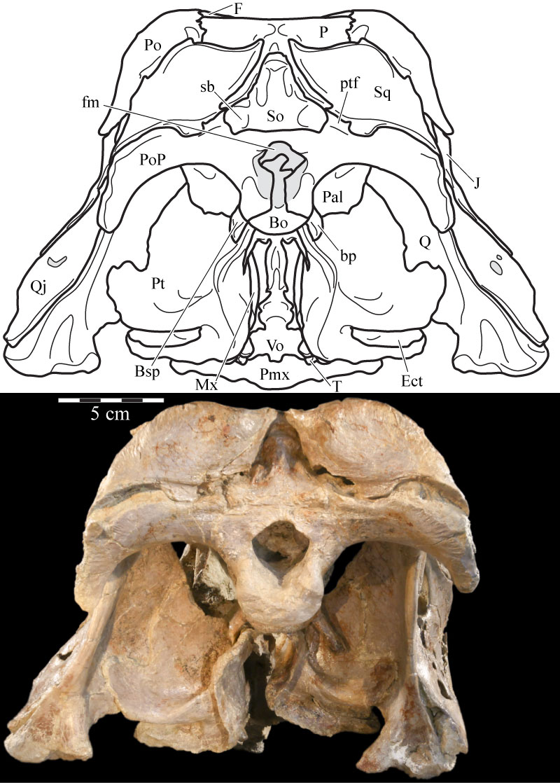
FIGURE 2. Left lateral skull schematic (above) and left skull photograph (below) of OMNH 58340. The skull is angled at the ‘alert position’ indicated by the horizontal semicircular canal. Natural fenestrae are shaded gray. Dashed outline denotes conjectural sclerotic ring. Anterior is to the left. Abbreviation: mf - maxillary foramen.
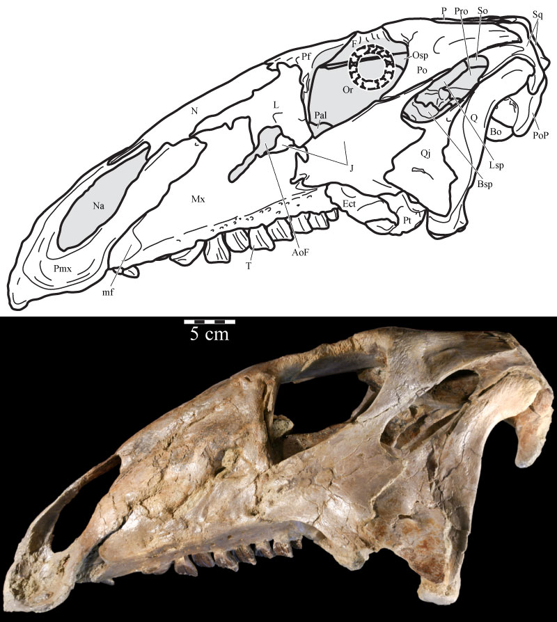
FIGURE 3. Photograph of the lateral side of the disarticulated right maxilla and other associated elements of OMNH 58340. The exclusion of the maxilla from contact with the nasal is apparent, although there is some separation, probably taphonomic, of the premaxilla and lacrimal. Dotted line indicates uncertain sutural placement between the nasal and maxilla.
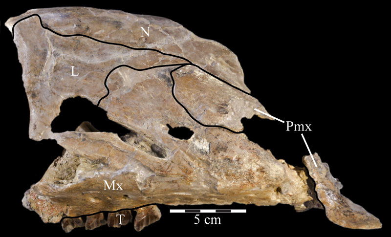
FIGURE 4. Ventral (4.1) and anterior (4.2) photographs of the right premaxilla of OMNH 34191. In 4.1, anterior is out of the page and lateral is left. In 4.2, lateral is to the left and anterior is up.
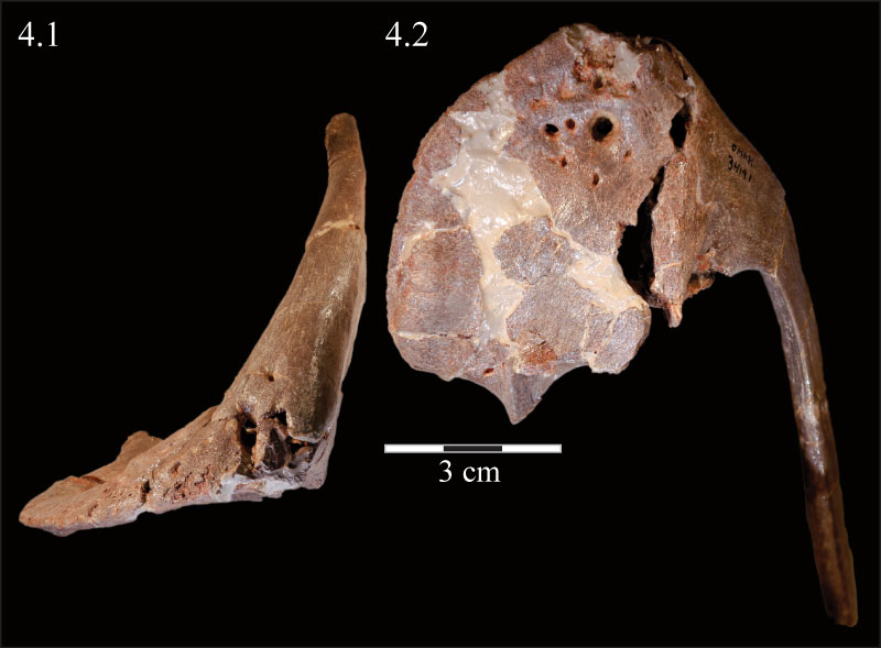
FIGURE 5. Dorsal skull schematic (above) and photograph (below) of OMNH 58340. The schematic was reconstructed by digitally mirroring the left side of the rostrum and suspensorium in order to approximate the actual appearance of the skull. Dotted line at the anterior edge of the frontals indicates uncertain sutural placement. Natural fenestrae are shaded gray. Anterior is to the right.
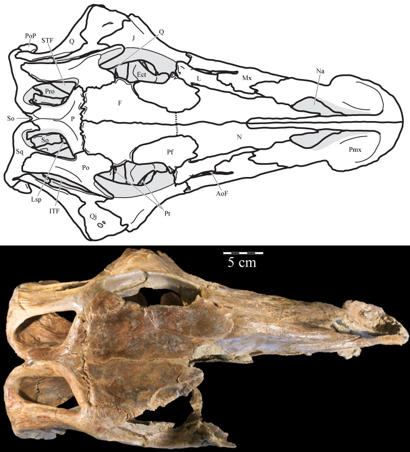
FIGURE 6. Ventral skull schematic (above) and photograph (below) of OMNH 58340. The schematic was reconstructed by digitally mirroring the left side of the rostrum and suspensorium in order to approximate the actual appearance of the skull. Natural fenestrae are shaded gray. Anterior is to the right. Abbreviations: bt - basal tubera of the basisphenoid; lt - lateral tubercle of the basioccipital; mt - median tubercle of the basioccipital; ? - possible second lateral tubercle of the basioccipital.
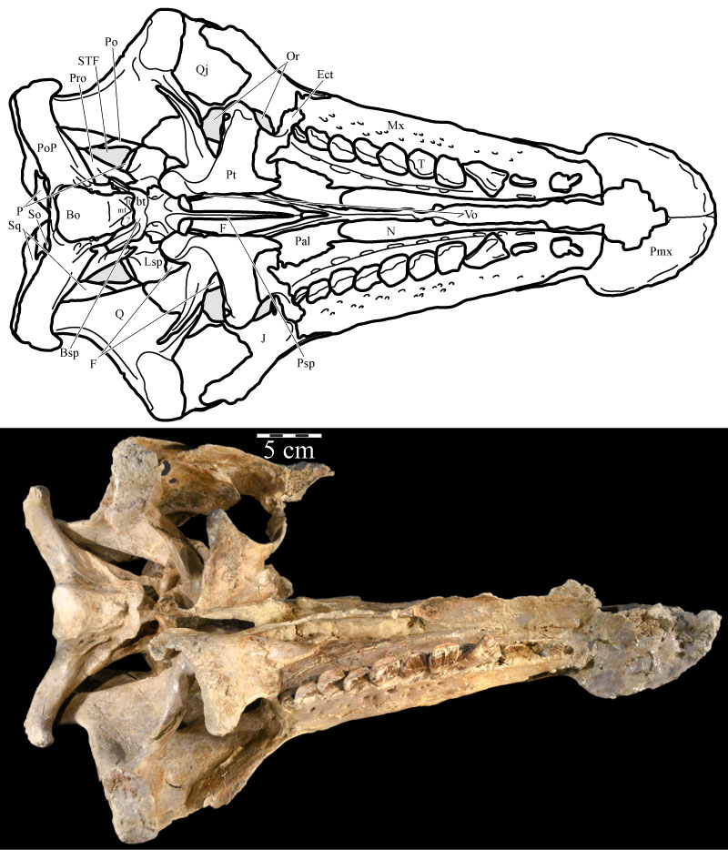
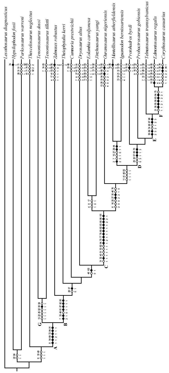
FIGURE 7. Strict component consensus tree of selected members of Ornithopoda included in this analysis. Lesothosaurus diagnosticus is the outgroup. Bremer support values are located below, while Bootstrap and Group Present/Contradicted (GC) support values are located above the branches immediately preceding the node to which they refer. GC values are in bold. Certain nodes are noted with letters referenced in the text. Optimization was performed in PAUP* version 4.0b10 (Swofford, 2002) and checked in TNT (Goloboff et al., 2008).
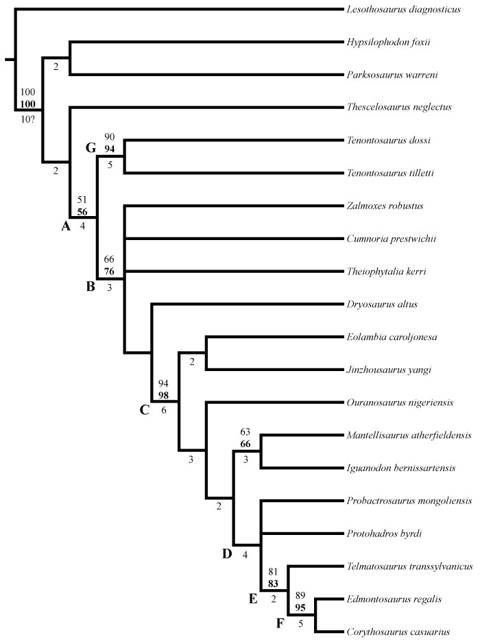
FIGURE 8. Coronal CT image, taken at the level of the open naris, of the left premaxilla (green, left), maxilla (pink, middle), and the vomer (cream, bottom) of OMNH 58340 showing the nature of their articulation. The maxilla likely fitted in closer articulation with the premaxilla. Faint green lines are an artifact of the program used to define the element on each image. Anterior is into the page.
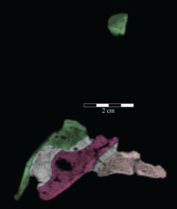
FIGURE 9. Medial skull schematic (above), right skull photograph (middle), and right skull schematic (below) of OMNH 58340. Parts of the vomer, frontal, parietal, laterosphenoid, squamosal, supraoccipital, exoccipital/opisthotic, and prootic, as well as the entirety of the right quadrate, quadratojugal, and jugal, have been removed from the medial skull schematic in order to show the structure of the braincase and its orientation with the rest of the skull. The broken texture in gray in the upper schematic indicates the sectioning of elements necessary to view the endocranium. The skull is angled at the ‘alert position’ indicated by the horizontal semicircular canal. Natural fenestrae are shaded gray. Anterior is to the right. Abbreviations: ap - alveolar parapet; s - sulcus.
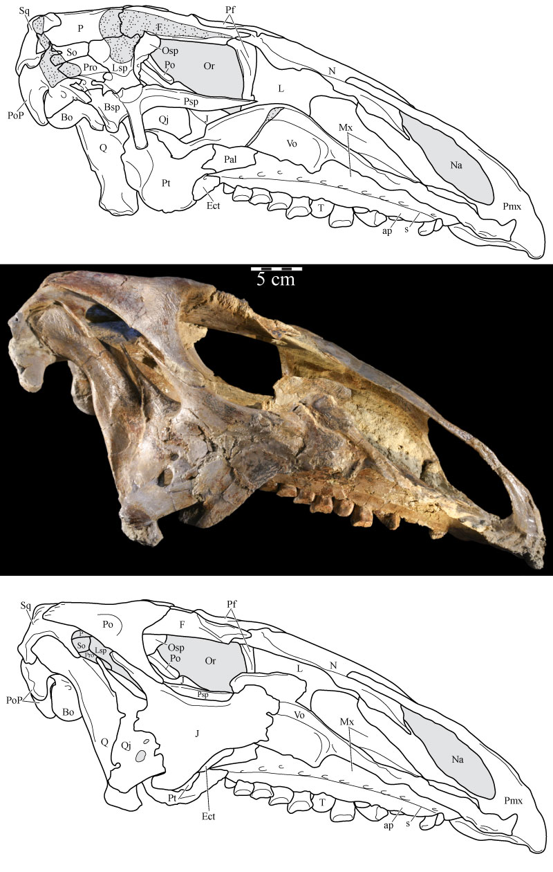
FIGURE 10. Medial view of the left side of the virtual skull of OMNH 58340 with the vomer present (10.1), allowing a view of the articulation of the vomer with the pterygoid, and with the palatine and vomer removed (10.2), allowing a view of the joints between the maxilla, lacrimal, prefrontal, jugal, ectopterygoid, and pterygoid. The vertically striated texture present on the visible surfaces of many elements, notably the lacrimal, maxilla, and premaxilla, is an artifact of the process used to isolate CT images of each element from the remainder of the data set. Abbreviations: f - flange; pp - posterior processes; tp - triangular processes.
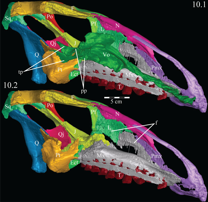
FIGURE 11. Photographs of the medial aspect of the vomer (foreground) and dental ridge of the maxilla (background, just below matrix) of OMNH 58340 (11.1) and showing the fine grain on the dorsal surface of the maxilla (11.2). Single headed arrow indicates the median ridge of the vomer. Double headed arrows indicate the paired sharp ridges of the same element. The area figured in 11.2 is indicated by the shaded area in 11.1. Anterior is to the right.
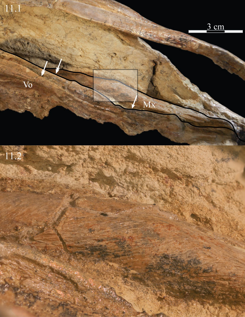
FIGURE 12. Coronal CT image, taken at the level of the posterior termination of the antorbital fenestra, of the paired posterior processes of the vomer of OMNH 58340. This image shows the disarticulated nature of the two halves of the posterior part of the element, as well as their shape just anterior to the opening of the orbit, between the paired palatines. Faint green lines are an artifact of the program used to define the element on each image. Anterior is into the page.
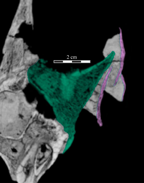
FIGURE 13. Medial (13.1) and ventral (13.2) photographs of the disarticulated right palatine of OMNH 58340. Anterior is to the left. Abbreviations: ap - anterior process; apt - articulation with pterygoid; l - lamina; lp - lateral process.
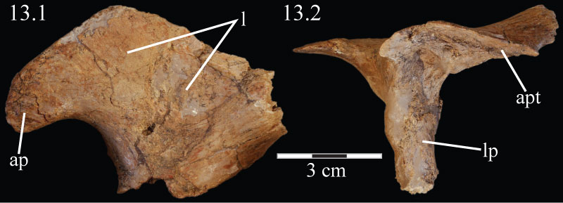
FIGURE 14. Dorsal view of the elements of the virtual palate of OMNH 58340, including the palatine, maxilla, pterygoid, and ectopterygoid, as well as the jugal, detailing the means of articulation between the various elements. Anterior is to the right.
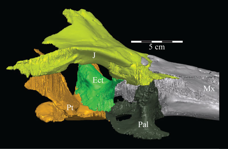
FIGURE 15. Posterolaterodorsal view of the left orbit of the virtual skull of OMNH 58340, presenting a view of the joint between the prefrontal, lacrimal, jugal, and palatine. The parallel contour lines on the posterior surfaces of the parietal, prefrontal, quadrate, and squamosal, are an artifact of the CT scanning process, and represent the faces of individual slice images. These lines occur on any surface angled near parallel with the scanner. Anterior is into the page, toward the top left. Abbreviation: lt - lateral tubercle; pg - pterygoid groove.
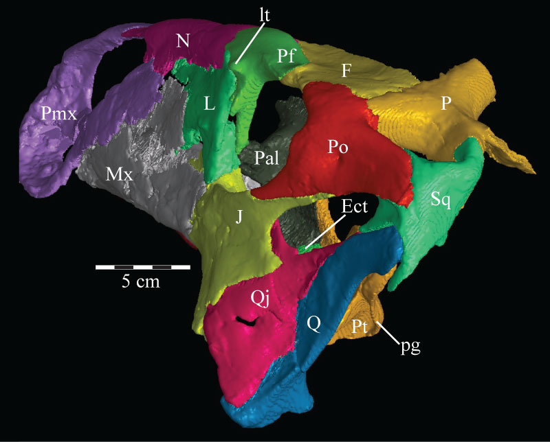
FIGURE 16. Lateral view (16.1) of the virtual ectopterygoid (green) and pterygoid (yellow) of OMNH 58340. Dorsal view (16.2) of the virtual pterygoid only. Anterior is to the left. Abbreviations: ep - ectopterygoid process; lf - lateral fossa; pf - pterygoid flange; pp - palatine process; mp - medial process; qp - quadratic process.
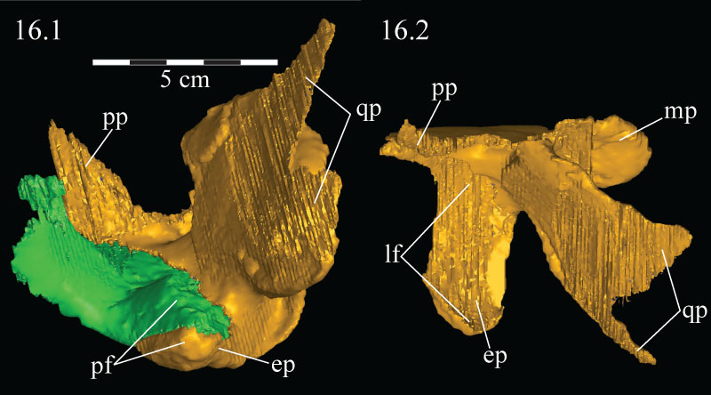
FIGURE 17. Anteromediodorsal view of the virtual skull of OMNH 58340, showing the features surrounding the maxillary groove of the maxilla, including the dental ridge and the possibility of a soft-tissue roof covering it in life. Abbreviations: dr - dental ridge of the maxilla; la - lamina of the maxilla; mf - medial flange of the lacrimal; mg - maxillary groove; mp - maxillary projection.
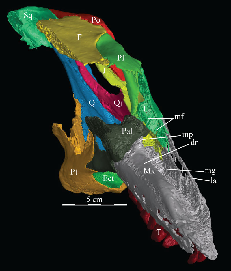
FIGURE 18. Anterior skull schematic (above) and photograph (below) of OMNH 58340. The two images are set to the same scale, demonstrating the amount of displacement in the right side of the skull. The schematic was reconstructed by digitally mirroring the left side of the rostrum and suspensorium in order to approximate the actual appearance of the skull. Natural fenestrae are shaded gray. Anterior is out of the page.
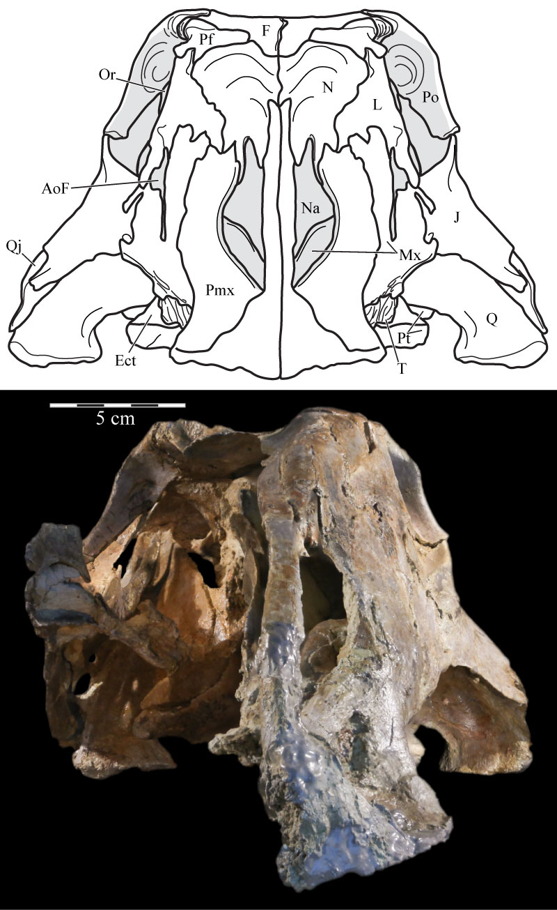
FIGURE 19. Medial view of the virtual left premaxilla, nasal, lacrimal, and maxilla of OMNH 58340 showing the nasal arch of the nasal just below its articulation with the nasal process of the premaxilla. Anterior is to the right. Abbreviation: na - nasal arch.
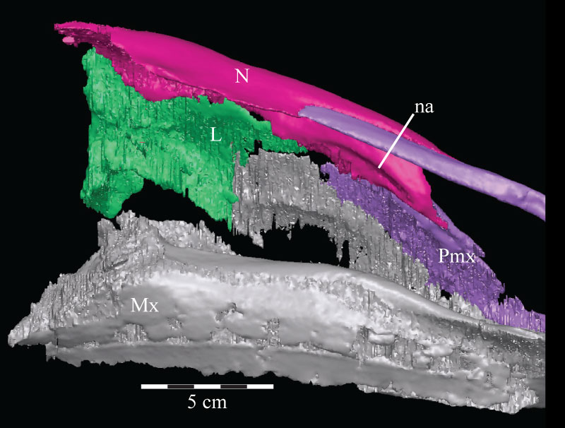
FIGURE 20. Coronal CT image, taken at the anterior edge of the left orbit, of the prefrontal (violet), nasal (dark green), and frontal (yellow) of OMNH 58340 showing their articulation. Faint green lines are an artifact of the program used to define the element on each image. Anterior is into the page.
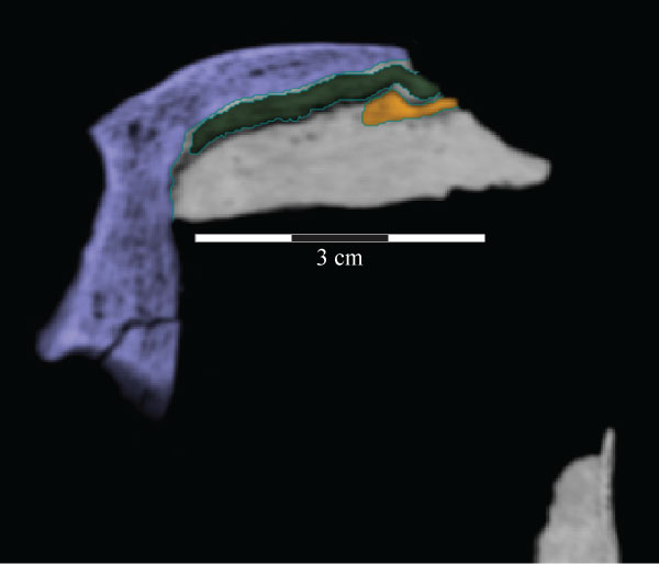
FIGURE 21. Ventral view of the skull roof of OMNH 58340. The palate and the lateral facial elements have been removed to allow for the viewing of the associations of the elements depicted. Location of endocast indicated by black outline. Note the nasal and prefrontal ending at nearly the same point posteriorly. Anterior is to the right. Abbreviations: la - area of articulation with capitate process of laterosphenoid; pp - pedicles of partietal; ar - arcuate ridge; sa - area of articulation with supraoccipital.
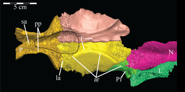
FIGURE 22. Coronal CT image of the left maxilla (pink) of OMNH 58340, taken midway along the axial length of the element, showing its facial lamina (left) and dental ridge (right, surrounding two teeth, lighter, due to higher density). Faint green lines are an artifact of the program used to define the element on each image. Abbreviations: a - alveolus; c - canal; lg - lamellar gap.
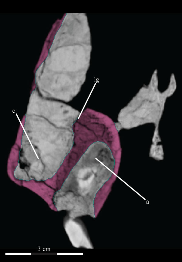
FIGURE 23. Labial photograph of the left maxillary dentition (23.1) and lingual photograph of the left dentary dentition (23.2) of OMNH 58340. Note the finely ornamented surface of the alveolar parapet of the dentary between the lower exposed surfaces of the teeth and the dorsally arched sulcus.
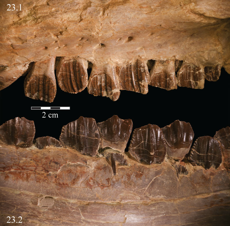
FIGURE 24. Coronal CT image, taken just anterior of the left orbit, of the lacrimal (light blue) and its shelf (lower right) of OMNH 58340. Faint green lines are an artifact of the program used to define the element on each image. Anterior is into the page.
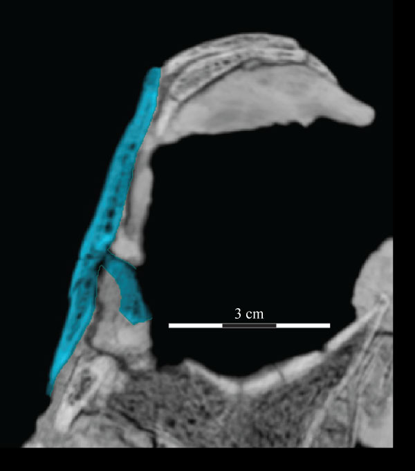
FIGURE 25. Ventral (25.1) and medial (25.2) photographs of the disarticulated right prefrontal of OMNH 58340. Anterior is left. Arrows indicate undulating texture on the ventral and medial surfaces of the element, which indicate the area of articulation with the frontal. The space between two broken ends of the ventral process in 25.2 is indicated by a dashed gray line. Abbreviation: vp - ventral process.
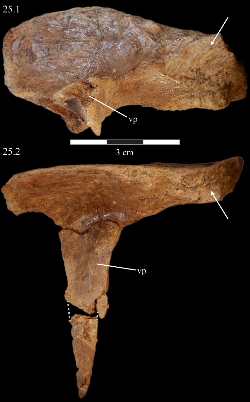
FIGURE 26. Photograph of the left prefrontal, frontal, and postorbital of OMNH 58340, showing the high degree of rugosity above the orbit. Anterior is to the left.
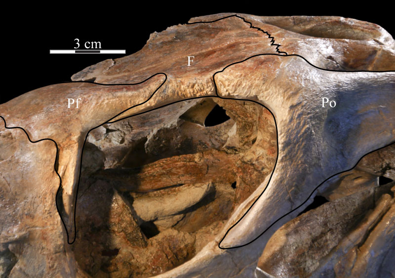
FIGURE 27. Photograph of the prefrontal fossa (indicated by arrows) of the right frontal of OMNH 58340. The pyramidal corner of the disarticulated prefrontal fits into this fossa. Anterior is to the right and out of the page.
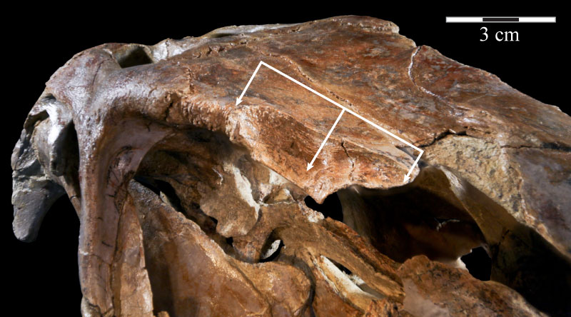
FIGURE 28. Coronal CT image, taken just anterior of the orbit, of the left prefrontal and its ventral process (violet), lacrimal (light blue), and palatine and its lateral process (brown) of OMNH 58340 showing the nature of their articulation. Faint green lines are an artifact of the program used to define the element on each image. Anterior is into the page.
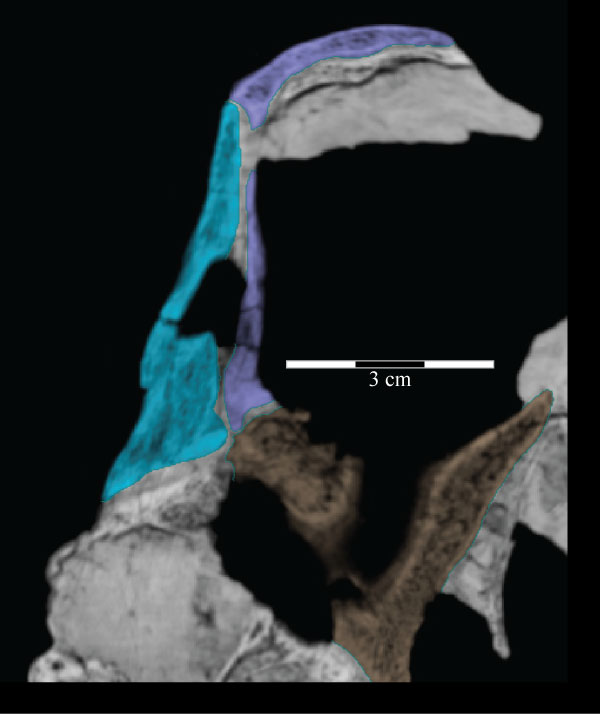
FIGURE 29. Photograph of the left squamosal of OMNH 58340, showing the deep texture on the lateral surface of the body of the bone, continued forward onto the lateral surface of the squamosal process of the postorbital. Anterior is to the upper left and into the page. Abbreviations: vt - ventral tab of the squamosal.
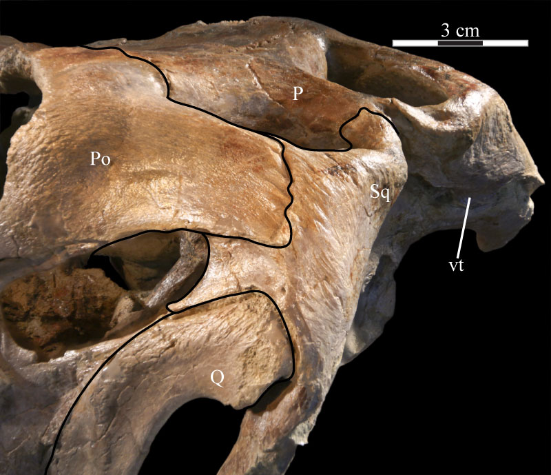
FIGURE 30. Lateral view of the virtual left squamosal of OMNH 58340 showing the deep articular fossae and the processes of the element. Anterior is to the left. Abbreviations: ap - anterior process; ah - articulation for head of quadrate; apo - articulation for postorbital; poq - postquadratic process; preq - prequadratic process.
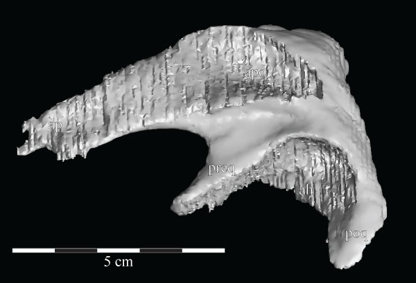
FIGURE 31. Lateral view of the virtual left quadratojugal of OMNH 58340 showing the rhomboidal shape of the element. The corrugated area on the left half of the element (outlined in black) is an artifact of the sectioning process and represents the articular surface for the jugal. Anterior is to the left.
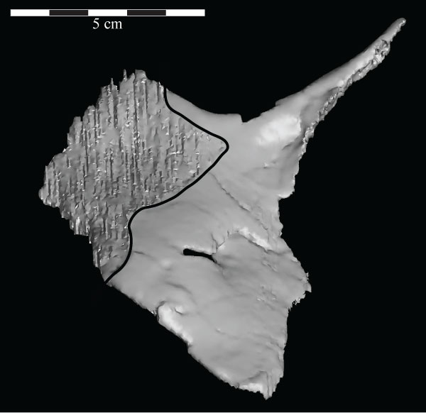
FIGURE 32. A lateral view of the virtual skull (lower left) and braincase (inlay, upper right) of OMNH 58340 with a high transparency, making the endocast (blue), cranial nerves (yellow), and semicircular canals (magenta) visible. The skull is at the ‘alert position’ indicated by the horizontal semicircular canals. Abbreviations: bt - basal tubera; bpt - basipterygoid process; oc - occipital condyle.
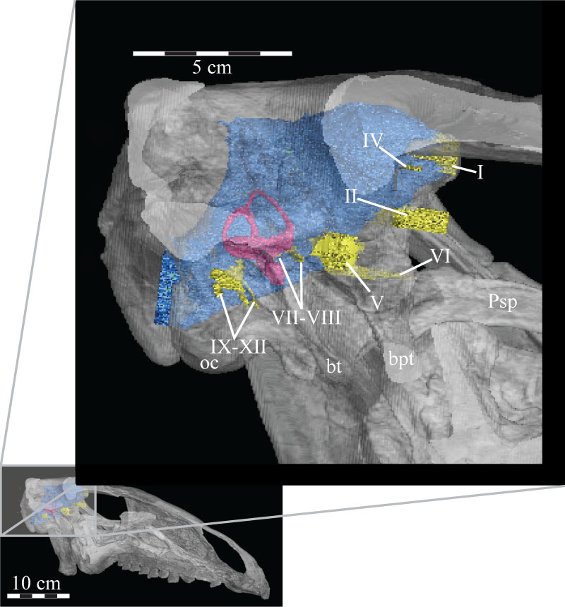
FIGURE 33. Photograph of the posterior portion of the dorsal surface of the skull of OMNH 58340, showing the small waist of the parietal (indicated by arrow). This groove may have been for the passage of an artery or may mark the separation between two muscles. There is an identical feature on the opposite side of the skull. Anterior is to the left.
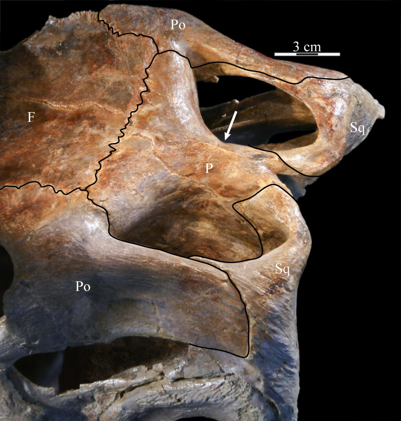
FIGURE 34. Anterior view of the virtual braincase of OMNH 58340. The supraoccipital is indicated by the pink that shows through the foramina for CN I–III. The smooth area across the front of the parasphenoid and the left basipterygoid process, as well as the regular curvature of the outer edge of the right laterosphenoid, prootic, and opisthotic (to the left), are artifacts of CT scanning and represent the limits of the data set. Note the irregular anterior surface of the parietal, showing the interdigitate nature of the suture of that element with the frontals. Black lines are used to denote particular features. Anterior is out of the page and ventral is down. Abbreviations: bt - basal tubera; bpt - basipterygoid process; ct - crista terminalis; eV - excavation for trigeminal nerve (CN V); hf - hypophysial fossa; pp - preotic pendant; st - sella turcica; vt - ventral tubercle of the orbitosphenoid.
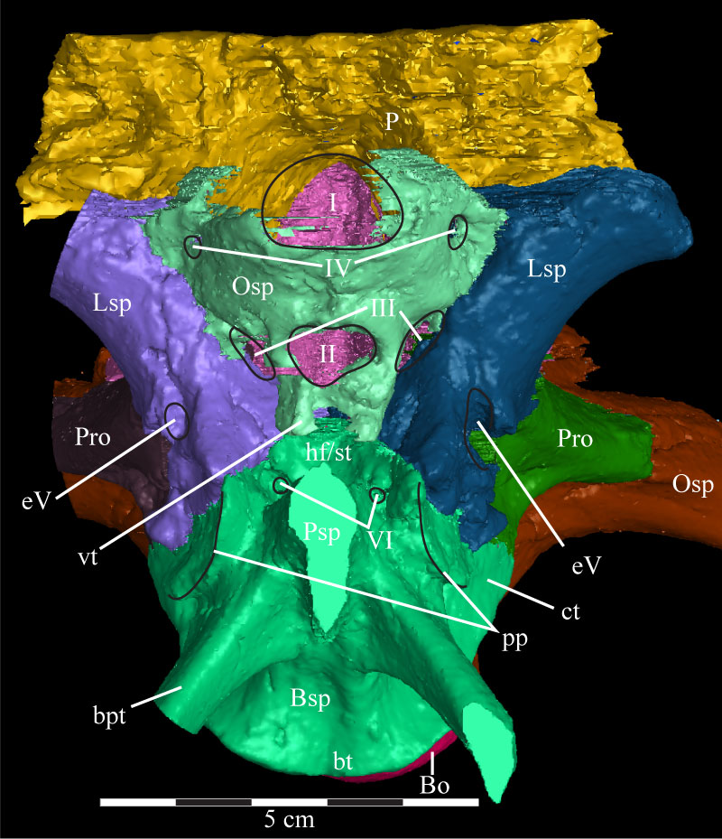
FIGURE 35. Lateral view of the virtual braincase of OMNH 58340. Black lines are used to denote particular features. The straight lines at the front of the basisphenoid to the left and the exoccipital/opisthotic to the right are artifacts of the CT scanning and represent the limits of the data set. The frontal and parietal extend beyond these levels because they were transplanted from the full cranial scan. Note that the basioccipital is visible in two locations. Anterior is to the left. Abbreviations: bt - basal tubera; bpt - basipterygoid process; cc - internal carotid canal (obscured); ci - crista interfenestralis; cm - crista metotica; cp - crista prootica; ct - crista tuberalis; eV - excavation for trigeminal nerve (Cranial Nerve V); fm - fenestra metotica; fo - fenestra ovalis; g - groove of crista tuberalis; pp - preotic pendant; sr - stapedial recess; ? - unnamed ridge.
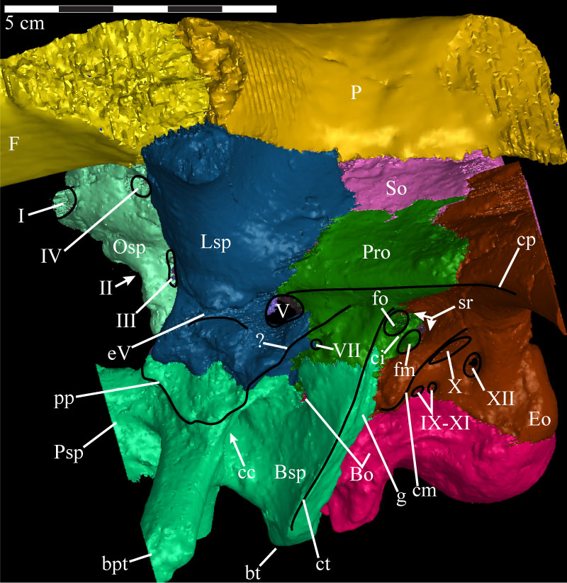
FIGURE 36. Dorsolateral view of the virtual braincase of OMNH 58340 with the parietal, left laterosphenoid, left prootic, and left sections of the supraoccipital and exoccipital/opisthotic removed. The flat edges of the parasphenoid and of the supraoccipital and exoccipital to the left and right, respectively, are artifacts of the CT scanning, and represent the limits of the data set. The supraoccipital (pink), exoccipital (brown), and orbitosphenoid (light green) were sectioned to allow for viewing of structures that would normally be covered by these elements. The computer program used to do this randomly assigns interior colors to the models, hunter green in the case of the supraoccipital, light blue in the case of the exoccipital, and maroon for the orbitosphenoid. Anterior is to the left. Abbreviations: bpt - basipterygoid process; dVIII - dorsal ramus of the acoustic nerve; ed - endolymphatic duct; mp - median process of the basioccipital; pf - area of the pontine flexure; vp - ventral process of the supraoccipital; vVIII - ventral ramus of the acoustic nerve.
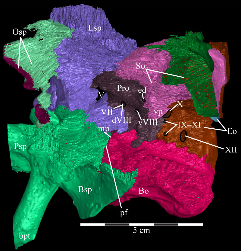
FIGURE 37. Posterior view of the virtual braincase of OMNH 58340 with the exoccipital/opisthotic removed. The regularly curved borders along the right side, as well as the flat squamosal bosses of the supraoccipital, are an artifact of the CT scanning and represent the limits of the data set. Anterior is into the page and dorsal is up. Abbreviations: ab - auditory bulla; ap - ascending process; bf - basioccipital furrow; bt - basal tubera; bpt - basipterygoid process; dt - dorsal tubercle; ns - nuchal shelf; sb - squamosal boss; ss - supraoccipital shelf; vp - ventral processes.
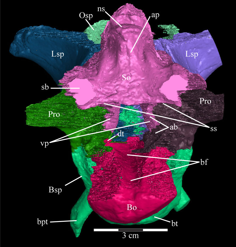
FIGURE 38. Lateral view of the virtual supraoccipital (purple) and the left endosseous labyrinth (pink) of OMNH 58340 showing the common crus of the semicircular canals entering the ventral process of the supraoccipital. Anterior is to the left. Abbreviations: asc - anterior semicircular canal; cc - common crus; nc - nuchal crest; psc - posterior semicircular canal; ss - supraoccipital shelf; vp - ventral process.
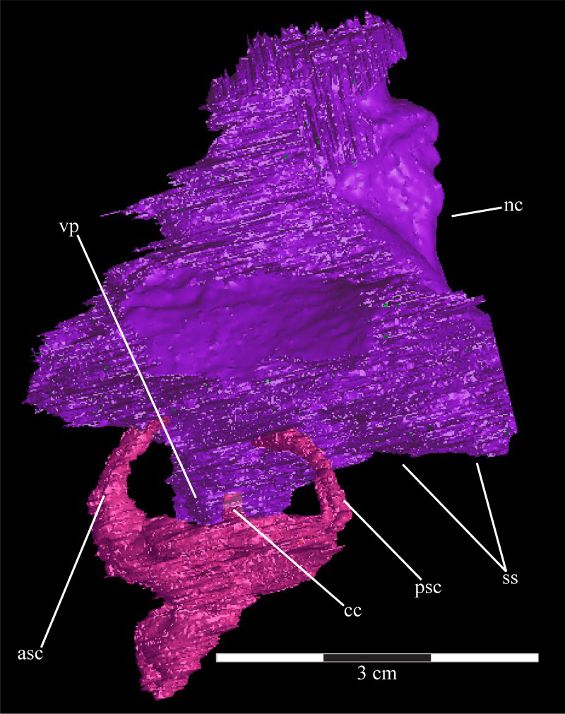
FIGURE 39. Dorsal view of the virtual braincase of OMNH 58340 with the parietal removed, showing the composition of the floor of the braincase. The straight posterior edge of the opisthotic, as well as the straight anterior edge of the basisphenoid and lateral edge of the laterosphenoid, are artifacts of the CT scanning and represent the limits of the data set. Anterior is down. Abbreviations: ap - area of articulation with parietal; i - infundibular canal; pp - posterior process of the orbitosphenoid.
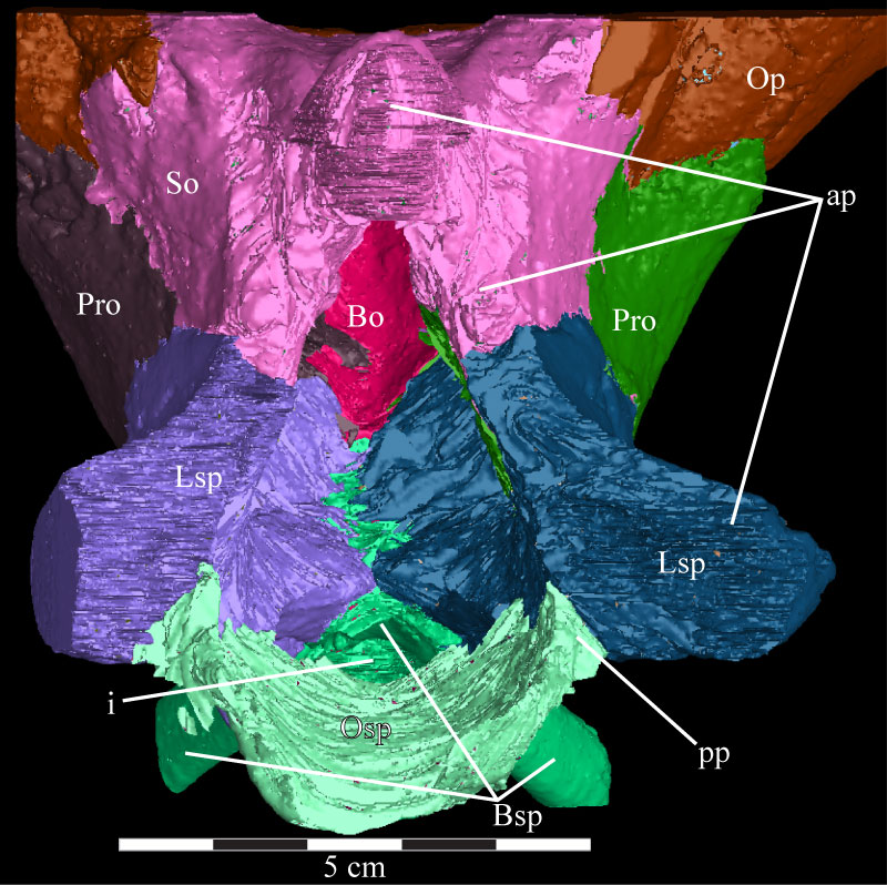
FIGURE 40. Horizontal CT image, taken at the level of the trigeminal foramina, of the basisphenoid and parasphenoid (orange), laterosphenoids (pink and light blue), trigeminal nerve foramina (yellow), prootics (teal and red), horizontal semicircular canals (purple), and exoccipital/opisthotic (green) of OMNH 58340. The endocast is the black and gray area in the middle of the image. The ventral processes of the supraoccipital can also be seen, as well as their peculiar density. Faint green lines are an artifact of the program used to define the element on each image. Anterior is down. Abbreviations: sc - horizontal semicircular canals; vp - ventral processes.
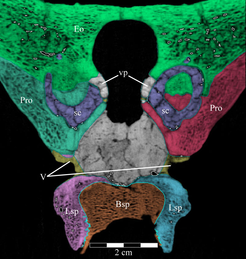
FIGURE 41. Anterior view of the virtual braincase of OMNH 58340 with the parietal, orbitosphenoid, left laterosphenoid, and basisphenoid/parasphenoid removed. The curvature along the left border is an artifact of the CT scanning and represents the limit of the data set. Black lines are used to denote particular features. Anterior is out of the page and ventral is down. Abbreviations: conical fossa; ds - dorsal sagittal sinus; fm - foramen magnum.
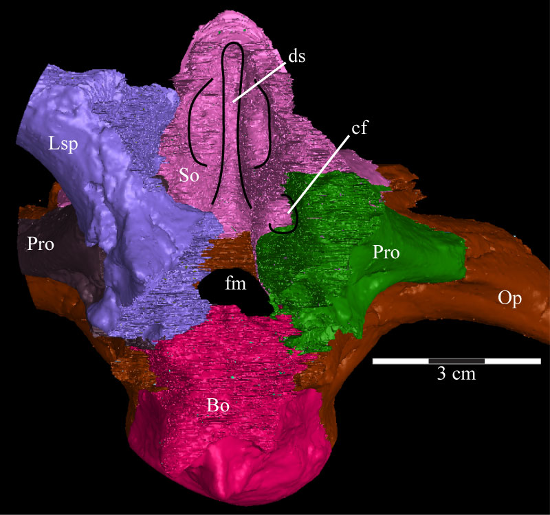
FIGURE 42. Posterodorsal view of the virtual braincase of OMNH 58340 with the parietal, supraoccipital, and exoccipital/opisthotic removed and the paths of the cranial nerves highlighted. Anterior is into the page. Abbreviations: i - infundibulum; mp - median process.
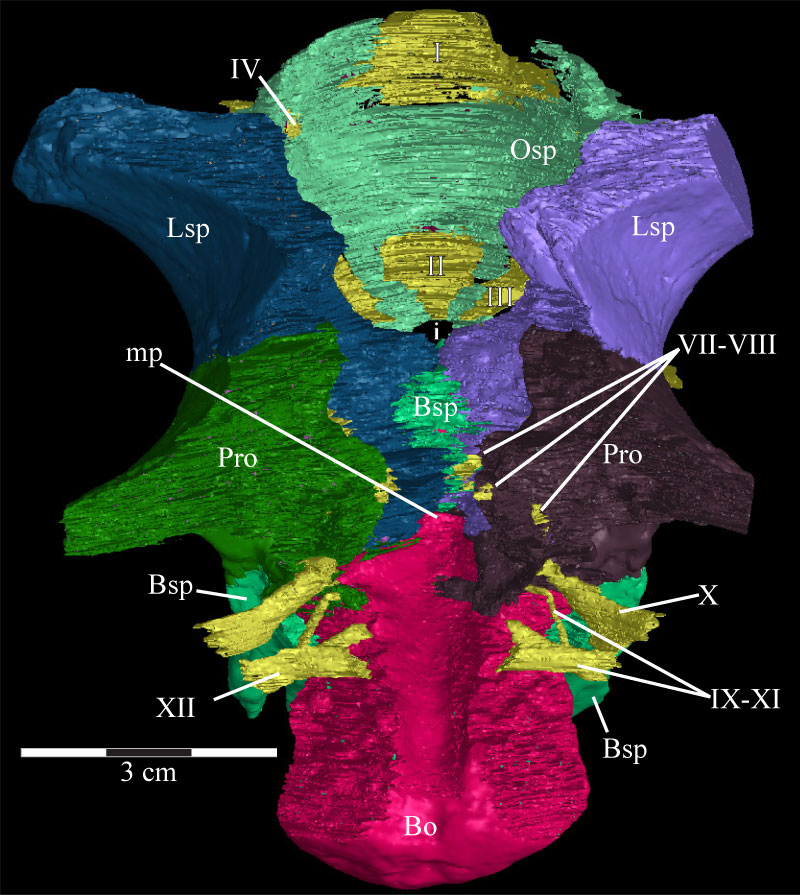
FIGURE 43. Photograph of the parasphenoid and basisphenoid complex and the anterior braincase of OMNH 58340. The parasphenoid extends forward (right) from the body of the basisphenoid and, at this angle, disappears behind the displaced right posterior process of the vomer. The trench running along the dorsal surface of the parasphenoid is visible (indicated by arrows). The ventral outline of the preotic pendant is indicated by the dashed line. Anterior is to the right and out of the page. Abbreviations: ct - possible crista trabecularis; pp - preotic pendant.
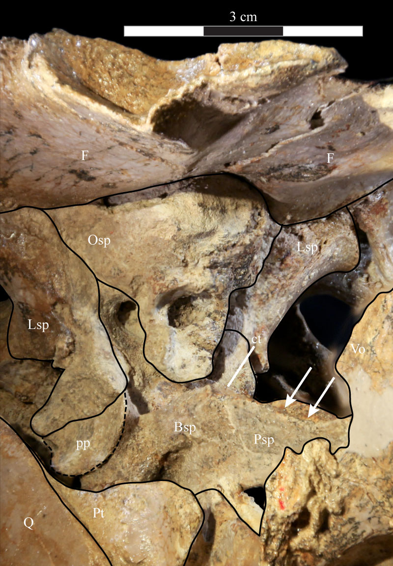
FIGURE 44. Right lateral (44.1), anteroventral (44.2), anterior (44.3), and posterior (44.4) views of the virtual endocast of OMNH 58340. 44.1 shows various landmarks for the passage of venous sinuses and cranial nerves. 44.2 highlights the waists between the cerebrum and cerebellum anterodorsally and the cerebellum and medulla posteroventrally. 44.3 shows the faint lobes present on the dorsal surface of the endocast, separated by a small trough (indicated by arrow). 44.4 shows the dorsal sagittal sinus in the middle and the flocculi of the endocast, as well as the dorsal bulbous expansions. Abbreviations: ad - anterodorsal ridge; av - anteroventral ridge; be - bulbous expansions; die - diencephalon; ds - dorsal sagittal sinus; dvs - dorsal venous sinus; f - flocculus; i - beginning of infundibular stalk; mes - mesencephalon; met - metencephalon; my - myelencephalon; pi - pineal process; pro - prosencephalon; rho - rhombencephalon; rV - ridge running to trigeminal nerve (CN V); rVI - ridge running to abducens nerves (CN VI) tel - telencephalon.
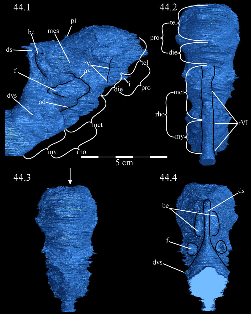
FIGURE 45. Dorsal (45.1), right lateral (45.2), anterior (45.3), and posterior (45.4) views of the virtual endosseous labyrinths of OMNH 58340. The canals are oriented in 45.1 such that anterior is above and posterior below. Ventral is down in all other images. Abbreviations: aa - anterior ampulla; asc - anterior semicircular canal; au - anterior utricle; ca - cavum capsularis; cc - common crus; fo - fenestra ovalis; lr - lagenar recess; lsc - lateral semicircular canal; pa - posterior ampulla; psc - posterior semicircular canal; pu - posterior utricle; v - vestibule.
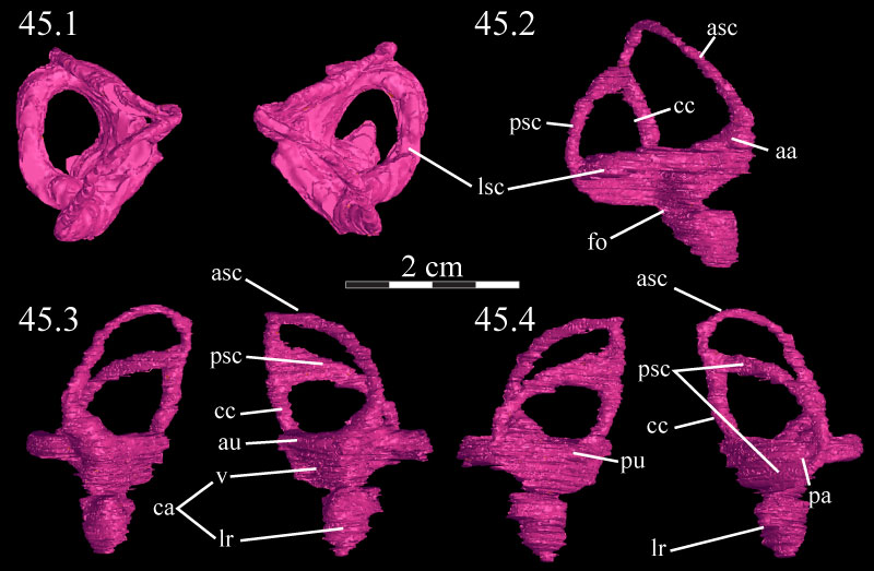
FIGURE 46. Lateral schematic of the left (above) and photographs of the left (middle) and right (below) mandibles of OMNH 58340. Note the amount of dorsoventral compression of the right mandible when compared with the left. Anterior is to the left in the first two images and to the right in the third. Abbreviation: sf - surangular foramen.
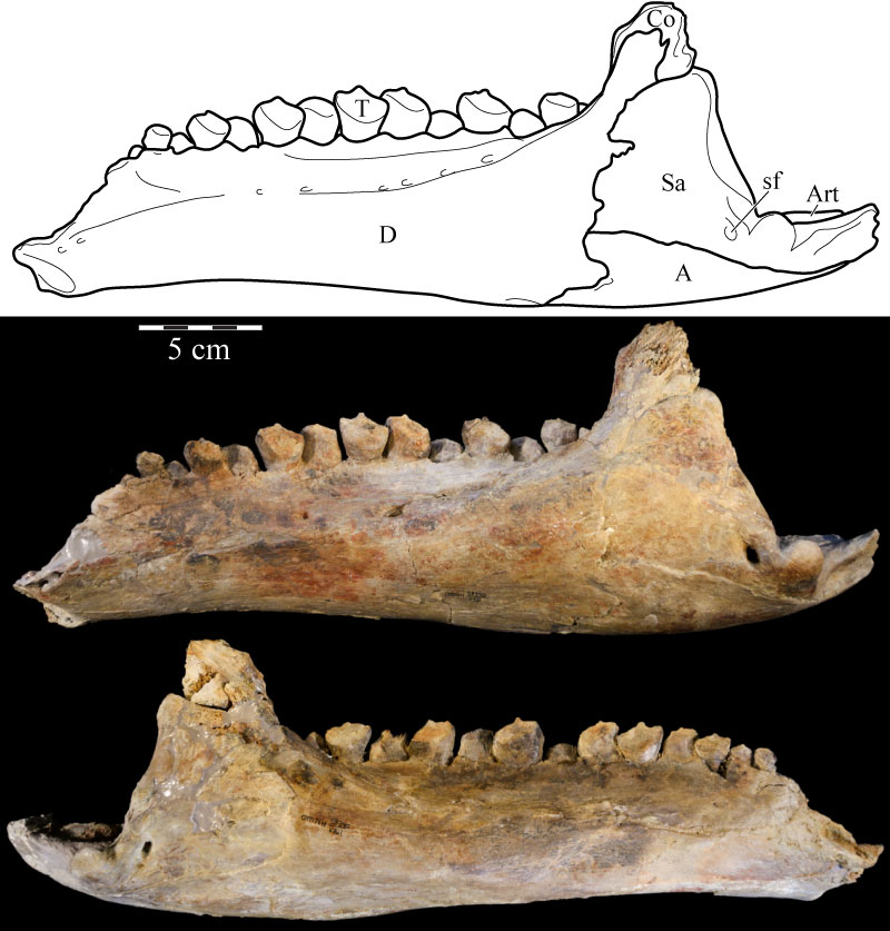
FIGURE 47. Medial schematic of the left (above) and photographs of the left (middle) and right (below) mandibles of OMNH 58340. Note the amount of dorsoventral compression of the right mandible when compared with the left. Anterior is to the right in the first two images and to the left in the third. Abbreviations: ap - alveolar parapet; imc - internal mandibular foramen; mc - Meckelian canal.
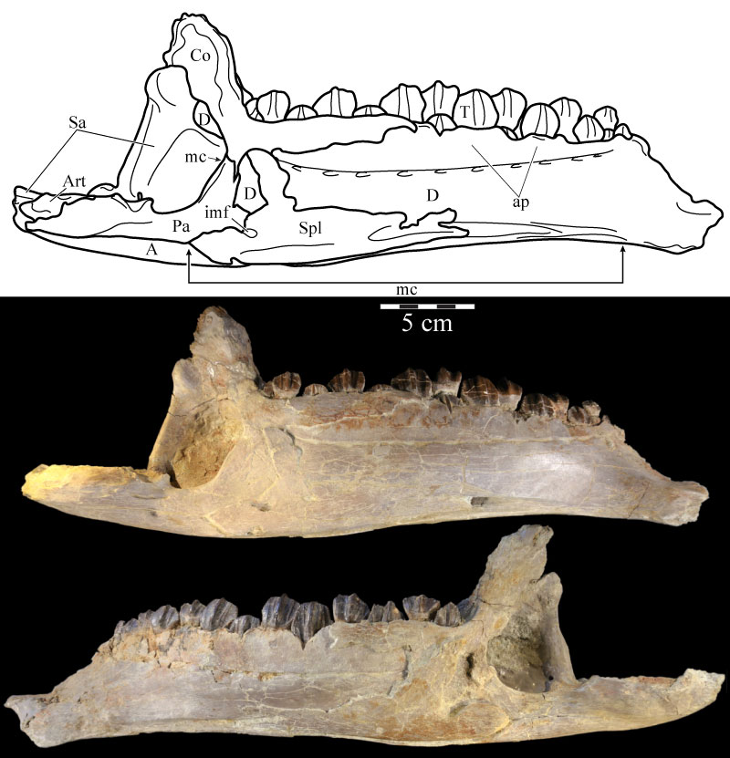
FIGURE 48. Photographs of the predentary of OMNH 58340 in dorsal (48.1), anterior (48.2), lateral (48.3), posterior (48.4), and ventral (48.5) views. Abbreviations: dp - dentary process; mt - median tab; pd - primary denticle; vp - ventral process; vs - ventral sulcus.
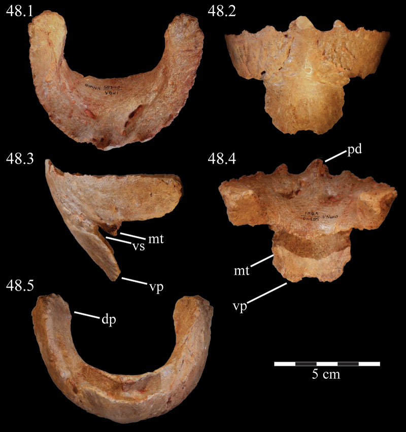
FIGURE 49. Dorsal (49.1), lateral (49.2), and medial (49.3) views of the virtual left dentary of OMNH 58340, disarticulated. Anterior is to the left in the first two images and to the right in the third. Abbreviations: at - alveolar trench; au - alveolar undulations; ca - area for articulation with coronoid; Mc - Meckelian canal; pf - predentary foramina; sa - area for articulation with surangular.
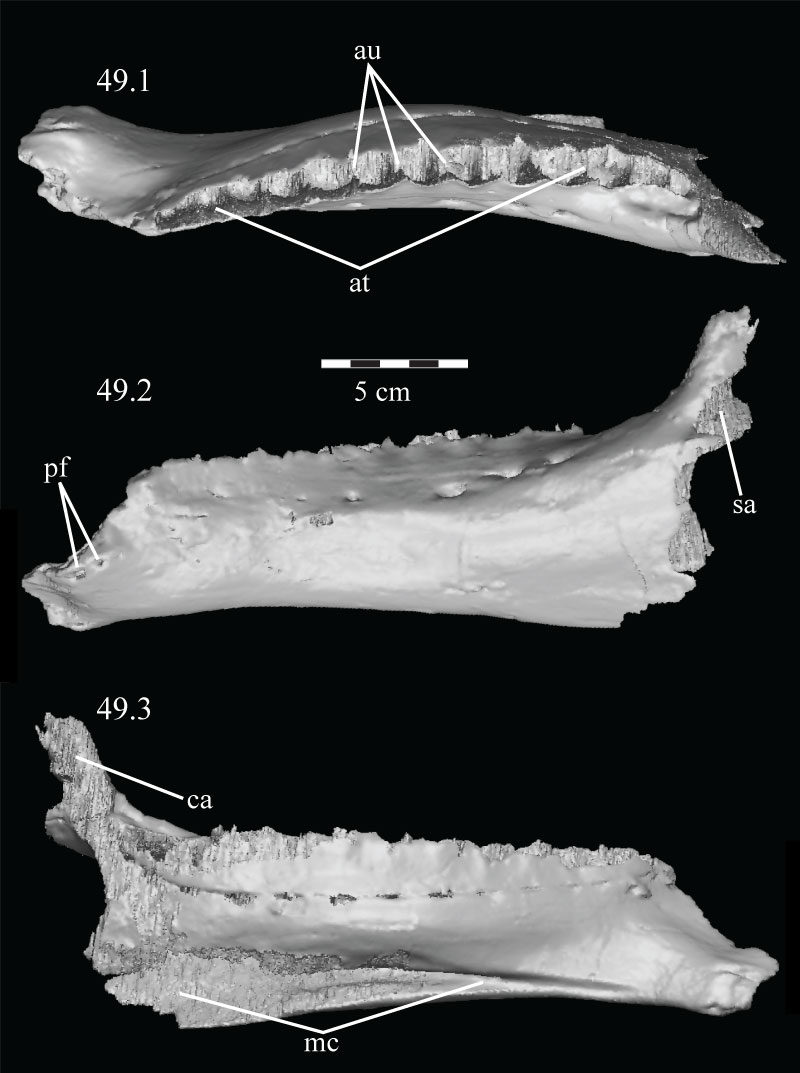
FIGURE 50. Lateral (50.1) and medial (50.2) views of the disarticulated virtual left coronoid of OMNH 58340. The parallel texturing covering most of 50.1 is an artifact of the sectioning process used to isolate the element and represents the articular surface for the surangular and dentary. Anterior is left in 50.1 and right in 50.2. Abbreviations: ap - anterior process; cp - coronoid process; da - articular surface for dentary; sa - articular surface for surangular; vt - ventral tab.
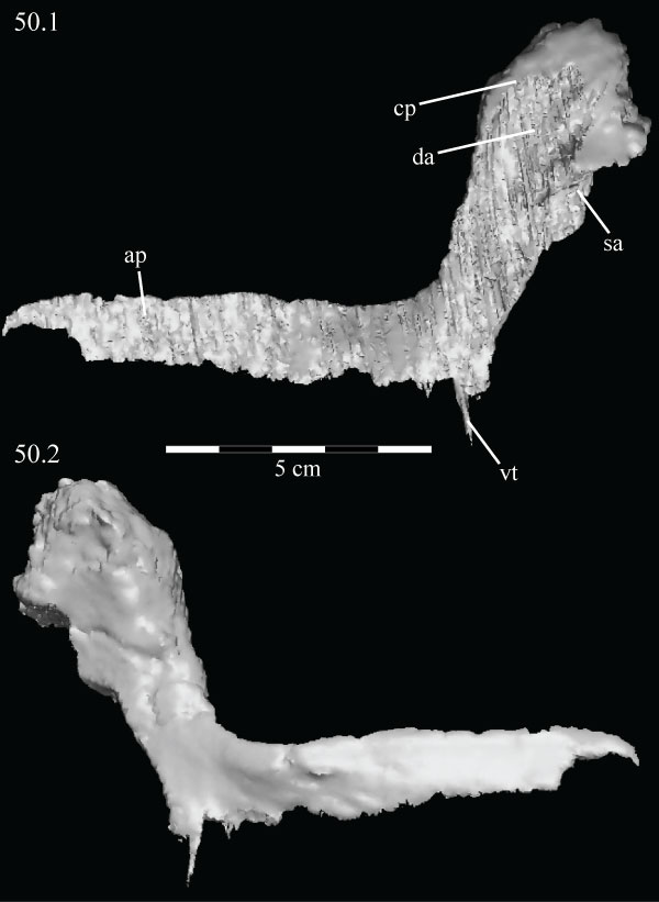
FIGURE 51. Medial view of the virtual left mandible of OMNH 58340 with the splenial and prearticular removed to show the extent of the Meckelian canal as it traverses the mandible. Anterior is right. Abbreviation: mc - Meckelian canal.
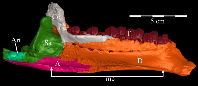
FIGURE 52. Lingual (above) and labial (below) views of the complete virtual tooth set of the left dentary of OMNH 58340, demonstrating the method of replacement, with groups of teeth beginning to be replaced in alternating succession from posterior to anterior. Individual Zahnreihen are indicated by dots of the same color placed at the occlusal tips of the teeth. Anterior is to the left in the upper image and to the right in the lower image.
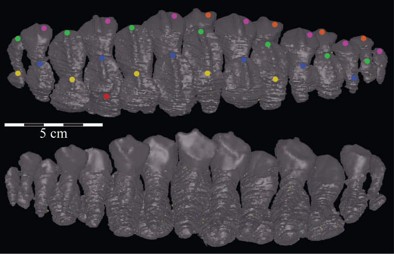
FIGURE 53. Lateral view of the virtual left splenial of OMNH 58340, disarticulated. Anterior is left. Abbreviations: ar - axial ridge; dp - dorsal process; imf - internal mandibular foramen; pr - posterior ridge.
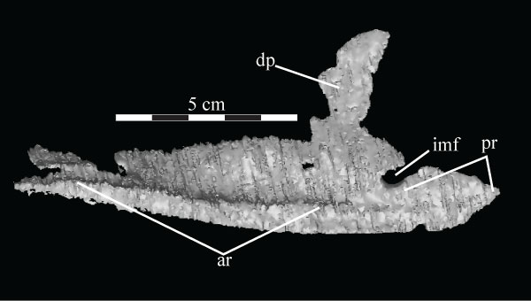
FIGURE 54. Lateral (54.1) and medial (54.2) views of the virtual left angular of OMNH 58340. Anterior is left in the upper image and right in the lower image. Abbreviations: da - articular surface for the dentary; mr - medial ridge; vr - ventral ridge.
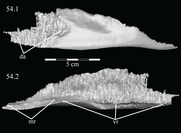
FIGURE 55. Medial view with the dorsal tip rotated lingually (55.1) of the virtual left angular (blue) and surangular (pink) and medial view (55.2) of the disarticulated virtual left surangular of OMNH 58340. Anterior is to the right. Abbreviations: aa - anterior angle; aw - anterior wing; da - dorsal angle; sf - surangular foramen; rp - retroarticular process; va - ventral angle.
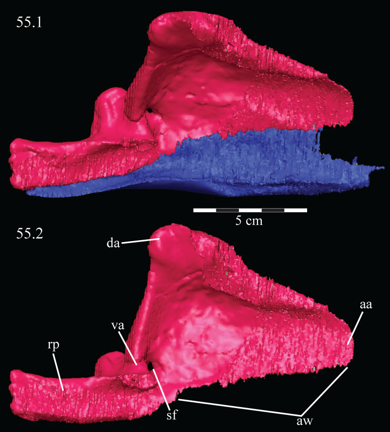
FIGURE 56. Photograph of the articular surface of the left mandible of OMNH 58340, detailing the facets present on the dorsal surfaces of the surangular and prearticular. The dashed lines circumscribe the articular fossae of the prearticular. Anterior is up. Abbreviations: lf - lateral articular fossa; m - matrix; mf - medial articular fossa.
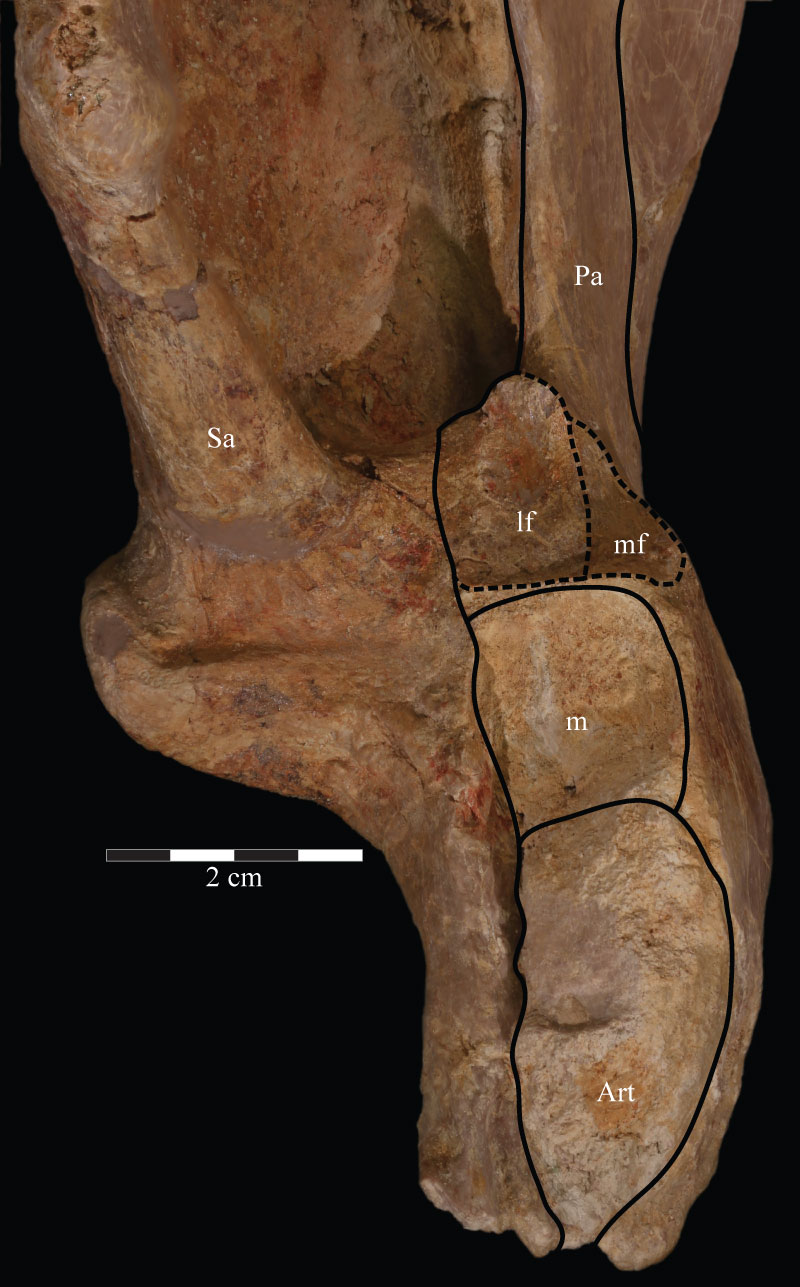
FIGURE 57. Dorsal schematic of the left (above) and photographs of the left (middle) and right (below) mandibles of OMNH 58340. Anterior is to the right. Abbreviation: df - dental foramina.
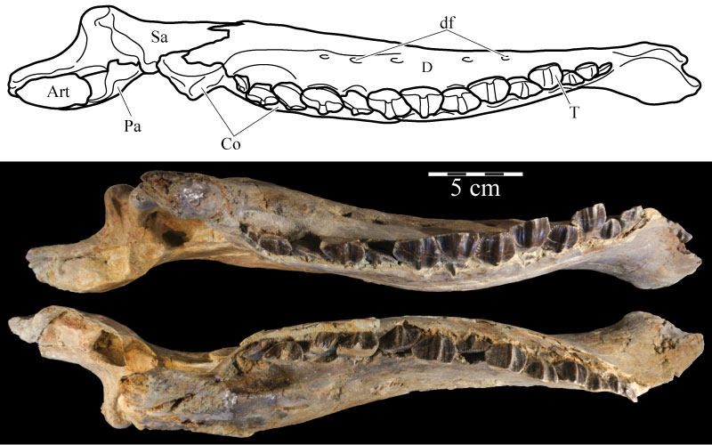
FIGURE 58. Posterodorsal view (58.1) of the virtual left surangular (pink) and prearticular (green) and medial view (58.2) of the virtual left prearticular of OMNH 58340. The left image shows the nature of the articulation of the two elements at the point of articulation of the mandible with the quadrate (indicated by arrow). Anterior is into the page in 58.1 and to the right in 58.2. Abbreviations: ap - anterior process; rp - retroarticular process; sa - area of articulation with the splenial.
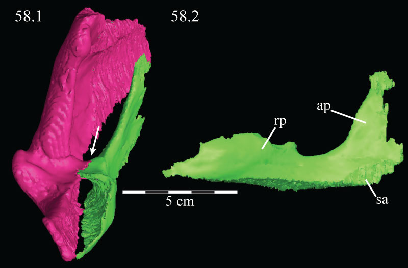
FIGURE 59. Ventral schematic (above) and photographs of the right (middle) and left (below) mandibles of OMNH 58340. Anterior is to the left.
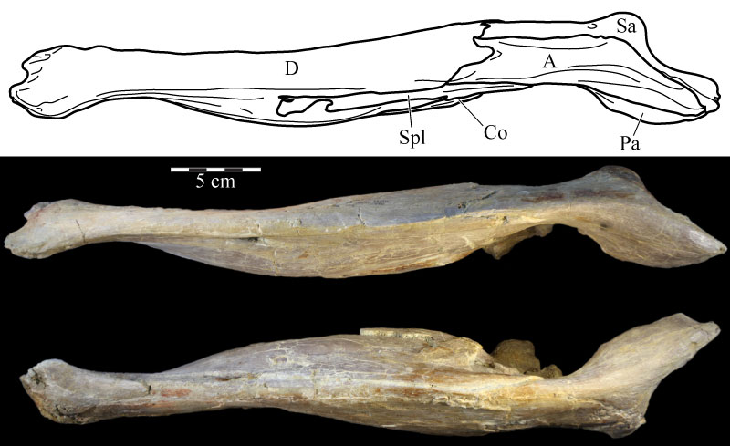
FIGURE 60. Coronal CT images of the maxillary (above) and dentary (below) teeth, taken midway along the length of the respective elements, of OMNH 58340 showing the method of lingual replacement and the concurrent resorption of the tooth being replaced. Dorsal is up and anterior is into the page. Abbreviations: ca - carina; ci - cingulum; e - enamel.
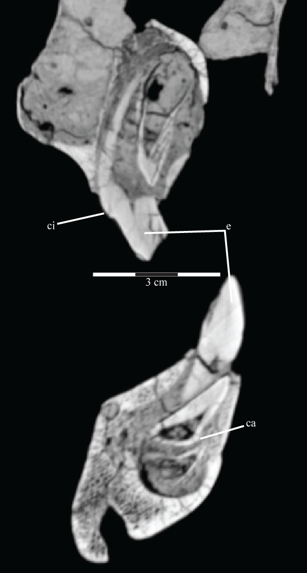
FIGURE 61. Photograph of the left (above, in lingual view) and right (below, in buccal view) hyoids of OMNH 58340. Anterior is to the left in the upper image and to the right in the lower image.
FIGURE 62. Optimized character distribution showing unambiguous character state changes on one of 12 most parsimonious trees (MPTs) recovered in this analysis. Character number and state present are indicated by the number above and below each line, respectively. Synapomorphic character states are indicated by black circles, while white circles denote homoplastic character states. Certain nodes are noted with letters referenced in the text. Optimization was performed in PAUP* version 4.0b10 (Swofford, 2002) and checked in TNT (Goloboff et al., 2008).
 D. Andrew Thomas
Department of Biology
Sam Noble Oklahoma Museum of Natural History
University of Oklahoma
2401 Chautauqua Avenue
Norman, Oklahoma 7307
USA
david.a.thomas-1@ou.edu
Andrew Thomas is a Ph.D. student at the University of Oklahoma, in the town where he was raised. He lives there with his wife and son. Andrew became fascinated with paleontology at an early age on regular field trips to outcrops in Oklahoma, Arkansas, and Texas with his father and brother, collecting the usual marine critters: brachiopods, echinoderms, and gastropods. He never grew out of that early delight in ancient life, and decided to turn that passion into a career in vertebrate paleontology. His research is now focused on the ornithopod dinosaurs of the Jurassic and Cretaceous, especially in the evolutionary trends in basal forms that led to the wildly successful duck-billed hadrosaurs, as well as the ability of ornithopod species of different sizes and shapes to coexist.
The cranial anatomy of Tenontosaurus tilletti Ostrom, 1970 (Dinosauria, Ornithopoda)
Plain Language Abstract
Tenontosaurus tilletti is a species of ornithopod, a two-legged herbivore related to the duck-billed dinosaurs, from the Lower Cretaceous (120–110 m.y.a.). Previous descriptions of the anatomy of the species have consisted exclusively of a short account of specimens collected in the Cloverly Formation of the Bighorn Basin of Montana, as well as a more detailed description of the postcranial skeleton. To date, the skull of T. tilletti remains poorly described. The present study is an attempt to rectify the situation using Tenontosaurus tilletti material collected from southeastern Oklahoma. In particular, an especially well preserved skull (OMNH 58340) of T. tilletti was CT-scanned and virtually separated into its individual skull bones. These elements, as well as reconstructions of the internal spaces for soft tissues, such as the braincase and cranial nerve passages, are described and illustrated in detail. This description is used to conduct a new analysis of the relationships of Tenontosaurus with its fellow ornithopods. The analysis strongly supports the genus Tenontosaurus, which includes another species, T. dossi from Texas, as well as its position relative to more basal ornithopods and derived iguanodontians. This new analysis largely agrees with previous studies.
Resumen en Español
La anatomía craneal de Tenontosaurus tilletti Ostrom, 1970 (Dinosauria, Ornithopoda)
Tenontosaurus tilletti Ostrom, 1970, históricamente asignado a 'Iguanodontia basales', es una especie de herbívoro bípedo del Cretácico Temprano (Aptiano–Albiano). Las publicaciones previas sobre la anatomía de la especie han consistido de breves informes de especímenes colectados en la Formación Cloverly de la Cuenca Bighorn de Montana, y una descripción más detallada del esqueleto postcraneal hecha por Forster (1990). Al día de hoy, el cráneo de T. tilletti permanece pobremente descripto debido a falta de investigación y especímenes mal preservados.
El presente estudio es un intento de rectificar la situación con material referible a Tenontosaurus tilletti, colectado en el sudeste de Oklahoma. Un cráneo particularmente bien preservado (OMNH 58340) de T. tilletti fue escaneado mediante TC y se aislaron sus elementos constituyentes. Estos elementos, así como las reconstrucciones de los espacios internos ocupados por tejidos blandos, tales como el endomolde y forámenes de nervios craneales, son aquí descriptos e ilustrados en detalle. Esta descripción se utiliza para conducir un análisis sistemático novedoso. El análisis respalda al género Tenontosaurus, así como a su posición relativa respecto de los 'hipsilofodontes' y los iguanodontianos, y coincide en gran parte con análisis previos.
Palabras clave: anatomía craneal; descripción; tomografía computarizada (TC); ornitópodo; Iguanodontia; Tenontosaurus
Traducción: Diana Elizabeth Fernández
Résumé en Français
L'anatomie crânienne de Tenontosaurus tilletti Ostrom, 1970 (Dinosauria, Ornithopoda)
Tenontosaurus tilletti Ostrom, 1970, historiquement attribué aux "Iguanodontia basaux", est une espèce de dinosaure herbivore bipède du Crétacé inférieur (Aptien-Albien). Les publications précédentes sur l'anatomie de cette espèce comprennent un bref rapport sur les spécimens collectés dans la formation de Cloverly du bassin du Bighorn dans le Montana et une description plus détaillée du squelette post-crânien par Forster (1990). Jusqu'à présent, le crâne et la mandibule de T. tilletti sont encore mal décrits, et ce en raison du peu de recherches qui leur sont consacrées et de la mauvaise préservation des spécimens.
Cette étude tente de rectifier la situation en décrivant du matériel attribué à Tenontosaurus tilletti, collecté au sud-est de l'Oklahoma. Un spécimen de T. tilletti, comprenant le crâne et la mandibule associés et particulièrement bien préservés (OMNH 58340), a notamment été scanné par tomographie assistée par ordinateur et les éléments le composant ont été séparés virtuellement. Ces éléments, ainsi que la reconstruction des espaces internes abritant les tissus mous, tels que le moulage endocrânien et les foramens des nerfs crâniens, sont ici décrits et illustrés en détail. Cette description est utilisée pour effectuer une nouvelle analyse phylogénétique. Cette dernière soutient fortement la monophylie du genre Tenontosaurus, de même que sa position relativement aux autres Iguanodontia et aux 'hypsilophodontes', et est en grande partie en accord avec les analyses précédentes.
Mots-clés : anatomie crânienne ; description ; tomographie assistée par ordinateur (CT) ; ornithopode ; Iguanodontia ; Tenontosaurus
Translator: Antoine Souron
Deutsche Zusammenfassung
Die Cranial-Anatomie von Tenontosaurus tilletti Ostrom, 1970 (Dinosauria, Ornithopoda)
Tenontosaurus tilletti Ostrom, 1970, historisch den 'basalen Iguanodontia' zugeordnet, ist eine Art bipeder Herbivoren aus der unteren Kreide (Apt–Alb). Vorangehende Veröffentlichungen zur Anatomie dieser Art bestanden aus einem oberflächlichen Bericht über die Stücke aus der Cloverly Formation des Bighorn Beckens von Montana ebenso wie aus einer detaillierteren Beschreibung des Postcranialskeletts von Forster (1990). Bis heute verbleibt der Schädel von T. tilletti wegen fehlender Untersuchungen und schlecht erhaltener Stücke unzureichend beschrieben.
Die vorliegende Untersuchung möchte mit Material, das Tenontosaurus tilletti zugeschrieben werden kann und das im Südosten von Oklahoma gesammelt wurde, die Situation verbessern. Insbesondere ein sehr gut erhaltener Schädel (OMNH 58340) von T. tilletti wurde CT-gescannt und virtuell in seine Einzelkomponenten zerlegt. Diese Elemente wurden hier zusammen mit Rekonstruktionen der internen Weichteil-Zwischenräume wie dem Schädelausguss und cranialen Nervenöffnungen beschrieben und detailliert abgebildet. Diese Beschreibung wird für die Umsetzung einer neuen systematischen Analyse verwendet. Die Untersuchung unterstützt nachdrücklich die Gattung Tenontosaurus, sowie deren Position relativ zu den 'Hypsilophodonten und Iguanodontinen und stimmt weitestgehend mit vorangegangenen Analysen überein.
Schlüsselwörter: Cranial-Anatomie; Beschreibung; Computertomografie (CT); Ornithopode; Iguanodontia; Tenontosaurus
Translator: Eva Gebauer
Arabic
Translator: Ashraf M.T. Elewa
-
-
-
Review: The Princeton Field Guide to Mesozoic Sea Reptiles
 The Princeton Field Guide to Mesozoic Sea Reptiles
Article number: 26.1.1R
April 2023

 Poster Winners 2024