

Article Search
Volume 27.1
January–April 2024
Full table of contents
ISSN: 1094-8074, web version;
1935-3952, print version
Recent Research Articles
See all articles in 27.1 January-April 2024
See all articles in 26.3 September-December 2023
See all articles in 26.2 May-August 2023
See all articles in 26.1 January-April 2023
APPENDIX 1.
List of characters used in the phylogenetic analysis. Characters were mainly taken from Carrano et al. (2012), often with modifications. A complete documentation of the changes in character definition and character codings will be presented elsewhere (Rauhut and Pol, in prep.).
Cranial skeleton
1. Premaxilla, inter-premaxillary suture in adults: open (0), fused (1).
2. Premaxilla, height/length ratio ventral to external naris (length measured along ventral border of bone): < 0.5 (0), 0.5-0.99 (1), 1-1.49 (2), 1.5 or higher (3)
3. Premaxilla, subnarial process and ventral border of naris: contacts nasals, excluding maxilla from narial margin (0), reduced and separate from nasals by maxillary contribution to narial margin (1).
4. Premaxilla, posterior extent of nasal process relative to posterior tip of subnarial process: considerably more anterior (0); even (1); posterior (2).
5. Premaxilla, form of premaxilla-nasal suture: V-shaped (0), W-shaped (1).
6. Position of anterior end of external nares: over anterior half of ventral margin of premaxillary body (0); over the posterior half of the ventral margin of the premaxillary body (1); posterior to the ventral margin of the premaxillary body (2).
7. Premaxilla, diastema (‘subnarial gap’) adjacent to maxilla along dentigerous margin: absent (0), present (1).
8. Premaxilla, mediolateral constriction of posterior portion: absent (0), present (1).
9. Premaxilla, development of subnarial (maxillary) process: well-developed, rod or plate-like (0), reduced to a short, stout triangle, as long as or shorter than its basal width (1).
10. Subnarial foramen on the premaxilla-maxilla suture: absent (0), present (1).
11. Premaxilla, articulation with maxilla: planar (0), interlocking (1).
12. Anterior ramus of the maxilla: absent, anterior margin of maxillary body confluent with anterior margin of the ascending process (0); present but very short (length/height ratio less than 1) (1); present moderately long (1-1.35) (2); present, long (1.35-1.7) (3); present, very long (more than 2) (4).
13. Maxilla, orientation of anteriormost alveolus: vertical (0), angled anteriorly (1).
14. Anterodorsal margin of the ascending process of the maxilla: straight or gently curved (0), with pronounced kink at about mid-length, with a more anteriorly facing margin ventral and a more dorsally facing margin dorsal to the kink (1).
15. Maxilla, morphology of palatal process: long, ridged or fluted prong (0), long and plate-shaped (1).
16. Maxilla, position of palatal process: ventral, immediately dorsal to paradental plates (0), dorsal, immediately ventral to dorsal surface of maxillary anterior ramus (1).
17. Maxilla, horizontal ridge (prominent ‘lingual bar’) between palatal process and antorbital fenestra: absent (0), present (1).
18. Maxilla, depth of paradental plates relative to anteroposterior width: low, < 1.8 (0); tall > 1.8 (1).
19. Maxilla, ventral extent of paradental plates relative to lateral wall: as far ventral (0); fall short (1).
20. Maxilla, arrangement of nutrient foramina on lateral surface: single row or no distinct pattern (0); a second, more dorsally placed row is present anteriorly and converges with the ventral row posteriorly (1); a second, dorsally placed row is present and extends posteriorly more or less parallel to the ventral row (2).
21. Maxilla, anteroventral border of antorbital fossa: graded or stepped (0), demarcated by raised ridge (1).
22. Maxilla, anterior margin of antorbital fossa: rounded (0); squared (1).
23. Maxilla, ventral extent of antorbital fossa (as measured from the rim of the antorbital fenestra to the highest point of the ridge marking its border) at the level of about the half length of the antorbital fenestra: small or absent, less than 1/3 the height of the maxillary body at this level (0); moderate, between 1/3 and half the height (1), dorsoventrally deep, more than half the height (2).
24. Maxilla, position of anterior end of antorbital fossa: posterior to or level with posterior rim of external nares (0), ventral to external nares (1).
25. Medial wall of the anterior end of the maxillary antorbital fossa: lacking depressions or foramina (0); with a large depression without sharply defined margins (1); with a sharply rimmed maxillary fenestra (2).
26. Development of maxillary fenestra: opens medially into a small maxillary antrum with a robust medial wall (0); opens medially into a large maxillary antrum that is medially open or only covered by a very thin bony wall (1); opens anteriorly into a large antrum within the ascending process of the maxilla (2) This character is inapplicable in taxa that lack a maxillary fenestra.
27. Promaxillary foramen: absent (0), present and opens anteriorly into pneumatic recesses in the ascending process of the maxilla (1).
28. Size of the promaxillary foramen in relation to the maxillary fenestra: smaller (0); larger (1). This character is inapplicable in taxa that lack a maxillary fenestra.
29. Maxilla, development of pneumatic fossa (excavatio pneumatica) in ascending process: absent (0), present (1).
30. Maxilla, pneumatic region on medial side of maxilla posteroventral to maxillary fenestra: absent (0); present (1).
31. Maxilla, posterior end of tooth row relative to orbit: beneath (0), anterior (1).
32. Maxilla and nasal, external surface texture: smooth (0), sculptured (1).
33. Nasal, inter-nasal contact in adults: separate (0), partly or fully fused (1).
34. Nasal, posterior narial margin: absent or weak fossa (0), large fossa (1), laterally splayed hood (2).
35. Posteriorly pointed, sharply rimmed depression on the lateral side of the nasal posterodorsal to the external nares: absent (0), present (1).
36. Nasal, participation in antorbital fossa: absent or at edge (0), present (1).
37. Nasal, antorbital fossa in lateral view: visible (0); occluded by ventrolaterally overhanging lamina (1).
38. Nasal, pneumatic foramina: absent (0), present (1).
39. Nasal, development of dorsolateral surfaces: none, nasals low and dorsally convex (0), pronounced dorsolateral rims, sometimes with lateral crests (1), tall, parasagittal crests (2), inflated and forming a hollow midline crest (3).
40. Nasal, sculpturing: low rugosity (0), deeply rugose, bears large excresences (1) [inapplicable in taxa that lack craniofacial rugosity].
41. Antorbital fossa and dorsal rim on the anterior process of the lacrimal: present, but fossa is largely hidden in lateral view by an overhanging lateral lamina and only exposed anteriorly (0); present, widely exposed laterally and confluent with the antorbital fossa of the dorsal part of the ventral process (1); dorsal rim absent, no differentiation between fossa and rim on the lateral surface of anterior process (2).
42. Lacrimal, morphology of lateral lamina of ventral process of lacrimal: forming a continuous sheet of bone between the ventral and anterior processes (0); invaginated dorsally and convex anteriorly, anteriormost point situated dorsal to midheight of ventral process (1); anteriormost point situated around midheight of ventral process (2).
43. Lacrimal, dorsal and ventral portions of antorbital fossa: separated by anterior projection of lateral lamina (0), continuous, lateral lamina does not project far anteriorly (1).
44. Lacrimal fenestra: absent (0); present (1).
45. Lacrimal, openings in lacrimal recess: single (0), multiple (1).
46. Lacrimal horn: absent (0); small dorsal rugosity (1); low, broad, rugose bar (2); large triangular horn (3).
47. Lacrimal, suborbital process: absent (0), present (1).
48. Lacrimal, angle between anterior and ventral rami: ~90° (0), < 75° (1).
49. Lacrimal, length of anterior process relative to ventral process: more (0), or less than 80% (1).
50. Jugal, position of anterior end: posterior to internal antorbital fenestra, but reaching its posterior rim (0), excluded from internal antorbital fenestra (1), expressed at rim of internal antorbital fenestra, with distinct anterior process extending beneath it (2).
51. Jugal, pneumatisation: absent (0), internally hollowed and transversely inflated by foramen in posterior rim of antorbital fossa (1).
52. Jugal, antorbital fossa: absent (0), present (1).
53. Anterior end of jugal: slender and not or only slightly expanded (0); strongly expanded, and expansion forms at least a small part of the anterior margin of the orbit (1).
54. Jugal, orientation of orbital margin: angled posterodorsally (0), vertical (1).
55. Dorsoventral height of the posterior process of jugal: less than (0) or subequal or more than dorsoventral height of suborbital part (1).
56. Postorbital, articulation with jugal: planar (0), grooved, ventral process with U-shaped cross-section (1).
57. Postorbital, suborbital flange: absent (0), present (1).
58. Development of suborbital flange of postorbital: small, triangular eminence (0), dorsoventrally elongate, large rounded flange (1), jugal process curved anteroventrally and suborbital process developed as large, sharply angled, triangular flange (2) This character is not applicable to taxa that do not have a suborbital flange.
59. Postorbital, ventral extent relative to ventral margin of orbit: substantially above (0), approximately same level (1).
60. Medial side of posterior process of the postorbital: straight or concave, dorsal margin forms a sharp rim (0); convex, medial margin curves into dorsal margin (supratemporal fossa extends onto process) (1).
61. Supratemporal fossa on the anterior process of the postorbital: forms a large shelf on the dorsal surface of the process (0); reduced, restricted to the posteriormost part of the anterior process and faces more posterodorsally than dorsally (1).
62. Supraorbital brow: absent, anterior end of postorbital tapers (0); preset as a dorsoventrally expanded, anteriorly rounded, rugose swelling over the posterior part of the orbit (1); large, strongly rugose supraorbital brow (possible formed by a separate palprebal ossification fused to the postorbital) that connects the postorbital with the lacrimal present (2).
63. Posterior process of the postorbital: Pointed and transversely narrow (0); broadened transversely posteriorly and wider than high (1).
64. Parietal-postorbital contact at the anterior end of the supratemporal fenestra: absent, parietal and postorbital separated by posterior process of frontal that reaches the laterosphenoid (0); present, frontal excluded from laterosphenoid (1).
65. Prefrontal in adult individuals: Exposed on the anterodorsal margin of the orbit (0), reduced, not exposed at the anterior margin of the orbit, might only be visible at the dorsal margin of the orbit (1), partially or completely fused to the lacrimal (2).
66. Prefrontal, articulation with frontal: planar (0), peg-and-socket (1).
67. Frontal, exposure along orbital rim: broad (0), narrow or absent (1).
68. Parietal, articulation with supraoccipital: abuts (0), overlaps (1).
69. Parietal, development of median skull table: flat and broad (0), narrow with sagittal crest (1), very broad, widely separating upper temporal fenestrae (2).
70. Parietal, size and elevation of nuchal wedge and alae: moderate (0), tall and expanded (1).
71. Supratemporal fossa, anteromedial corner: open dorsally (0); partially roofed over by a small shelf of the frontalparietal (1).
72. Squamosal, constriction of lower temporal fenestra: absent (0), present (1).
73. Squamosal, anterodorsal lamina: emarginated by upper temporal fenestra (0); continuous (1).
74. Squamosal, flange covering quadrate head laterally: absent (0), present (1).
75. Squamosal, articulation with quadratojugal: at tip (0), absent (1), broad (2).
76. Quadratojugal, anteriormost point of ventral process relative to lower temporal fenestra: ventral (0), anterior (1).
77. Quadrate, pneumatisation: absent (0), present (1).
78. Quadrate, height of dorsal ramus relative to orbit height: less (0), greater (1).
79. Quadrate, axis in posterior view: vertical (0), oblique (1).
80. Quadrate, height of pterygoid flange relative to complete bone: 2/3 (0) subequal (1).
81. Quadrate foramen: present (0), absent (1).
82. Quadrate, axis in lateral view: vertical (0), anterior (1), posterior (2).
83. Quadrate, head shape in dorsal view: oval (0), subrectangular (1).
84. Quadrate, medial foramina adjacent to condyles: absent (0), present (1).
85. Paroccipital process, position of ventral rim of base relative to occipital condyle: at same level (0), below (1).
86. Paroccipital process, position of ventral edge of distal end relative to occipital condyle: at or above dorsal border of condyle, process approximately horizontal or dorsolaterally inclined (0), at or below mid-height of condyle, process ventrolaterally oriented (1).
87. Supraoccipital, anteroposterior depth of median ridge relative to occipital condyle length: less (0), greater (1).
88. Supraoccipital, width of knob relative to foramen magnum diameter: equal (0), 1.5x (1).
89. Supraoccipital, participation in foramen magnum: absent, exoccipitals contact dorsally (0), narrow, separating exoccipitals on dorsal edge of foramen (1), wide, supraoccipital extends ventrolaterally around foramen magnum (2).
90. Basioccipital, ventrolateral pair of pneumatic cavities invading neck of occipital condyle and joining medially: absent (0), present (1).
91. Basioccipital, sharp dorsoventrally oriented lamina situated immediately ventral to occipital condyle: absent (0), present (1).
92. Basioccipital, fossa ventral to occipital condyle in basioccipital apron: narrow and groove-like, one-half or less the width of the occipital condyle (0), broad depression approximately two-thirds the width of occipital condyle (1).
93. Basioccipital, notch along contact with exoccipital/opisthotic: absent (0), present (1).
94. Basioccipital, width of basal tubera relative to occipital condyle width: ≥ (0), < (1).
95. Basisphenoid, location of basipterygoid processes relative to basal tubera: anterior or slightly anteroventral, basisphenoid recess opens ventrally (0), anteroventrally, basisphenoid recess opens posteroventrally (1), almost directly ventral, basisphenoid recess anteroposteriorly narrow and opens more posteriorly than ventrally (2).
96. Basisphenoid, depth of basisphenoid recess: shallow (0), very deep (1).
97. Basisphenoid, shape of opening for basisphenoid recess: ovoid (0), teardrop-shaped (1).
98. Basisphenoid, depth of indentation between basal tubera and basipterygoid processes: deep notch (0), shallow embayment (1).
99. Basisphenoid, proportions of basipterygoid processes: elongate (0), broad (1).
100. Braincase, number of foramina (representing cranial nerves XII, XI and X) exiting ventrolateral to occipital condyle: two (0); three (1).
101. Braincase, ventral extension of subcondylar recess: pronounced (0); shallow/absent (1); narrow incisure (2).
102. Braincase, shape of ventral margin of paroccipital process and stapedial groove/foramen ovale: open curve (0); acute/closed curve (1).
103. Braincase, anteroposterior angle of occiput in lateral view: vertical (0), sloping anterodorsally-posteroventrally (1).
104. Braincase, morphology of trigeminal foramen: single (0), partly split (1), fully split (2).
105. Braincase, median ridge separating exits of left and right sixth cranial nerves: present (0), absent (1).
106. Braincase, number of tympanic recesses: two (0), three (1).
107. Braincase, internal carotid pneumatization: absent (0), fossa (1), opening (2).
108. Braincase, ossification of interorbital region: weak or absent (0), extensive, ossified sphenethmoid and interorbital septum (1).
109. Palatine, shape: triradiate (0), tetraradiate, well-developed jugal process (1).
110. Palatine, anteroposterior extent of maxillary flange: short (0), extended (1).
111. Palatine, morphology of jugal process: tapered process (0), expanded process (1).
112. Palatine, orientation of maxillary contact: lateral (0), ventral (1).
113. Palatine, pneumatic recess: absent (0), present (1).
114. Pterygoid, pocket on ectopterygoid flange: absent (0), present (1).
115. Ectopterygoid, dorsoventral depth: narrow (0), deep (1).
116. Ectopterygoid, ventral fossa: absent (0), present (1).
117. Ectopterygoid, lateral depth of ectopterygoid fossa: shallow (0), deep (1).
118. Mandible, size of external mandibular fenestra: small to moderate (0), large (1).
119. Mandible, position of anterior end of external mandibular fenestra relative to last dentary tooth: posterior (0), ventral (1).
120. Dentary, shape of anterior end in lateral view: blunt and unexpanded (0), dorsoventrally expanded, rounded, and slightly upturned (1), ‘squared off’ in lateral view via anteroventral process (2).
121. Dentary, size of mesialmost alveoli: subequal (0), third alveolus circular and enlarged (1).
122. Dentary, shape in dorsal view: straight (0), curves anteromedially (1).
123. Dentary, paradental groove: narrow along entire length (0), wide anteriorly defining a distinct gap between medial dentary wall and paradental plates (1).
124. Dentary, longitudinal groove housing dorsally situated row of neurovascular foramina on lateral surface: absent or weak (0), present and well-defined (1).
125. Dentary, number of Meckelian foramina: one (0), two (1).
126. Dentary, morphology of posterior end: notched by external mandibular fenestra (0), straight or slightly concave (1).
127. Dentary, morphology of surangular articulation just above external mandibular fenestra: small notch (0), large socket (1).
128. Splenial, contour of posterior edge: straight (0), curved (1), notched (2).
129. Splenial, size of splenial (‘mylohyoid’) foramen: small (0), large (1).
130. Splenial, foramen in ventral part: completely enclosed by bone (0), open anteroventrally (1).
131. Surangular, horizontal ridge on lateral surface below mandibular joint: weak or absent (0), strong (1).
132. Surangular, number of posterior surangular foramina: one (0), two (1).
133. Mandibular glenoid, morphology of medial edge: flat or rounded (0), projecting (1).
134. Mandibular glenoid, development of anterior wall: weak (0), tall (1).
135. Retroarticular process, length: long (0), blunt (1).
136. Retroarticular process, mediolateral width relative to posterior width of dentary: ≤ (0), > (1).
137. Retroarticular process, orientation of attachment surface: posterodorsal (0), posterior (1).
138. Paradental plates, continuity and replacement groove: separated, groove present (0), forming a continuous medial lamina (‘fused’), groove absent (1).
139. Paradental plates, visibility in medial view: widely exposed, subpentagonal and moderate-tall (0), obscured by medial wall of dentary, triangular apices only may be visible (1).
140. Paradental plates, surface texture: smooth (0), vertically striated or ridged (1).
141. Teeth, curvature: present, marked (0), reduced or absent (1).
142. Teeth, crown striations: absent (0), present (1).
143. Teeth, enamel wrinkles: absent (0), present, extending as bands across labial and lingual tooth surfaces (1), pronounced marginal enamel wrinkles (2).
144. Teeth, mid-crown cross-section: elliptical (0), circular (1).
145. Teeth, root shape: broad (0), tapered (1).
146. Teeth, maxillary and dentary, serrations: present (0), absent (1).
147. Teeth, maxillary and dentary, extent of anterior carina: to base of crown (0), at mid-height of crown or more dorsally (1).
148. Premaxillary teeth, arrangement of carinae: nearly symmetrical, on opposite sides (0), more asymmetrical, both on lingual side (1).
149. Premaxillary teeth, serrations: present (0), absent (1).
150. Premaxillary teeth, number: four (0), three (1), five (2), six/seven (3).
151. Premaxillary teeth, spacing: even (0), paired and spaced (1).
152. Premaxillary teeth, size of tooth 1 relative to others: subequal (0), smaller (1).
153. Maxillary teeth, number: > 17 (0), 11-17 (1), < 11.
154. Maxillary teeth, mid-tooth spacing: adjacent (0), with diastemata (1).
155. Dentary teeth, size and number relative to maxillary teeth: approximately equal (0), smaller and approximately 1.5 times as numerous (1).
Axial Skeleton
156. Presacral vertebrae, anterior face of anterior elements: flat (0), convex (1).
157. Presacral vertebrae, pleurocoel posterior to parapophysis (anterior pleurocoel) in anterior elements: absent (0), present (1).
158. Presacral vertebrae, posterior pleurocoel in anterior elements: absent (0), present (1).
159. Presacral vertebrae, extent of anterior pleurocoel: to D4 (0), to sacrum (1).
160. Vertebrae, internal structure of pneumatic centra: absent, ‘pleurocoels’ if present, form fossae, not foramina (0), camerate (1), camellate (2).
161. Atlas, length of epipophyses: moderate (0), elongate (1).
162. Axis, spinous process shape: dorsal end expanded transversely (0), tapers mediolaterally (1).
163. Axis, orientation of intercentrum ventral surface: horizontal or slightly anteroventral (0), tilted anterodorsally (1).
164. Axis, length of epipophyses: moderate (0), long (1), short (2).
165. Axis, morphology of spinopostzygapophyseal lamina: broad, well-developed (0), invaginated (1).
166. Axis, development of parapophyses: moderate/large (0), reduced/absent (1).
167. Axis, development of diapophyses: moderate (0), reduced or absent (1).
168. Axis, pleurocoels: absent (0), present (1).
169. Cervical vertebrae, morphology of anterior pleurocoel: single opening (0), two openings oriented anteroventralposterodorsal or very plastic morphology (1).
170. Cervical vertebrae, middle, shape of anterior pleurocoel: round (0), anteroposteriorly elongate (1).
171. Cervical vertebrae, anterior, ventral keel: present (0), absent or weak ridge (1).
172. Cervical vertebrae , anterior, demarcation of dorsal surface of neural arch from diapophyseal surface: gently sloping (0), ridge (prominent prezygapophyseal-epipophyseal lamina) (1).
173. Cervical vertebrae, position of parapophysis on centrum: anterior (0), middle (1).
174. Cervical vertebrae, articular surface of prezygapophyses: planar (0), flexed (1).
175. Cervical vertebrae, perimeter of anterior articular surface: not rimmed by a flattened peripheral band (0), flat, forming a distinct rim (1).
176. Cervical vertebrae, anterior, transverse distance between prezygapophyses relative to width of neural canal: < (0), >, prezygapophyses situated lateral to neural canal (1).
177. Cervical vertebrae, anterior, morphology of epipophyses: low, blunt (0), long, thin (1), long, robust (2).
178. Cervical vertebrae, anteroposterior length of neural spines: nearly as long as centrum (0), ≤ 75% centrum length (1).
179. Cervical vertebrae, longest post-axial elements: first five (0), last five (1).
180. Cervical vertebrae, middle, length/height ratio of centra: less than 3 (0), more than 3 (1).
181. Dorsal vertebrae, pneumaticity/webbing at base of neural spines: absent (0), present (1).
182. Dorsal vertebrae, accessory centrodiapophyseal lamina: absent (0), present (1).
183. Dorsal vertebrae, size of infraprezygapophyseal fossa: small (0), expanded (1).
184. Dorsal vertebrae, anterior, ventral keel: absent or developed as a weak ridge (0), pronounced, around 1/3 the height of centrum and inset from lateral surfaces (1).
185. Dorsal vertebrae, anterior, size of pneumatic foramen in centrum: small (0); enlarged (1).
186. Dorsal vertebrae, elevation of parapophyses: slightly elevated from centrum (0), project far laterally, more than half the diapophyseal length (1).
187. Dorsal vertebrae, orientation of hyposphene laminae: diverge ventrolaterally (0), parallel and sheet-like (1).
188. Dorsal vertebrae, position of parapophyses in posteriormost elements: on the same level as transverse process (0); distinctly below transverse process (1).
189. Dorsal vertebrae, distinct step-like ridge lateral to hyposphene, running posterodorsally from dorsal border of neural canal to posterior edge of postzygapophyses: absent (0); present (1); ridge present and is developed into a prominent lamina that bisects the infrapostzygapophyseal fossa in posterior dorsal vertebrae (2).
190. Dorsal vertebrae, middle and posterior, postzygapophyses with tab-like lateral extensions of articular facets: absent (0); present (1).
191. Dorsal vertebrae, morphology of neural spines: transversely compressed sheets (0), transversely broad anteriorly and posteriorly, central regions of lateral surface embayed by deep vertical troughs (1).
192. Dorsal vertebrae, posterior, inclination of neural spines: vertical or posterior (0), anterior (1).
193. Dorsal vertebrae, height of neural spines relative to centrum height: low, ≤ 1.3x (0), moderate, 1.4-1.8x (1); tall, ≥ 2.0x (2).
194. Dorsal vertebrae, posterior, centrum constriction: weak (0), strong (1).
195. Dorsal vertebrae, centrum length relative to height: more than 2 (0), less than 2 (1).
196. Sacral vertebrae, centrum pneumaticity: absent (0), pleurocoelous fossae (1); pneumatic foramina (2).
197. Sacral vertebrae, number: 2 [primordial sacrals only] (0), 5 [1 dorsosacral, 2 caudosacrals] (1), 6 [2 dorsosacrals, 2 caudosacrals] (2).
198. Sacral vertebrae, transverse dimensions of middle centra relative to other sacrals: equivalent (0), constricted (1).
199. Sacral vertebrae, orientation of ventral margin of middle centra: approximately horizontal (0), strongly arched (1).
200. Sacral vertebrae, dorsal edge of neural spines: as thin as remainder of spine (0), transversely thickened (1).
201. Sacral vertebrae, pneumaticity of neural arches: weak or absent (0), paired fossa ventral to diapophyses (1).
202. Caudal vertebrae, anterior, morphology of ventral surface: flat (0), groove (1), ridge (2).
203. Caudal vertebrae, L-shaped neural spines: absent (0), present (1).
204. Anterior to mid-caudal vertebrae, depressions or pneumatic foramina in centrum: absent (0), large, pronounced pleurocentral depressions on the dorsal part of the lateral side (1), pneumatic foramina (2).
205. Caudal vertebrae, anterior, centrodiapophyseal laminae on neural arch: weak or lacking (0), as prominent as in dorsal vertebrae, defining deep infradiapophyseal fossa that penetrates neural arch (pneumatic) (1).
206. Caudal vertebrae, anterior, proportions of neural arch base relative to centrum proportions: < (0), ≥ (1).
207. Caudal vertebrae, middle, morphology of neural spines: rod-like and posteriorly inclined (0), subrectangular and sheet-like (1), rod-like and vertical (2).
208. Cervical ribs, articulation with cervical vertebrae in adults: separate (0), fused (1).
209. Cervical ribs, length of anterior process: short (0), long (1).
210. Gastralia, posteriormost gastral segments: separate (0), united into single, boomerang-shaped elements (1).
211. Sacral ribs, articulations in adults: separate (0), fused together (1).
212. Sacral ribs, position of posterior attachment to ilium: ventral (0), posterodorsal (1).
213. Sacral ribs, depth relative to ilium height: < 85% (0), ≥ 90% (1).
214. Chevrons, morphology in middle caudal vertebrae: rodlike or only slightly expanded ventrally (0), L-shaped (1).
215. Chevrons, proximal articular surface: divided into anterior and posterior facets by distinct transverse ridge (0), no ridge, but low lateral mounds may be present, one on each side (1).
216. Chevrons, curvature: straight or gently curved (0), strongly curved (1).
217. Chevrons, anterior process: absent (0); present (1).
218. Chevrons, morphology of distal end in anterior and middle elements: expanded anteroposteriorly (0), unexpanded, tapers ventrally (1).
Appendicular Skeleton
219. Scapula, angle between blade and acromion: gradual, oblique (0), abrupt, perpendicular (1).
220. Scapula, size of acromion process: moderate (0), marked (1).
221. Scapula, midshaft expansion of blade: absent (0), present (1).
222. Scapula, distal expansion of blade: marked (0), weak/absent (1).
223. Scapula, length:width ratio of blade: ≤ 7 (0), 7.5-9 (1), > 10 (2).
224. Scapulocoracoid, shape of anterior margin: indented or notched between acromial process and coracoid suture (0), smoothly curved and uninterrupted across scapula-coracoid contact (1).
225. Scapulocoracoid, glenoid lip: moderate (0), marked (1).
226. Coracoid, development of posteroventral process: low, rounded posteroventral eminence (0), pronounced, posteroventrally tapering process (1).
227. Coracoid, development of biceps tubercle (= acrocoracoid process): absent or poorly developed (0), conspicuous and well developed as tuber (1), developed as a posteroventrally oriented ridge (2).
228. Coracoid, prominent fossa on ventral surface posteroventral to glenoid (subglenoid fossa): absent (0); present (1).
229. Humerus, shape of head: elongate (0), globular (1).
230. Humerus, longitudinal torsion of shaft: absent (0), present (1).
231. Humerus, size of trochanters relative to midshaft diameter: < (0), > 150% (1) > 250% (2).
232. Humerus, development of internal tuberosity: low/rounded (0), hypertrophied (1).
233. Humerus, length of deltopectoral crest relative to total bone length: < 0.4 (0), 0.43-0.49 (1) > 0.52 (2).
234. Humerus, development of deltopectoral crest: large rectangular crest (0), reduced to a low, rounded flange (1).
235. Humerus, orientation of deltopectoral crest apex: anteriorly (0), anterolaterally (1).
236. Humerus, relative orientation of proximal & distal condyles in anteroposterior view: parallel, humerus straight (0), distal canted (1).
237. Humerus, anterior surface of bone adjacent to ulnar condyle: smooth or gently depressed (0), bears well-defined fossa (1).
238. Humerus, shape of distal condyles: rounded (0), flattened (1).
239. Radius and ulna, development of radial external tuberosity and ulnar internal tuberosity: low, rounded (0), hypertrophied distal ends of radius and ulna broadened (1).
240. Radius, shaft: straight (0); curves laterally (1).
241. Radius, development of medial biceps tubercle: small or indistinct (0), hypertrophied (1).
242. Ulna, olecranon process: absent (0), present (1).
243. Ulna, morphology of olecranon process: transversely robust (0); transversely compressed and ‘blade-like’ (1).
244. Ulna, crest extending distally along posterior surface from olecranon process: absent (0), present (1).
245. Ulna, hypertrophied medial and lateral processes on proximal end: absent (0), present (1).
246. Ulna, length relative to minimum circumference: stout, < 2.3 (0); gracile > 2.6 (1).
247. Carpus, morphology and articulations of distal carpals: separate dc1 and dc2 over separate metacarpals, flattened proximodistally (0), fused dc1 and dc2, dc1 overlaps metacarpals I and II, flattened proximodistally (1), fused dc1 and dc2, dc1 overlaps metacarpals I and II, strongly arched
proximodistally (2).
248. Manus, length of digit II relative to length of humerus: < (0), > (1) (modified from Carrano et al. 2012)
249. Manus, composition: digit IV and V present (0), digit IV present, digit V absent (1), MC IV present, IV phalanges and digit V absent (2), digits IV and V absent (3).
250. Manual digits, lengths: III longest (0), II longest (1).
251. Metacarpals, transverse width of proximal articular ends relative to minimum transverse shaft width: < (0), ≥ 2x (1).
252. Metacarpal I, length to minimum width ratio: 1.4-1.9 (0), ≥ 2.4 (1).
253. Metacarpal I, length relative to length of metacarpal II: > 50% (0), < 50% (1).
254. Metacarpal I, extent of contact with metacarpal II relative to shaft length: < 1/3 (0), 1/2 (1).
255. Metacarpal I, angle between facet for metacarpal II and proximal articular facet: perpendicular (0), obtuse (1).
256. Metacarpal III, position of base relative to those of other metacarpals: at same level (0), on palmar surface (1).
257. Metacarpal III, shape of proximal end: rectangular (0), triangular (1).
258. Metacarpal III, width relative to width of metacarpal II: > 50% (0), < 50% (1).
259. Manual ungual I, length:height ratio: < 2.5x (0), > 2.5x (1).
260. Manual unguals, proximal height:width ratio: transversely broad, < 2.0 (0), transversely narrow, > 2.4 (1).
261. Pelvic elements, articulations in adults: separate (0), fused (1).
262. Ilium, large external pneumatic foramina and internal spaces: absent (0), present (1).
263. Ilium, vertical ridge on lateral surface of blade dorsal to acetabulum: absent (0), low swollen ridge (1), low double ridge (2).
264. Ilium, posterior width of brevis fossa: subequal to anterior width, fossa margins subparallel (0), twice anterior width, fossa widens posteriorly (1).
265. Ilium, height of lateral wall of brevis fossa relative to medial wall: taller along whole length (0), shorter anteriorly, exposing medial wall in lateral view (1).
266. Ilium, morphology between supraacetabular crest and brevis shelf on lateral surface: gap (0), continuous ridge (1).
267. Ilium, ventrolateral development of supraacetabular crest: large/pendant ‘hood’ (0), reduced shelf (1).
268. Ilium, orientation of pubic peduncle: mostly ventral (0), mostly anterior or ‘kinked’ double facet with anterior and ventral components (1).
269. Ilium, shape of acetabular margin of pubic peduncle: transversely convex or flat (0); transversely concave (1).
270. Ilium, relative sizes of pubic and ischial articulations: subequal (0), pubic articulation ≥ 130% of iliac articulation (1).
271. Ilium, morphology of ischial peduncle: rounded (0), acuminate (1).
272. Ilium, pubic peduncle length to width ratio: ≤ 1 (0), 1.3-1.75 (1), > 2 (2).
273. Ilium, ridge on medial surface adjacent to preacetabular notch: absent (0), present (1), strongly developed, forming a shelf (2).
274. Ilium, preacetabulum length relative to anterior edge of pubic peduncle: reaches anteriorly to same point as (‘brachyiliac’) (0), or well past (‘dolichoiliac’) (1).
275. Ilium, depth of preacetabular process: shallow (0), deep (1).
276. Ilium, anteroventral lobe of preacetabular process: absent (0), present (1).
277. Ilium, shape of dorsal margin: convex (0), straight (1).
278 Ilium, postacetabulum length relative to ischial peduncle length: ≤ (0), > (1).
279. Ilium, depth of postacetabular process: shallow (0), deep (1).
280. Ilium, shape of posterior margin of postacetabular process: convex (0), concave (1), straight (2), with prominent posterodorsal process but lacking posteroventral process (3).
281. Puboischiadic plate, morphology and foramina/notches: fully closed along midline, 3 fenestrae (0), open along midline, 1 fenestra (obturator foramen of pubis) and 1-2 notches (1), open along midline, 0 fenestrae, 1-2 notches (2).
282. Pubis, shaft orientation: straight (0), ventrally curved (1).
283. Pubis, articulation between apices in adults: unfused (0); fused (1).
284. Pubis, contact between distal portions: separate distally (0), contacting (1), contacting with slit-like opening proximal to distal expansion (interpubic fenestra) (2).
285. Pubis, angle between long axes of shaft and boot: 75-90° (0), < 60° (1).
286. Pubis, morphology of symphysis: marginal (0), broad (1).
287. Pubis, morphology of obturator foramen: small and subcircular (0), large and oval (1).
288. Pubis, anterior expansion of distal end: absent (0), present (1).
289. Pubis, boot length relative to shaft length: < (0), > 30% (1), > 60% (2).
290. Pubis, shape of boot in ventral view: broadly triangular (0), narrow, with subparallel margins (1).
291. Pubis, articulation with ilium: planoconcave (0), peg-and-socket (1).
292. Ischium, length relative to pubis length: 75-80% (0), ≤ 70% (1), > 80% (2).
293. Ischium, shaft orientation: straight (0), ventrally curved (1).
294. Ischium, articulation with ilium: planoconcave (0), peg-and-socket (1).
295. Ischium, morphology of antitrochanter: large and notched (0), reduced (1).
296. Ischium, notch ventral to obturator process: absent (0), present (1).
297. Ischium, morphology of symphysis: unexpanded (0), expanded as apron (1).
298. Ischium, cross-sectional shape of paired midshafts: oval (0), heart-shaped, medial portions of shafts extend posteriorly as midline flange (1).
299. Ischium, morphology of distal end: rounded (0), expanded, triangular (1).
300. Ischium, articulation at distal end in adults: separate (0), fused (1).
301. Femur, head orientation: 45° anteromedial (0), 10-30° anteromedial (1), medial (2).
302. Femur, head angle: ventromedial (0), horizontal (medial) (1), dorsomedial (2).
303. Femur, groove on proximal surface of head oriented oblique to long axis of head (‘articular groove’): absent (0), present (1).
304. Femur, oblique ligament groove on posterior surface of head: shallow, groove bounding lip does not extend past posterior surface of head (0), deep, bound medially by well-developed posterior lip (1).
305. Femur, placement of lesser trochanter relative to femoral head: does not reach ventral margin (0), rises past ventral margin (1), rises to proximal surface (2).
306. Femur, morphology of anterolateral muscle attachments at proximal end: continuous trochanteric shelf (0), distinct lesser trochanter and attachment bulge (1).
307. Femur, development of fourth trochanter: prominent semioval flange (0), very weak or absent (1).
308. Femur, distinctly projecting accessory trochanter (derived from lesser trochanter): weak, forms slightly thickened margin of lesser trochanter (0), present as triangular flange (1).
309. Femur, M. femorotibialis externus origin medially on anterodistal surface: faint, small rugose patch (0), pronounced rugose depression that extends to distal femur (1).
310. Femur, development of medial epicondyle: rounded (0), ridge (1).
311. Femur, distal extensor groove: absent (0), present (1).
312. Femur, morphology and orientation of tibiofibularis crest: broad (0), narrow, longitudinal (1), lobular, oblique (2).
313. Femur, infrapopliteal ridge connecting medial distal condyle and crista tibiofibularis: absent (0), present (1).
314. Femur, orientation of long axis of medial condyle in distal view: anteroposterior (0), posterolateral (1).
315. Femur, projection of lateral and medial distal condyles: approximately equal (0), lateral projects distinctly further than medial, distal surface of medial is gently flattened (1).
316. Femur, morphology of distal end: central depression connected to crista tibiofibularis by a narrow groove (0), anteroposteriorly oriented shallow trough separating medial and lateral convexities (1).
317. Tibia, lateral malleolus: backs astragalus (0), overlaps calcaneum (1).
318. Tibia, shape of edge of lateral malleolus: smoothly curved (0), tabular notch (1).
319. Tibia, morphology of distal cnemial process: rounded (0), expanded proximodistally (1).
320. Tibia, morphology of lateral (fibular) condyle: large (0), small and lobular (1).
321. Tibia, anterolateral process of lateral condyle: absent or horizontal projection (0), prominent, curves ventrally (1).
322. Tibia, anteromedial buttress for astragalus: absent (0), ventral (1), marked oblique step-like ridge (2), reduced oblique ridge (3), bluntly rounded vertical ridge on medial side (4).
323. Tibia, morphology of fibular crest: narrow (0), bulbous (1).
324. Tibia, development of fibular crest: extends to proximal end of tibia as high crest (0), extends to proximal end of tibia as low ridge (1), does not extend to proximal end of tibia (2).
325. Groove or depression on the medial side of the proximal end of the fibula: absent or only shallow concavity present (0), deep groove on the posterior half of the medial side of the proximal end, offset from anterior margin and opening at least partially posteromedially (1), large, deep depression that opens medially and is offset from the anterior margin only by a thin ridge or lip (2)
326. Ridge on the medial side of the proximal end of the fibula that extends anterodistally from the posteroproximal part: absent (0), present (1).
327. Fibula, size of iliofibularis tubercle: faint scar (0), large (1), anterolaterally curving flange (2).
328. Fibula, size of proximal end relative to width of proximal tibia: < 75% (0), ≥ 75% (1).
329. Astragalus, articulation between ascending process and fibula in adults: separate (0), fused (1).
330. Astragalus, orientation of distal condyles: ventral (0), 30-45° anterior (1).
331. Astragalus, ascending process morphology: blocky (0), laminar (1).
332. Astragalus, angle of dorsal margin of ascending process: low and oblique (0), high and oblique (1).
333. Astragalus, ascending process height relative to depth of astragalar body: less (0), subequal (1), > 1.6 times (2).
334. Astragalus, prominent proximolateral extension: absent (0); present (1).
335. Astragalus, round fossa at base of ascending process: absent (0), small (1), large (2).
336. Astragalus, development of articular surface for distal end of fibula: large, dorsal (0), reduced, lateral (1).
337. Astragalus, posterolateral crest: absent (0), present (1).
338. Astragalus, posteromedial crest: absent (0), present (1).
339. Astragalus, articulation with calcaneum in adults: separate (0), fused (1).
340. Metatarsal I, length relative to length of metatarsal II: ≥ 50% (0), < 50% (1).
341. Metatarsal III, shape of proximal end: rectangular (0), shallow notch (1), deep notch (2).
342. Metatarsal III, midshaft cross-sectional shape: rectangular (0), wedge-shaped, plantar surface pinched (1).
343. Metatarsal III, relative proportions of shaft: short and thick, length:transverse width ratio < 12.0 (0), long and gracile, ratio > 12.5 (1).
344. Metatarsal IV, proportions of distal end: broader than tall (0), taller than broad (1).
345. Metatarsal V, morphology of distal end: articular (0), non-articular (1).
346. Metatarsal V, length relative to length of metatarsal IV: > 50% (0), < 50% (1).
347. Antarctometatarsus: absent (0), present (1).
348. Pedal unguals, morphology of lateral and medial grooves: single (0), double (1).
349. Pedal unguals, digits III and IV, cross-sectional shape: triangular (0), elliptical (0).
350. Pedal unguals, digit II, mediolateral symmetry: symmetrical (0), asymmetrical (1).
351. Pedal digit phalanges, length of I-1 + I-2 relative to III-1: greater (0), less than or equal (1).
APPENDIX 2.
Middle and Late Jurassic theropod-bearing localities (PDF file).
 Oliver W.M. Rauhut. Staatliche naturwissenschaftliche Sammlungen Bayerns (SNSB), Bayerische Staatssammlung für Paläontologie und Geologie, Department for Earth and Environmental Sciences and GeoBioCenter, Ludwig-Maximilians-University, Richard-Wagner-Str. 10, D-80333 München, Germany. o.rauhut@lrz.uni-muenchen.de
Oliver Rauhut mainly works on dinosaur anatomy and evolution, but is interested in everything to do with Mesozoic, and especially Jurassic, terrestrial ecosystems. He got a diploma from the Free University of Berlin in 1995 and a PhD from the University of Bristol in 2000. As postdoc at the Museo Paleontológico Egidio Feruglio in Trelew, Argentina, he initiated an extensive fiedlwork project in Jurassic rocks of Chubut Province, Argentina, which has yielded abundant new data on Jurassic terrestrial ecosystmens in South America. Since taking on the job as curator of lower vertebrates at the Bavarian State Collection for Palaeontology in Munich in 2004, he has increasingly also worked on Jurassic vertebrates from Germany. Since 2007 he furthemore holds a post as adjunct professor at the Ludwig-Maximilians-University in Munich.

 Tom R. Hübner. Posener Str. 10, 30659 Hannover, Germany. Current address: paläon - Forschungs- und Erlebniszentrum Schöninger Speere, Paläon 1, 38364 Schöningen, Germany. Tom-Ray@gmx.net
Tom Hübner got his Diploma from the University of Leipzig in 2005 and finished his PhD at the Ludwig-Maximilians-University in Munich in 2011. He is mainly interested in the biology and evolution of dinosaurs, especially ornithopods. He focuses his work on the growth, life history, and ecology of this group by studying their anatomy, bone histology, and taphonomy. The comparison with modern animals, especially large mammals, is an important aspect of his work. In addition, he is also interested in the evolution of terrestrial ecosystems during the Mesozoic, especially the Jurassic. A minor part of the research of Tom Hübner is also the study of elasmobranch teeth of various ages. In recent years, he was involved in exhibition projects for the Senckenberg Naturhistorische Sammlungen Dresden, the LWL-Museum für Naturkunde in Münster, and currently for the Palaeon - Forschungs- und Erlebniszentrum Schoeninger Speere.
 Klaus-Peter Lanser. Landesverband Westfalen-Lippe (LWL), Museum für Naturkunde, Sentruper Str. 285, D-48161 Münster, Germany . Current address: Langebusch 948159 Münster, Germany. klaus.peter.lanser@gmx.de
Klaus Peter Lanser, born 1947, studied at the University of Cologne and finished his PhD there in 1983. Topic of his thesis were ice-age mammals from the younger terraces of the lower rhine valley. Since 1979, he he worked as scientific assistant at the Ruhrlandmuseum in Essen. In 1985, he moved to become researcher in charge of geological and palaeontological patrimony at the LWL Museum für Naturkunde in Münster, until his retirement in January 31st, 2016. He is currently still carrying out excavations in Lower Cretaceous fissure fills in Devonian limestones at Balve, Hönnetal, which he initiated in 2002. Apart from the reptiles and mammals found in this excavation, he is also interested in Middle and Late Jurassic dinosaurs, especially from localities in Westphalia. He furthermore also works on elephants, hippos and rhinoceroses of the Late Pliocene and Early Pleistocene of Westphalia.
FIGURE 1. Geographic and stratigraphic position of the locality where the new theropod was found. 1, Overview map of Germany, indicating the area of the locality of the new theropod in north-eastern Northrhine-Westphalia. 2, locality of the disused Pott quarry at Lutternsche Egge in the Wiehengebirge. 3, Simplified stratigraphic column of the rocks that crop out in the Wiehengebirge (E = east; W = west). Modified from Riegraf (1994). 4, Geological map of the area between Bünde and Minden. Middle Jurassic units, including the Ornatenton, are represented by the dullish dark green that follows the course of the Wiehengebirge; light blue-grey colours represent Upper Jurassic units; light green marks the Lower Cretaceous (‘Wealden’) outcrops; orange colours represent Upper Triassic rocks. From the Northrhine-Westphalian Geological Survey (www.gd.nrw.de).
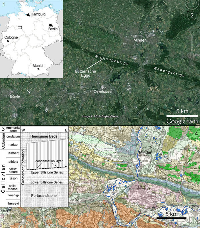
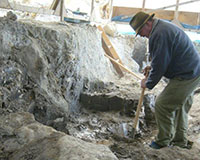
FIGURE 2. Quarry map of the excavation at Lutternsche Egge, showing the position of the different elements of the new taxon in situ. Numbers refer to the specimen numbers of the separate elements (see text). Scale is in 50 cm increments.
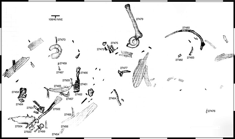
FIGURE 3. Outline reconstruction of Wiehenvenator albati n. gen., n. sp., indicating recovered elements. Based on the reconstruction of Torvosaurus by Scott Hartman; used with permission. Scale bar equals 1 m.
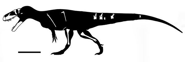
FIGURE 4. Tentative reconstruction of the skull of Wiehenvenator albati, with the recovered elements shown in their approximate relation to each other. Scale bar equals 10 cm.
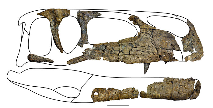
FIGURE 5. Right premaxilla of Wiehenvenator albati in lateral (1), medial (2), anterior (3) and ventral (4) views. Abbreviations: en, external nares; f, foramen; ips, interpremaxillary suture facet; np, nasal process; numbers A1 to A4 indicate alveoli. Scale bar equals 50 mm.
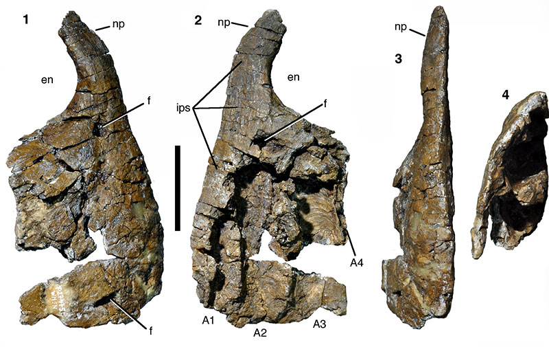
FIGURE 6. Right maxilla of Wiehenvenator albati in lateral (1) and medial (2) views. Abbreviations: af, antorbital fossa; afo, alveolar foramen; amp, base of anteromedial process; ap, ascending process; ep, excavatio pneumatica; f, foramen; iv, invertebrate; jf, jugal facet; pdr, paradental ridge; pmr, promaxillary recess; pro, promaxillary foramen. Scale bar equals 100 mm.
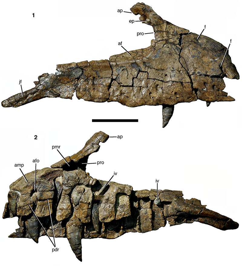
FIGURE 7. Stereophotographs of the right maxilla of Wiehenvenator albati in medial (1) and lateral (2) views.
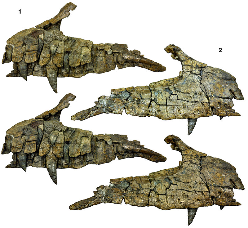
FIGURE 8. Stereophotographs of the promaxillary foramen in the right maxilla of Wiehenvenator albati in posterodorsal view. Abbreviations as in Figure 6. Scale bar equals 50 mm.
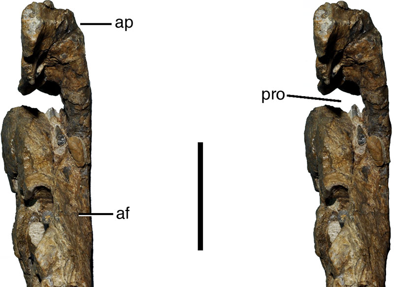
FIGURE 9. Right lacrimal of Wiehenvenator albati in lateral (1; stereophotographs), medial (2), anterior (3; stereophotographs), posterior (4; stereophotographs), and dorsal (5) views. Abbreviations: af, antorbital fossa; d, depression; fo, foramen; lf, lacrimal fenestra; prf, facet for prefrontal; r, ridge. Scale bar equals 50 mm.
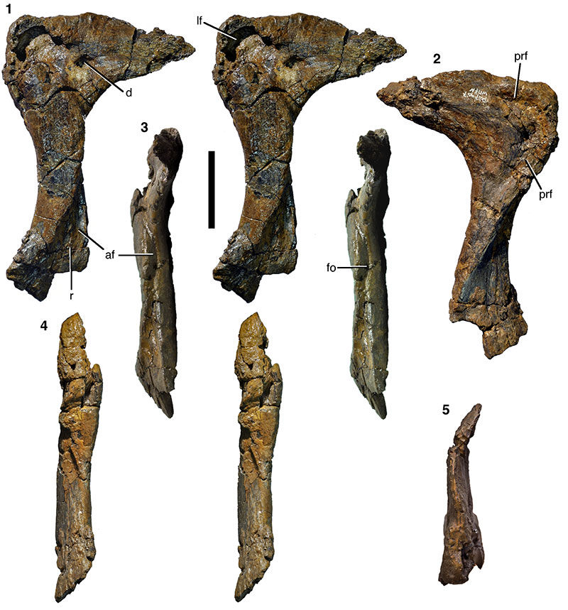
FIGURE 10. Right postorbital of Wiehenvenator albati in lateral (1), anterior (2), medial (3), posterior (4), and dorsal (5) views (all stereophotographs). Abbreviations: d, depression, ff, frontal facet; jf, jugal facet; lf, laterosphenoid facet; of, orbital facet; sob, supraorbital brow; stf, supratemporal fossa. Scale bar equals 100 mm.
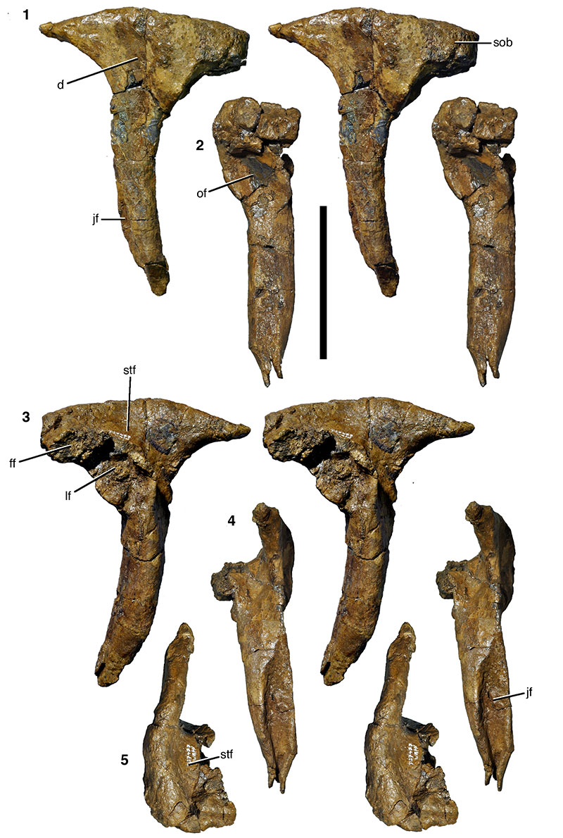
FIGURE 11. Anterior (jugal) process of the right quadratojugal(?) of Wiehenvenator albati in lateral (1), medial (2), dorsal (3), and ventral (4) views. Abbreviations: djp, facet for the dorsal posterior prong of the jugal; vjp, facet for the ventral posterior prong of the jugal. Scale bar equals 100 mm.
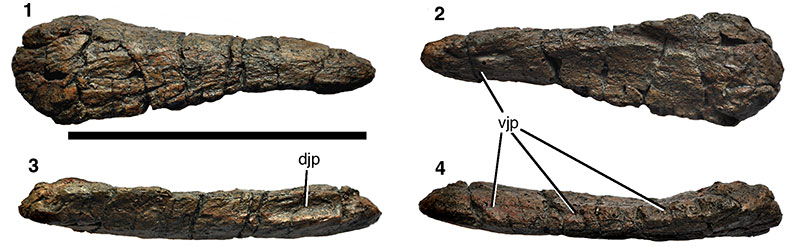
FIGURE 12. Right dentary of Wiehenvenator albati in lateral (1), ventral (2), and medial (3) views. Scale bar equals 100 mm.
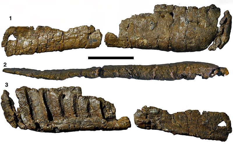
FIGURE 13. Dentition of Wiehenvenator albati. 1, replacement tooth in the 2nd maxillary alveolus; 2, functional tooth in the 4th maxillary alveolus; 3, probable maxillary tooth with partially preserved root (WMN P27459) in lingual view; 4-6, probable maxillary tooth with complete root (WMN P27483) in distal (4), lingual (5) and mesial (6) views; 7, mesial (posterior premaxillary or anterior dentary) tooth (WMN P27467) in labial(?) view; 8, maxillary or dentary tooth (WMN P27473) in labial(?) view; 9, detail of crown apex of WMN P27373, showing the carina that is continuous across the tip (mesial is to the left); 10-12, mesial (probably premaxillary) tooth (WMN P27456) in distal (10), labial (11) and mesial (12) views. Abbreviations: idp, interdental plates; pdl, paradental lamina. Scale bars equal 10 mm (1, 2) and 50 mm (3-12; not 9).
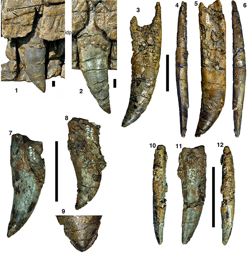
FIGURE 14. Anterior mid-caudal vertebra of Wiehenvenator albati in lateral (1; stereophotographs), dorsal (2), anterior (3), and ventral (4) views. Abbreviations: nc, neural canal; ns, neural spine; pcd, pleurocentral depression; poz, postzygapophysis, prz, prezygapophysis; r, ridge; tp, transverse process. Scale bar equals 100 mm.
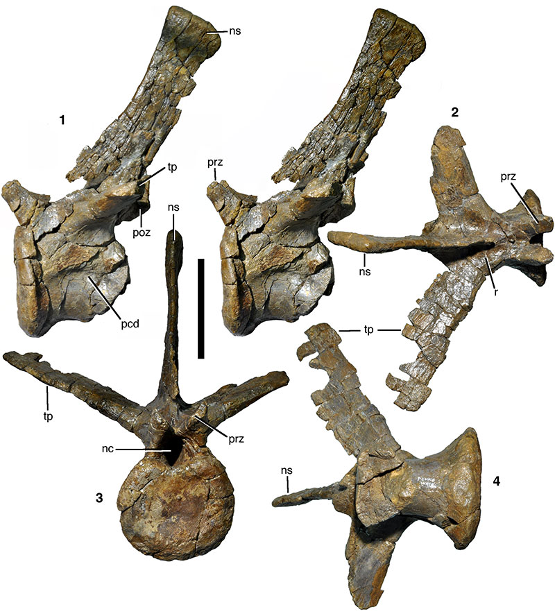
FIGURE 15. Mid-caudal vertebra of Wiehenvenator albati in anterior (1) and lateral (2; stereophotographs) views. Abbreviations as in Figure 14, and: as, anterior spur. Scale bar equals 100 mm.
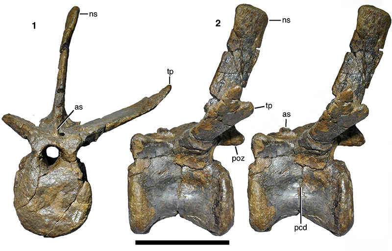
FIGURE 16. Posterior mid-caudal vertebra of Wiehenvenator albati in anterior (1), lateral (2), and ventral (3; stereophotographs) views. Abbreviations as in Figures 14 and 15, and: vg, ventral groove. Scale bar equals 100 mm.
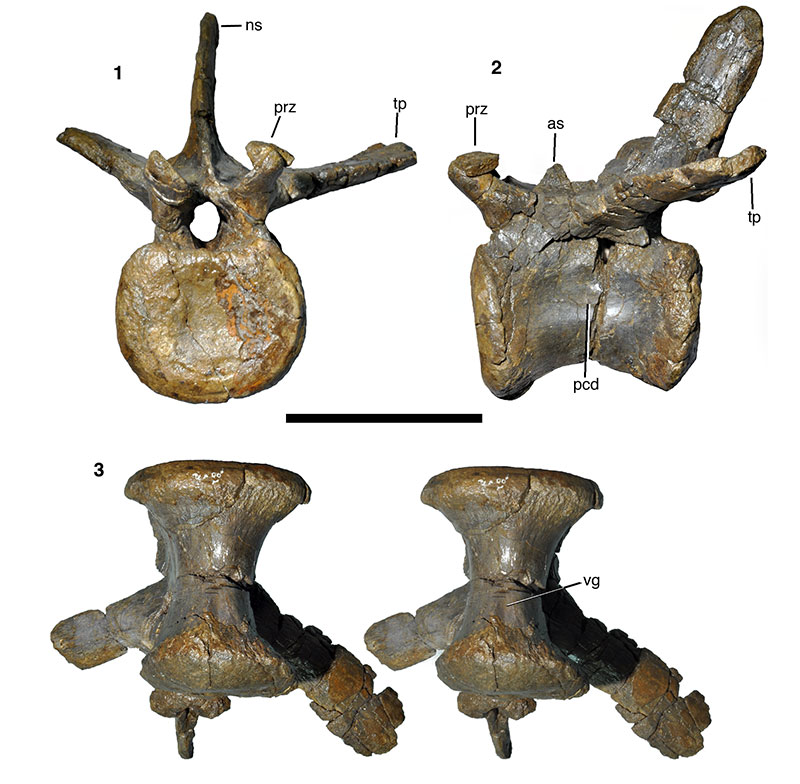
FIGURE 17. Dorsal ribs and gastralia of Wiehenvenator albati. 1, thoracic rib. 2, abdominal rib. 3-5, fused posterior medial gastralia in dorsal (3), ventral (4; stereophotographs), and anterior (5) views. Scale bar equals 100 mm.
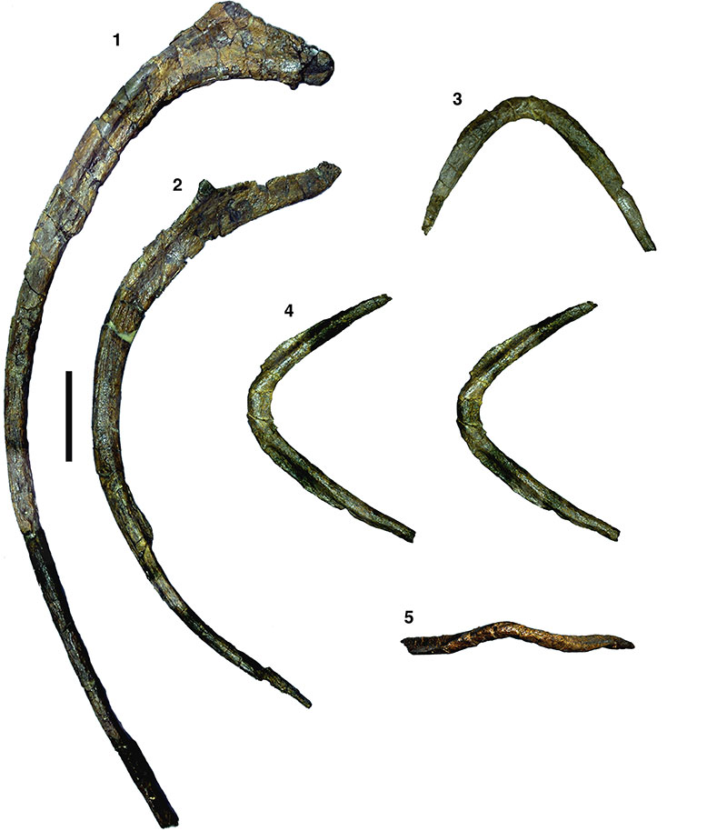
FIGURE 18. Manual phalanx (probably right phalanx III-1) of Wiehenvenator albati in medial (1), dorsal (2) and distal (3) views. Scale bar equals 50 mm.
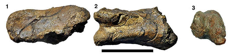
FIGURE 19. Left fibula of Wiehenvenator albati in lateral (1), proximal (2), medial (3), anterior (4), and distal (6) views. 5, detail of proximal shaft of the fibula in medial view (stereophotographs). Abbreviations: d, depression; or, oblique ridge; re, raised edge; sw, swelling; tu, tubercle. Scale bar equals 100 mm (not for 5).
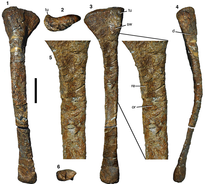
FIGURE 20. Right astragalus of Wiehenvenator albati in anterior (1; stereophotographs), medial (2), proximal (3; stereophotographs), and posterior (4) views. Abbreviations: af, articular facet for ridge on anterior side of tibia; ap, ascending process; d, depression; ff, fibular facet; tf, facet for articulation with tibia. Scale bar equals 50 mm.
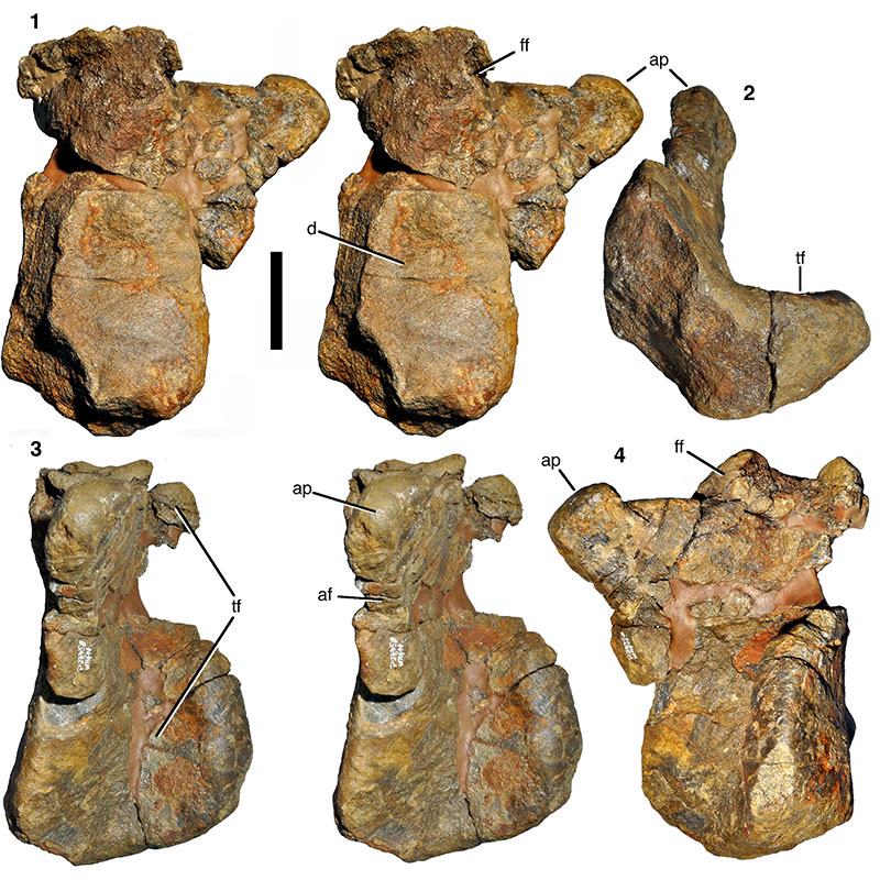
FIGURE 21. Detail of the right astragalus of Wiehenvenator albati: Base of the ascending process and fibular facet in anteroproximal view, showing large foramen anterior to the ascending process. Abbreviations as in Figure 20, and: f, foramen.
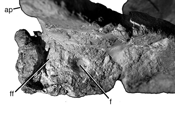
FIGURE 22. Right calcaneum of Wiehenvenator albati in anterior (1) and lateral (2) views. Scale bar equals 50 mm.
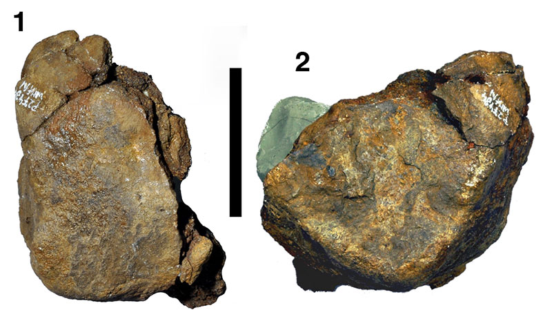
FIGURE 23. Overview of transverse thin section of the left fibula of Wiehenvenator albati. Most of the primary tissue is secondarily remodelled, but growth marks are still preserved anteriorly and posteriorly (white arrows). Abbreviations: lat, lateral; post, posterior. Scale bar equals 5 mm. Inset: Slightly clockwise rotated and magnified image of the posterior outer bone cortex (see frame in overview image). Growth marks are marked by black arrows. Scale bar equals 400 µm.
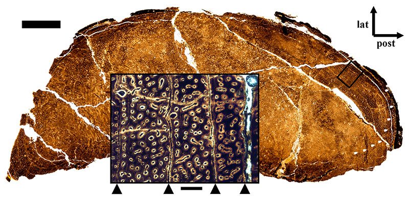
FIGURE 24. Phylogenetic position of Wiehenvenator albati, based on an analysis of 62 taxa and 351 characters (see text for details). 1, strict consensus tree. 2, reduced consensus tree after the a posteriori exclusion of Streptospondylus.
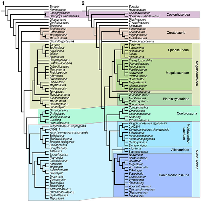
FIGURE 25. Time-calibrated cladogram of early tetanuran relationships, showing the earliest records of the distinct clades, and the rapid evolution of tetanurans in the early Middle Jurassic.
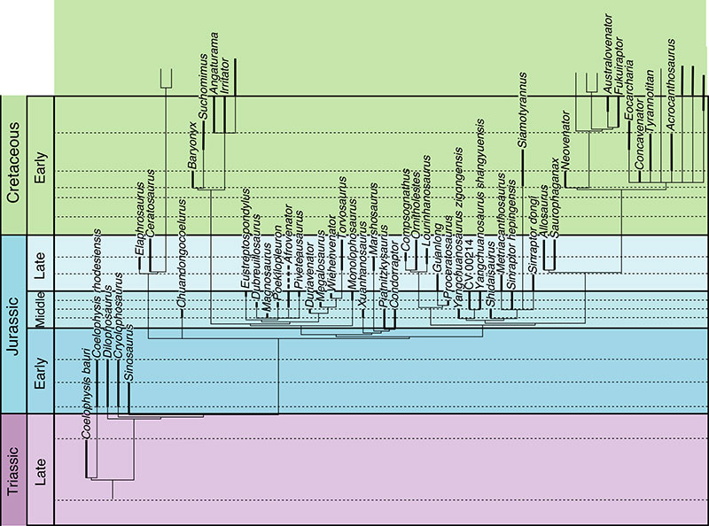
FIGURE 26. Time-calibrated informal supertree of theropod relationships, showing the explosive radiation of the clade in the latest Early and Middle Jurassic. Theropod interrelationships are based on Ezcurra (2012) and Langer et al. (2014) for non-averostran taxa, Rauhut and Carrano (2016) for Ceratosauria, the present study for basal tetanurans, Brusatte and Carr (2016) for tyrannosauroids, and Brusatte et al. (2014) and Foth et al. (2014) for other coelurosaurs.
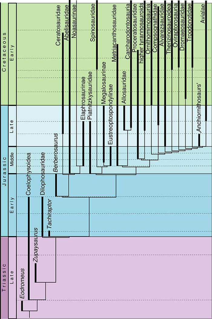
FIGURE 27. Representation of different clades of Middle (1) and Late Jurassic (2) nominal theropod taxa. Figures 3 and 4 show the same for large theropod taxa (body weight > 200 kg) only.

FIGURE 28. Composition of Middle and Late Jurassic theropod faunas, as percentage of localities in which certain clades have been recorded.
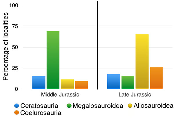
FIGURE 29. Number of Late Jurassic localities in which theropods have been recorded. The green portion of the column indicates how many of these occurrences are accounted for by the Morrison Formation.
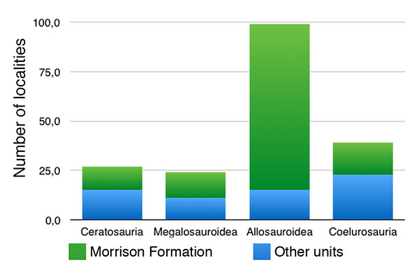
FIGURE 30. Possible biases that influence interpretations of the Jurassic theropod fossil record. Geographic distribution of Middle (1) and Late Jurassic (2) localities with identifiable theropod remains, and environments represented by Middle (3) and Late Jurassic (4) theropod localities.
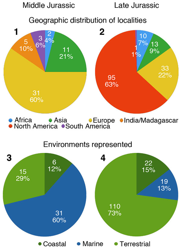
TABLE 1. Anteroposterior width of the maxillary alveoli of Wiehenvenator albati (in mm).
| Alveolus | 1 | 2 | 3 | 4 | 5 | 6 | 7 | 8 | 9 | 10 | 11 | 12 | 13 |
| Width | c. 35 | c. 39 | c. 45 | c. 40 | 37 | 35 | 30 | 25 | 23 | c. 18 | c. 20 | 13 | 10 |
TABLE 2. Measurements of caudal vertebrae of Wiehenvenator albati (in mm).
| WMN P27501 | WMN P27499 | WMN P27500 | |
| Centrum length | c. 110 | 114 | 114 |
| Anterior centrum height | 104 | 79 | 76 |
| Anterior centrum width | 113 | c. 82 | 85 |
| Posterior centrum height | -- | 80 | 72 + |
| Posterior centrum width | -- | 83 | 74 + |
| Minimal centrum width | 50 | c. 38 | c. 35 |
| Neural arch length | 143 | 125 + | 135 |
| Total height | c. 320 | 247 | 195 + |
TABLE 3. Theropod taxa from the Middle and Late Jurassic that are regarded as valid. Abbreviations: Allos, Allosauroidea; Cerat, Ceratosauria; Coel, Coelurosauria; Incert, Incertae sedis; Megal, Megalosauroidea.
| Name | Clade | Unit | Country | Age | ||||
| Condorraptor currumili | Megal. | Cañadón Asfalto Fm. | Argentina | Aalenian-Bajocian | ||||
| Piatnitzkysaurus floresi | Megal. | Cañadón Asfalto Fm. | Argentina | Aalenian-Bajocian | ||||
| Eoabelisaurus mefi | Cerat. | Cañadón Asfalto Fm. | Argentina | Aalenian-Bajocian | ||||
| Shidaisaurus jinae | Allos. | Upper Lufeng Fm. | China | Aalenian-Bajocian | ||||
| Duriavenator hesperis | Megal. | Inferior Oolite Fm. | UK | Bajocian | ||||
| Magnosaurus nethercombensis | Megal. | Inferior Oolite Fm. | UK | Bajocian | ||||
| Yangchuanosaurus zigongensis | Allos. | Xiashaximiao Fm. | China | Bajocian | ||||
| Gasosaurus constructus | Incert. | Xiashaximiao Fm. | China | Bajocian | ||||
| Xuanhanosaurus qilixiaensis | Megal. | Xiashaximiao Fm. | China | Bajocian | ||||
| Chuandongocoelurus primitivus | Incert. | Xiashaximiao Fm. | China | Bajocian | ||||
| Dubreuillosaurus valesdunensis | Megal. | Calcaires de Caen | France | Bathonian | ||||
| Poekilopleuron bucklandii | Megal. | Calcaires de Caen | France | Bathonian | ||||
| Cruxicheiros newmanorum | Incert. | Chipping Norton Limestone | UK | Bathonian | ||||
| Megalosaurus bucklandii | Megal. | Great Oolite | UK | Bathonian | ||||
| Proceratosaurus bradleyi | Coel. | Great Oolite | UK | Bathonian | ||||
| Kileskus aristocraticus | Coel. | Itat Fm. | Russia | Bathonian | ||||
| Yangchuanosaurus shangyouensis | Allos. | Shangshaximiao Fm. | China | Bathonian-Callovian | ||||
| Sinraptor hepingensis | Allos. | Shangshaximiao Fm. | China | Bathonian-Callovian | ||||
| Afrovenator abakensis | Megal. | Tiouraren Fm. | Niger | ?Bathonian-?Oxfordian | ||||
| MNN Tig6 (‚Spinostropheus‘) | Cerat. | Tiouraren Fm. | Niger | ?Bathonian-?Oxfordian | ||||
| Piveteausaurus divesensis | Megal. | Marnes de Dives | France | Callovian | ||||
| Eustreptospondylus oxoniensis | Megal. | Oxford Clay | UK | Callovian | ||||
| Monolophosaurus jiangI | Megal. | Shishugou Fm. | China | Callovian | ||||
| Wiehenvenator albati | Megal. | Ornatenton | Germany | Callovian | ||||
| Streptospondylus altdorfensis | Megal. | Marnes à Deltoideum delta | France | Callovian-Oxfordian | ||||
| Aurornis xui | Coel. | Tiaojishan Fm. | China | Oxfordian | ||||
| Eosinopteryx brevipenna | Coel. | Tiaojishan Fm. | China | Oxfordian | ||||
| Anchiornis huxleyi | Coel. | Tiaojishan Fm. | China | Oxfordian | ||||
| Epidexipteryx hui | Coel. | Tiaojishan Fm. | China | Oxfordian | ||||
| Epidendrosaurus ningchengensis | Coel. | Tiaojishan Fm. | China | Oxfordian | ||||
| Pedopenna daohugouensis | Coel. | Tiaojishan Fm. | China | Oxfordian | ||||
| Yi qi | Coel. | Tiaojishan Fm. | China | Oxfordian | ||||
| Metriacanthosaurus parkeri | Allos. | Oxford Clay | UK | Oxfordian | ||||
| Sinraptor dongi | Allos. | Shishugou Fm. | China | Oxfordian | ||||
| Guanlong wucaii | Coel. | Shishugou Fm. | China | Oxfordian | ||||
| Limusaurus inextricabilis | Cerat. | Shishugou Fm. | China | Oxfordian | ||||
| Zuolong salleei | Coel. | Shishugou Fm. | China | Oxfordian | ||||
| Haplocheirus sollers | Coel. | Shishugou Fm. | China | Oxfordian | ||||
| Allosaurus europaeus | Allos. | Lourinhã Fm. | Portugal | Kimmeridgian | ||||
| Aviatyrannis jurassica | Coel. | Alcobaça Fm. | Portugal | Kimmeridgian | ||||
| Juravenator starki | Coel. | Painten Fm. | Germany | Kimmeridgian | ||||
| Sciurumimus albersdoerferi | Megal. | Painten Fm. | Germany | Kimmeridgian | ||||
| Elaphrosaurus bambergi | Cerat. | Tendaguru Fm. | Tanzania | Kimmeridgian | ||||
| Veterupristisaurus milneri | Allos. | Tendaguru Fm. | Tanzania | Kimmeridgian | ||||
| Lourinhanosaurus antunesi | Coel. | Lourinhã Fm. | Portugal | Kimmeridgian-Tithonian | ||||
| Torvosaurus gurneyi | Megal. | Lourinhã Fm. | Portugal | Kimmeridgian-Tithonian | ||||
| Allosaurus fragilis | Allos. | Morrison Fm. | USA | Kimmeridgian-Tithonian | ||||
| Allosaurus ‚jimmadseni‘ | Allos. | Morrison Fm. | USA | Kimmeridgian-Tithonian | ||||
| Ceratosaurus nasicornis | Cerat. | Morrison Fm. | USA | Kimmeridgian-Tithonian | ||||
| Torvosaurus tanneri | Megal. | Morrison Fm. | USA | Kimmeridgian-Tithonian | ||||
| Ornitholestes hermanni | Coel. | Morrison Fm. | USA | Kimmeridgian-Tithonian | ||||
| Coelurus fragilis | Coel. | Morrison Fm. | USA | Kimmeridgian-Tithonian | ||||
| Marshosaurus bicentesimus | Megal. | Morrison Fm. | USA | Kimmeridgian-Tithonian | ||||
| Stokesosaurus clevelandi | Coel. | Morrison Fm. | USA | Kimmeridgian-Tithonian | ||||
| Tanycolagreus topwilsoni | Coel. | Morrison Fm. | USA | Kimmeridgian-Tithonian | ||||
| Saurophaganax maximus | Allos. | Morrison Fm. | USA | Kimmeridgian-Tithonian | ||||
| Compsognathus longipes | Coel. | Painten or Solnhofen Fm. | Germany | Kimmeridgian-Tithonian | ||||
| Compsognathus corallestris | Coel. | Calcaires blancs de Provence Fm. | France | Tithonian | ||||
| Juratyrant langhami | Coel. | Kimmeridge Clay | UK | Tithonian | ||||
| Archaeopteryx lithographica | Coel. | Solnhofen Fm. | Germany | Tithonian | ||||
| Archaeopteryx siemensii | Coel. | Solnhofen Fm. | Germany | Tithonian | ||||
| Archaeopteryx recurva | Coel. | Solnhofen Fm. | Germany | Tithonian | ||||
| Chilesaurus diegosuarezi | Incert. | Toqui Fm. | Chile | Tithonian | ||||
A new megalosaurid theropod dinosaur from the late Middle Jurassic (Callovian) of north-western Germany: Implications for theropod evolution and faunal turnover in the Jurassic
Plain Language Abstract
A fragmentary skeleton of a new theropod dinosaur was found in Middle Jurassic marine rocks of northern Germany. The new taxon, named Wiehenvenator albati, represents a large megalosaurid, adding to the diverse megalosaurid record known from the Middle Jurassic of Europe. An analysis of the Jurassic theropod fossil record and the relationships of the new taxon indicate a very rapid diversification of major theropod clades in the latest Early and the Middle Jurassic, probably following an extinction event in the late Early Jurassic. Furthermore, the available data indicates a change from megalosauroid dominated faunas in the Middle Jurassic to allosauroid dominated faunas in the Late Jurassic, although there are severe biases in the available data that make any such inference tentative.
Resumen en Español
Un nuevo dinosaurio terópodo megalosáurido del Jurásico Medio tardío (Calloviano) del noroeste de Alemania: implicancias para la evolución de los terópodos y reemplazo faunístico en el Jurásico
Se hallaron restos fragmentarios de un dinosaurio terópodo grande y robusto en la Formación Ornatenton, unidad marina del Calloviano medio del nordeste de Renania del Norte-Westfalia, Alemania. Este espécimen incluye: premaxila, maxila, lacrimal, postorbital, dentario, varias vértebras caudales, costillas, fíbulas, astrágalo y calcáneo parcial. Es descripto aquí como una nueva especie de megalosáurido, Wiehenvenator albati gen. n. sp. n., diagnosticado por una fosa anteorbital maxilar fuertemente reducida en la base del proceso ascendente de la maxila, una rama anterior del lacrimal muy corta con una depresión neumática adicional anteroventral a la fenestra lacrimal, una faceta orbital expandida transversalmente, y un extremo proximal del proceso ascendente del astrágalo flexionado lateralmente. Los análisis filogenéticos ubican a Wiehenvenator como un megalosáurido megalosaurino, taxón hermano del género Torvosaurus del Jurásico Tardío. Por ende aporta a la diversidad considerable de megalosauroideos del Jurásico Medio. Una filogenia de terópodos calibrada temporalmente indica una rápida radiación de los terópodos Averostra entre el Toarciano y el Bathoniano. Esta radiación fue probablemente disparada por el evento de extinción Pliensbachiano-Toarciano, el cual puede haber sido más importante para la evolución de los terópodos que la extinción del Triásico-Jurásico. El registro fósil indica un reemplazo faunístico de una fauna del Jurásico Medio dominada por megalosauroideos a una del Jurásico Tardío dominada por allosauroideos/coelurosaurios. Sin embargo, la existencia de diferencias en los registros fósiles de terópodos del Jurásico Medio y Tardío en cuanto a la distribución geográfica de localidades, así como también a los ambientes muestreados, hacen que esta inferencia sea problemática, al menos en lo referente a los allosauroideos. Un análisis de preferencias ambientales de allosauroideos y megalosauroideos indica que los primeros preferían ambientes más tierra adentro, mientras que los segundos son más comunes en ambientes de nearshore.
Palabras clave: Megalosauroidea; Formación Ornatenton; evolución de terópodos; Jurásico
Traducción: Diana Elizabeth Fernández
Résumé en Français
Un nouveau dinosaure théropode mégalosauridé du Jurassique moyen récent (Callovien) du nord-ouest de l'Allemagne : implications pour l'évolution des théropodes et les renouvellements fauniques pendant le Jurassique
Des restes fragmentaires d'un dinosaure théropode robuste et de grande taille ont été trouvés dans les dépôts marins du Callovien moyen de la formation d'Ornatenton au nord-est de Northrhine-Westphalia, Allemagne. Le spécimen inclut un prémaxillaire, un maxillaire, un lacrymal, un postorbitaire, un dentaire, plusieurs vertèbres caudales, des côtes, les péronés, un astragale, et un calcanéum partiel. Il est ici décrit comme appartenant à une nouvelle espèce de mégalosauroïde, Wiehenvenator albati n. gen. n. sp. Les caractères diagnostiques sont les suivants : une fosse anté-orbitaire du maxillaire fortement réduite et située à la base du processus ascendant du maxillaire ; un ramus antérieur du lacrymal très court, avec une dépression pneumatisée supplémentaire située antéroventralement à la fenêtre lacrymale ; une facette orbitaire de l'os postorbitaire développée transversalement ; et une extrémité proximale du processus ascendant de l'astragale fléchie latéralement. L'analyse phylogénétique a placé Wiehenvenator comme un mégalosauridé mégalosauriné, groupe-frère du genre Torvosaurus du Jurassique récent. Ce taxon s'ajoute donc à la diversité considérable des mégalosauroïdes au Jurassique moyen. Une phylogénie des théropodes calibrée temporellement indique une radiation rapide des théropodes Averostra entre le Toarcien et le Bathonien. Cette radiation a probablement été déclenchée par l'évènement d'extinction de la limite Pliensbachien-Toarcien. Ce dernier a pu être plus important pour l'évolution des théropodes que l'extinction de la limite Trias-Jurassique. Le registre fossile indique un renouvellement faunique entre les faunes du Jurassique moyen dominées par les mégalosauroïdes et celles du Jurassique récent dominées par les allosauroïdes et les coelurosaures. Cependant, les différences entre les registres fossiles des théropodes du Jurassique moyen et du Jurassique récent, à la fois en termes de distributions géographiques des localités et d'environnements échantillonnés, rendent cette conclusion problématique, au moins pour les allosauroïdes. Une analyse des préférences environnementales des allosauroïdes et des mégalosauroïdes indique que les premiers préféraient les environnements situés à l'intérieur des terres alors que les derniers étaient plus communs dans les environnements côtiers.
Mots-clés : Megalosauroidea ; formation d'Ornatenton ; évolution des théropodes ; Jurassique
Translator: Antoine Souron
Deutsche Zusammenfassung
In progress
Translator: Eva Gebauer
Arabic
Translator: Ashraf M.T. Elewa
-
-
-
Review: The Princeton Field Guide to Mesozoic Sea Reptiles
 The Princeton Field Guide to Mesozoic Sea Reptiles
Article number: 26.1.1R
April 2023

 Poster Winners 2024