

Article Search
Volume 27.1
January–April 2024
Full table of contents
ISSN: 1094-8074, web version;
1935-3952, print version
Recent Research Articles
See all articles in 27.1 January-April 2024
See all articles in 26.3 September-December 2023
See all articles in 26.2 May-August 2023
See all articles in 26.1 January-April 2023
 Julien Benoit. Evolutionary Studies Institute (ESI); School of Geosciences, University of the Witwatersrand, PO Wits, 2050, Johannesburg, South Africa; School of Anatomical Sciences, University of the Witwatersrand, 7 York Road, Parktown, 2193, Johannesburg, South Africa. julien.benoit@wits.ac.za
Researcher in vertebrate Palaeontology and Palaeobiology with special interest in the study of the evolution of endocranial structures and sense organs in extinct Mammalia and Therapsida using X-ray imagery on their fossolized skulls.

 Sandra C. Jasinoski. Evolutionary Studies Institute (ESI); School of Geosciences, University of the Witwatersrand, PO Wits, 2050, Johannesburg, South Africa. sandra_jas@hotmail.com
Research scientist with over 10 years of independent and collaborative research experience in Canada, South Africa, and the United Kingdom. My work experience includes analysing complex data, writing and presenting technical reports, collaborating across several disciplines, managing and supervising research projects, and chairing seminars at scientific conferences.
FIGURE 1. The label on the first page of the serial section drawings. It is written “Specimen N°1821 (FN). MN° 346. Complete Aneumogomphius ? skull therocephalian. Sections ½ mm. Magnification x4.” The document is signed “A.S. Brink”. Abbreviations: FN, Field Number; MN°, Museum Number.
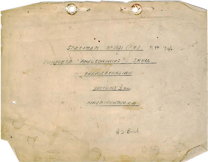
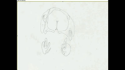
FIGURE 2. Comparison of the different reconstructions of BP/1/1821 in dorsal (left) and ventral (right) views. 1, the wax model; and 2, the original illustrations by Brink, found with the serial section drawings.
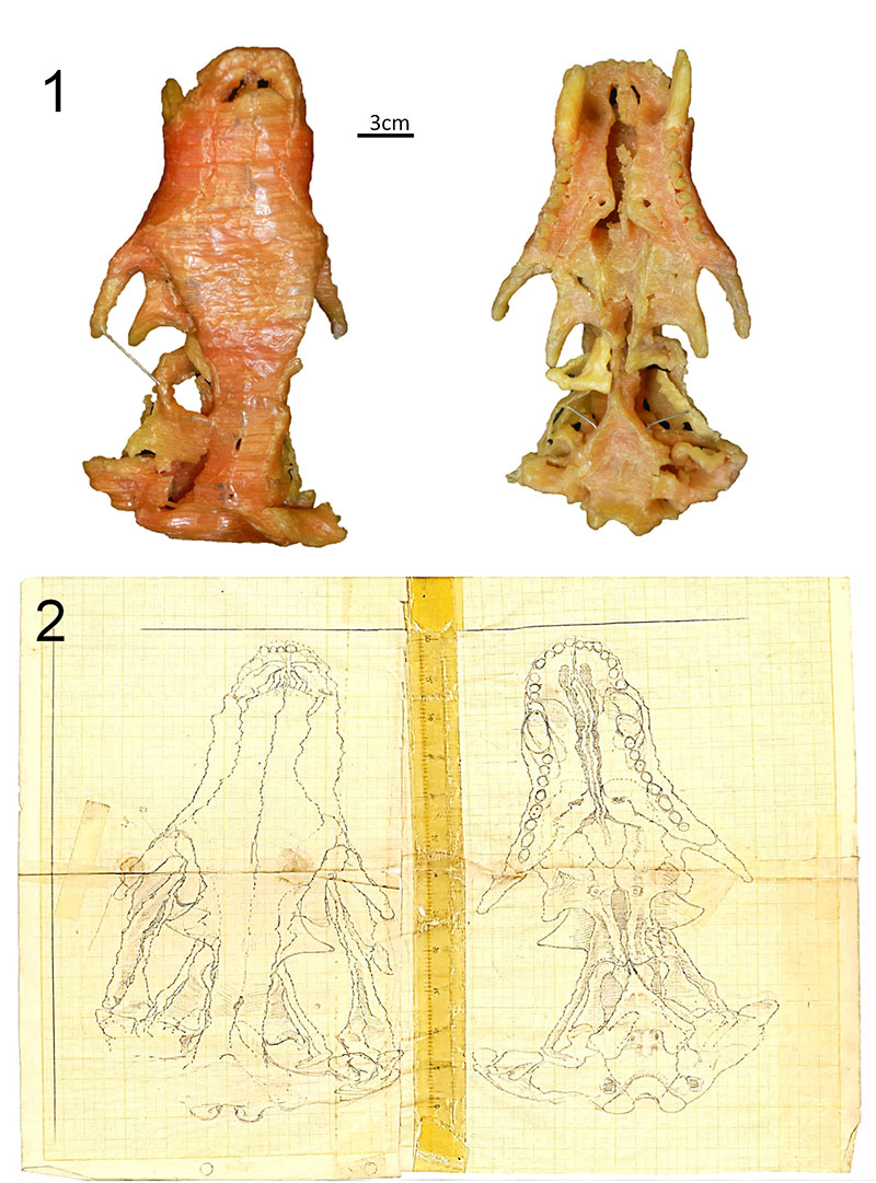
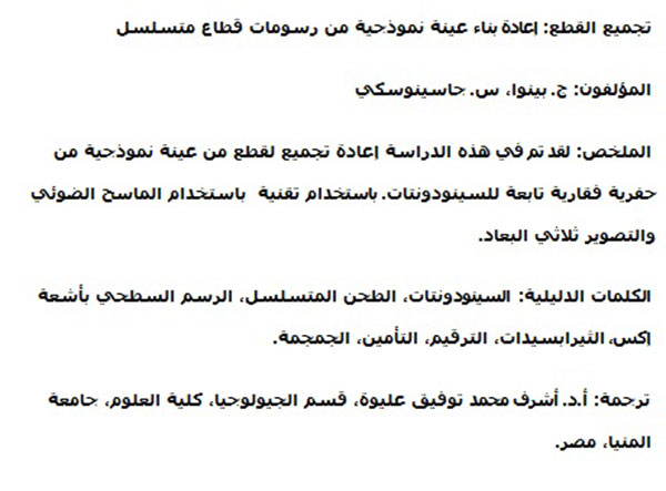
FIGURE 3. The digital model in (1) right lateral, (2) left lateral and (3) ventral view in stereopairs. Scale bar equals 10 mm. See also the supplementary video in Appendix 2.
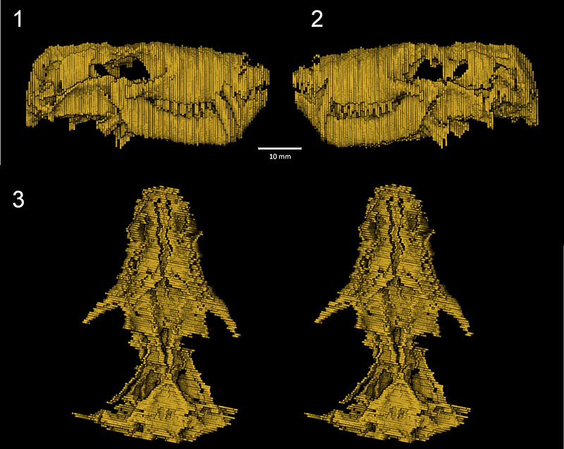
FIGURE 4. The slices of specimen BP/1/1821 finally put back together again, as seen in the hands of the CT scan facility technician K. Jakata (ESI). This 3D printed model is the first time in 55 years that BP/1/1821 has been accurately reconstructed at life-size.
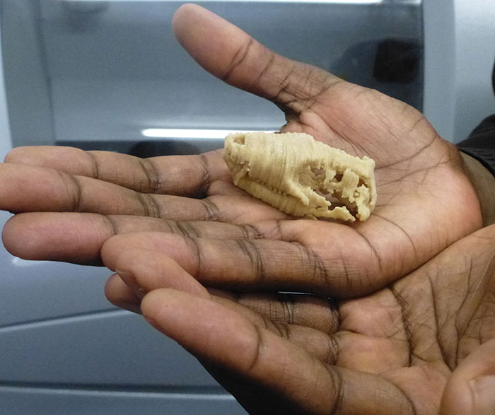
FIGURE 5. Segmentation of the skull bones of BP/1/1821, (1) in the cranium and (2) in the lower jaw. Scale bar equals 10 mm.
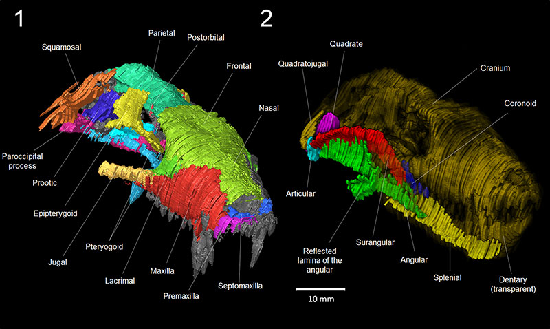
FIGURE 6. Comparison between the digital model (top) and the wax model (bottom), in rostral (left) and lateral views (right). Arrows indicate the direction of deformation of the wax model. Scale bar equals 10 mm.
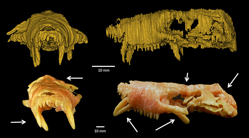
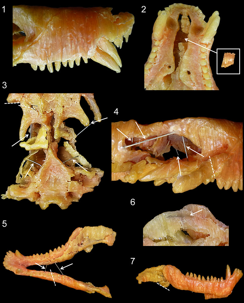
FIGURE 7. The state of preservation of the wax model of BP/1/1821. 1, the right side of the snout showing the deformation of the canine; 2, the palate showing the broken vomer; 3, the basicranium showing multiple cracks, nails and sticks around the pterygoid; 4, view of the right orbit showing multiple cracks, nails and sticks; 5, dorsal view of the of the mandible showing multiple cracks, nails and sticks; 6, detail of the inner side of the left ascending ramus of the mandible showing a stick embedded in the wax; and 7, lateral view of the mandible showing the incomplete reflected lamina of the angular. The arrows point to the nails, dotted lines demarcate the cracks. Not to scale.
APPENDIX 1
Movie of the aligned serial section drawings of BP/1/1821 made by A.S. Brink. The 116 sections represent the entire specimen. Note that Brink numbered the sections from 1 to 118 because he missed number 83 and duplicated 110; however Brink also labelled every section with the corresponding 0.5 mm interval so we are confident that no section is missing. Because of this mislabelling, the section numbers in the movie are shifted between 83 and 110 with respect to Brink’s numbered sections (see Brink, 1961, figure 35). Click on image to run animation.
APPENDIX 2
Movie of the virtual 3D model of BP/1/1821 that was reconstructed from the drawings made by A.S. Brink (1961). Click on image to run animation.
Picking up the pieces: the digital reconstruction of a destroyed holotype from its serial section drawings
Plain Language Abstract
When describing a new fossil species, palaeontologists designate a specimen to serve as landmark for future anatomical comparison and observations. This specimen, called the holotype, is thus a keystone to define a fossil species. For example, researchers who would like to refer a newly discovered fossil to a given species must compare it to the holotype to confirm its identity. As a consequence, the worst thing that could happen to the taxonomic process is for a holotype to be destroyed. This is exactly what happened to the holotype specimen of 'Scalopocynodon gracilis' (now synonymized with Procynosuchus delaharpeae). The irony is that it was destroyed by the very author of the species, A.S. Brink, in order to make a complete and thorough description through serial grinding. The holotype skull of 'Scalopocynodon gracilis' was the only known specimen of this species (although it has subsequently been recognised as a junior synonym of Procynosuchus delaharpeae of which there are multiple specimens). Fortunately, we re-discovered Brink's original drawings of the serial sections in the collections of the Evolutionary Studies Institute (University of the Witwatersrand, Johannesburg, South Africa) and were able to digitally put them back together to reconstruct an in silico model of Brink's 'Scalopocynodon gracilis' skull. A digital model was printed with a 3D printer, representing the first life-sized reconstruction of this specimen in 55 years.
Resumen en Español
Recogiendo los pedazos: la reconstrucción digital de un holotipo destruido a partir de dibujos de sus secciones seriadas
El corte pulido seriado, una técnica popular pero destructiva de comienzos del siglo XX, permite el estudio tomográfico detallado de fósiles de vertebrados. El espécimen BP/1/1821 (previamente BPI/1/346) es un cinodonte procinosúquido (Therapsida) que fue seccionado por A.S. Brink en 1961, resultando en un estudio detallado y revelador de su anatomía craneana. Sin embargo, BP/1/1821 también fue designado por Brink como el holotipo y único espécimen de una especie de cinodonte: 'Scalopocynodon gracilis'. Esta especie ha sido posteriormente reconocida como un sinónimo junior de Procynosuchus delaharpeae, pero la destrucción del holotipo sigue siendo una pérdida irreversible. A partir de las secciones seriadas, Brink construyó un modelo de cera agrandado, pero el mismo se encuentra degradándose rápidamente. En este artículo explicamos cómo conseguimos los dibujos originales de Brink de las secciones y cómo construimos un nuevo modelo digital de este espécimen usando un escáner, apilamiento virtual y toma de imagines 3D. La comparación con dibujos previamente publicados demuestra la precisión del modelo digital. Con una impresora 3D recreamos una replica más precisa de BP/1/1821 con resina. Esta réplica en tamaño real ahora ayuda a completar la colección del Instituto de Estudios Evolutivos (Universidad de Witwatersrand, Johannesburgo, Sudáfrica) y reemplaza al espécimen perdido. También se discute la posibilidad de reutilizar estos datos antiguos para la investigación paleontológica.
Palabras clave: Cinodonte; Tomografía de cortes seriados; Digitalización; Curación; Cráneo
Traducción: Diana Elizabeth Fernández
Résumé en Français
Rassembler les morceaux : un holotype détruit est reconstitué numériquement à partir des dessins des sections sériées
La tomographie par abrasion sériée, une technique destructive qui était très populaire au 20e siècle, permet l'étude détaillée de nombreux vertébrés fossiles. Le spécimen BP/1/1821 (autrefois BPI/1/346) appartient à un cynodonte procynosuchidé (Therapsida) qui a été étudié et détruit de cette façon par A. S. Brink en 1961, ce qui a donné lieu à une étude très complète de l'anatomie de sa tête osseuse. BP/1/1821 a aussi été désigné par Brink comme l'holotype et seul représentant d'une nouvelle espèce de cynodonte : 'Scalopocynodon gracilis'. Cette espèce est maintenant reconnue comme un synonyme de Procynosuchus delaharpeae, mais la destruction d'un spécimen holotype demeure une perte irremplaçable. Brink avait fabriqué, à partir des dessins des sections, un modèle en cire de la tête osseuse agrandie, mais cette réplique se dégrade rapidement. Dans cette article nous expliquons comment nous avons retrouvé les originaux des dessins des sections sériées réalisés par Brink et comment nous avons pu reconstituer un nouveau modèle numérique du spécimen original en scannant ces dessins, puis en les alignant virtuellement afin de leur appliquer des techniques classiques d'imagerie 3D. Une comparaison avec la description initiale indique que ce modèle numérique est précis. Il a ensuite été possible, à l'aide d'une imprimante 3D, de reconstituer en résine une réplique, plus fidèle que celle en cire, de BP/1/1821. Cette reconstitution grandeur nature vient compléter les collections du Evolutionary Studies Institute (Université du Witwatersrand, Johannesbourg, Afrique du Sud) et remplace le spécimen original détruit depuis longtemps. La possible réutilisation de ces anciennes données pour de nouvelles recherches en paléontologie est aussi discutée.
Mots-clés : cynodonte ; tomographie par abrasion sériée ; Therapsida ; numérisation ; conservation ; tête osseuse
Translator: Antoine Souron
Deutsche Zusammenfassung
Aufsammeln von Einzelteilen: digitale Rekonstruktion eines zerstörten Holotyps aus Serienschnitt-Zeichnungen
Dünnschliffe, eine populäre aber zerstörerische Technik im frühen 20. Jahrhundert, erlauben eine detaillierte tomographische Untersuchung von Wirbeltierfossilien. Das Stück BP/1/1821 (früher BPI/1/346) ist ein procynosuchider Cynodontier (Therapsida), der 1961 von A.S. Brink geschliffen wurde, was in einer detaillierten und aufschlussreichen Studie der Schädelanatomie resultierte. Allerdings wurde BP/1/1821 von Brink auch als Holotyp und einziges Belegstück der neuen Cynodontier-Art (jetzt Juniorsynonym von Procynosuchus delaharpeae) 'Scalopocynodon gracilis' bestimmt. Die Art wurde später als ein Juniorsynonym von Procynosuchus delaharpeae erkannt, jedoch bleibt die Zerstörung eines Holotys ein irreversibler Verlust. Brink stellte ein vergrößertes Wachsmodell aus den Serienschnitten her, was jedoch immer mehr zerfällt. In diesem Artikel erklären wir, wie wir die Originalzeichnungen von Brink wieder fanden und wie wir im Stande waren mit einem Scanner, virtuellem Stapel-Alignement und 3D Bildverarbeitung ein neues Modell zu erstellen. Ein Vergleich mit vorher publizierten Zeichnungen veranschaulicht die Genauigkeit dieses digitalen Modells. Mit einem 3D Drucker fertigten wird dann aus Kunstharz eine genauere Nachbildung von BP/1/1821 an. Die Lebensgroße Replik trägt nun dazu bei die Sammlung des Instituts für evolutionäre Studien der University of the Witwatersrand, Johannesburg, Südafrika zu komplettieren und ersetzt das verlorene Original. Die Möglichkeit zur Widerverwendbarkeit dieser alten Daten für paläontologische Untersuchungen wird ebenfalls angesprochen.
Schlüsselwörter: Cynodontier; Serienschnitt-Tomographie; Therapsida; Digitalisierung; Kuratieren; Schädel
Translator: Eva Gebauer
Arabic
Translator: Ashraf M.T. Elewa
-
-
-
Review: The Princeton Field Guide to Mesozoic Sea Reptiles
 The Princeton Field Guide to Mesozoic Sea Reptiles
Article number: 26.1.1R
April 2023

 Poster Winners 2024