

Article Search
Volume 27.1
January–April 2024
Full table of contents
ISSN: 1094-8074, web version;
1935-3952, print version
Recent Research Articles
See all articles in 27.1 January-April 2024
See all articles in 26.3 September-December 2023
See all articles in 26.2 May-August 2023
See all articles in 26.1 January-April 2023
 Michele Mazza. Dipartimento di Scienze della Terra "A. Desio", Università degli Studi di Milano, Via Mangiagalli 34, 20133, Milano (Italy); michele.mazza@unimi.it; mazza_michele@yahoo.it
Michele Mazza (Piacenza, 1978), took his Master Degree in Geology and Palaeontology in 2005 at the University of Milan and he obtained his PhD degree in Earth Sciences in 2010 at the same University. He worked as a PostDoc researcher in biostratigraphy and palaeontology for four years (from 2011 to 2015) at the Department of Earth Sciences "A. Desio" (University of Milan).
He is a conodont specialist and his main research interests are Middle and Late Triassic condont biostratigraphy, systematic, paleoecology (stable isotopes of Oxygen, Carbon and Strontium), phylogenesis and evolution. He is member of the International Working Groups for the definition of the GSSP of the Norian Stage from 2006 (candidate section Pizzo Mondello, Western Sicily, Italy) and of the Raethian Stage from 2016 (candidate section Pignola-Abriola, Southern Apennines, Italy).
In the last three years he focused his research activity on the correlation of the Late Triassic conodont faunas of the Tethys to the North American faunas and on the study of the ontogenetic processes of conodonts reconstructing traditional growth series and using innovative x-ray synchrotron microscopy. From 2012 he is Technical Editor of the ISI journal "Bollettino della Società Paleontologica Italiana".

 Carlos Martínez-Pérez. Departamento de Geología, Universidad de Valencia, Avda. Dr. Moliner, 50, 46100 Burjassot, Valencia (Spain); carlos.martinez-perez@uv.es; School of Earth Sciences, University of Bristol, Wills Memorial Building, Queen’s Road, Bristol BS8 1RJ, UK; carlos.martinez-perez@uv.es
Carlos Martinez-Perez (Valencia, 1978) obtained his PhD in Biology at the University of Valencia (Spain) in 2010. From 2011 to 2014 he was a Postdoctoral fellow at the University of Bristol working on condont palaeobiology, and since 2015 he is Assistant Professor at the University of Valencia. His main research interests are in the area of early vertebrate palaeobiology, using state of the art computational techniques, including tomographic, to shed light into the function and evolution of the vertebrate skeleton.
FIGURE 1. Cladogram representing the Epigondolella clade of Mazza et al. (2012b), illustrating the phylogenetic relationships between the Upper Carnian-Lower Norian (Upper Triassic) carnepigondolellids and epigondolellids and their main evolutionary trends. Only the species analysed in this work are figured. The specimens of E. vialovi, E. quadrata, E. uniformis, and E. triangularis are from Mazza and Martínez-Pérez (2015). Vertical bars beside the specimens indicate the platform (P)/blade (B) length ratio; white circles mark the cusp. All the specimens are at the same scale.
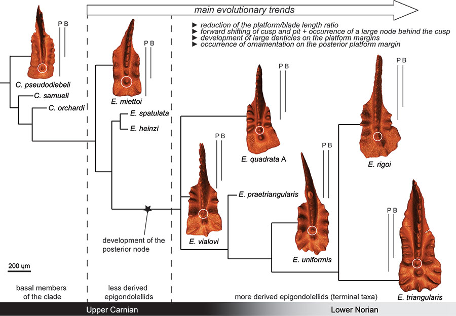
FIGURE 2. X-ray Synchrotron microtomography of Epigondolella quadrata specimen A. Three possible sections that can be obtained with the X-ray synchrotron microscopy are shown. 1, 3D model of the specimen: 2, Longitudinal section; 3, Horizontal section; 4, Cross section. Scale bar equals 200 µm.
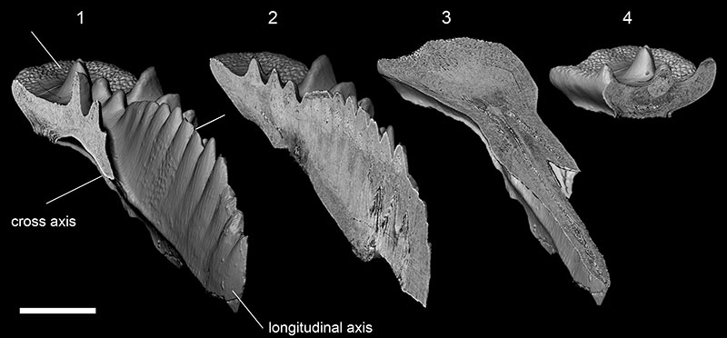
FIGURE 3. X-ray synchrotron microtomographic sections compared to SEM photos of artificially fractured conodont specimens. 1, Tomographic section of Epigondolella quadrata specimen A; 2-4, SEM photos of an artificially fractured specimen of Carnepigondolella carpathica (sample NA16). Both the specimens are sectioned in correspondence to the cusp; 5-6, Comparison between a tomographic section (5) and an artificially fractured (6) specimen of E. uniformis from the same sample (NA42). Legend: cu, cusp; wm, white matter; hp, hypocalcification; gl, growth lines.
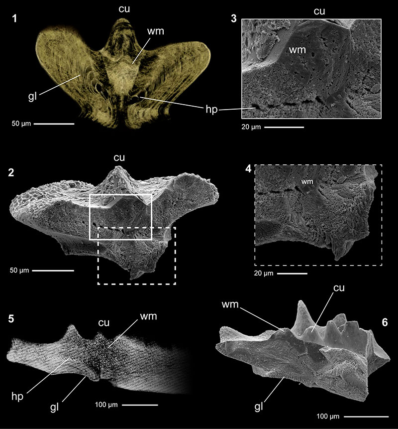
FIGURE 4. Microtomographic longitudinal sections focused on the posterior platform of Epigondolella quadrata and E. rigoi, aimed to show the ontogenesis of the posterior node growing behind the cusp. For each section a 3D model of the correspondent conodont is provided. Beside the sections, the outlines of selected growth lines are reported, in order to evidence some visible growth stages and show the different ontogenetic processes of E. quadrata and E. rigoi. Arrows and letters (a, b, c) mark the growth lines considered to draw the stages. 1, E. quadrata A; 2, juvenile specimen of E. quadrata from sample NA60 (from Mazza and Martínez-Pérez, 2015; repository number Micro-Unimi no. 2001); 3, E. quadrata B; 4, E. quadrata C; 5, E. rigoi A; 6, juvenile specimen of E. rigoi from sample NA68 (from Mazza and Martínez-Pérez, 2015; repository number Micro-Unimi no. 2003), showing that the posterior node is already occurring; 7, E. rigoi B. Scale bars equal 200 µm. Legend: cu, cusp; pn, posterior node.
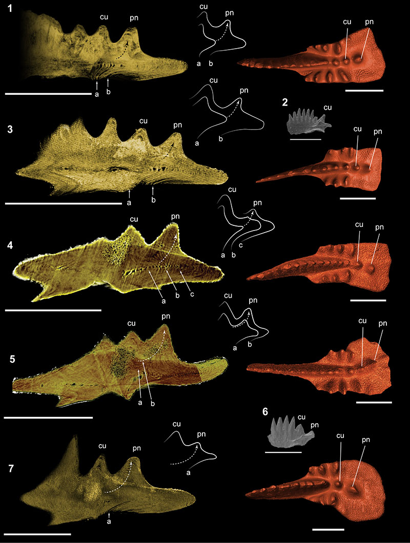
FIGURE 5. Microtomographic longitudinal sections focused on the posterior platform of Epigondolella vialovi, E. uniformis, and E. triangularis aimed to show the ontogenesis of the posterior node growing behind the cusp. For each section a 3D model of the correspondent conodont is provided. 1, E. vialovi ; 2, E. uniformis ; 3, juvenile specimen of E. uniformis from sample NA43 (from Mazza and Martínez-Pérez, 2015; repository number Micro-Unimi no. 2005); 4, E. triangularis ; 5, juvenile specimen of E. triangularis from sample NA43 (from Mazza and Martínez-Pérez, 2015; repository number Micro-Unimi no. 2004). Scale bars equal 200 µm. Legend: cu, cusp; pn, posterior node; wm, white matter.
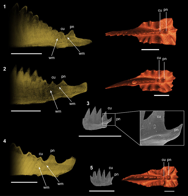
TABLE 1. List of the conodonts scanned at the X-ray Synchrotron Microscopy for analyses of the internal structure. Species for which more than one specimen was scanned are provided with a capital letter that, associated with the species name, identify the specimen in the text and in figures. All the specimens are from the Pizzo Mondello section (western Sicily, Italy).
| Species | Epigondolella vialovi | Epigondolella quadrata | Epigondolella quadrata | Epigondolella quadrata |
| Author and year | (Burij, 1989) | Orchard, 1991 | Orchard, 1991 | Orchard, 1991 |
| Specimen | A | B | C | |
| Range of the species | Upper Carnian - Lower Norian | Upper Carnian - Middle Norian | Upper Carnian - Middle Norian | Upper Carnian - Middle Norian |
| Sample | NA30 | NA60 | NA60 | NA66 |
| Age of the sample | Uppermost Tuvalian (Upper Carnian) | Upper Lacian (Lower Norian) | Upper Lacian (Lower Norian) | Upper Lacian (Lower Norian) |
| Repository number | Micro-Unimi no. 2010 | Micro-Unimi no. 2011 | Micro-Unimi no. 2012 | Micro-Unimi no. 2017 |
| CAI | 1.5 | 7 | 7 | 1.5 |
| Species | Epigondolella rigoi | Epigondolella rigoi | Epigondolella uniformis | Epigondolella triangularis |
| Author and year | Noyan and Kozur, 2007 | Noyan and Kozur, 2007 | (Orchard, 1991) | (Budurov, 1972) |
| Specimen | A | B | ||
| Range of the species | Lower - Middle Norian | Lower - Middle Norian | Lower - Middle Norian | Lower Norian |
| Sample | NA61 | NA60 | NA42 | NA44a |
| Age of the sample | Upper Lacian (Lower Norian) | Upper Lacian (Lower Norian) | Lower Lacian (Lower Norian) | Lower Lacian (Lower Norian) |
| Repository number | Micro-Unimi no. 2018 | Micro-Unimi no. 2014 | Micro-Unimi no. 2013 | Micro-Unimi no. 2015 |
| CAI | 7 | 1.5 | 1.5 | 1.5 |
Evolutionary convergence in conodonts revealed by Synchrotron-based Tomographic Microscopy
Plain Language Abstract
Conodonts are apatitic microfossils representing parts of the feeding apparatus of an extinct marine early vertebrate. They lived for 300 m.y., from the Cambrian to the end of the Triassic. Their fossil record is well known and documented, but the morphological evolution that characterizes this fossil group often fails to provide reliable phylogenetic relationships among species, because it is unclear if the diagnostic characters of the taxa are indicative of common ancestry (homology) or just evolutionary convergences (homoplasy). An homology is a character shared by two different species because it was inherited by the same ancestor, and it is thus indicative of a phylogenetic relation between species, while an homoplasy is a similar shared character but resulted from evolutionary convergence, and it is not indicative of common ancestry. In conodonts, the most reliable method to distinguish homology from homoplasy is by studying the ontogenesis (i.e., the growth process) of the single taxa, but, the only unequivocal way to describe the ontogenesis of a conodont is to reconstruct it from a single individual. We achieve this objective by using for the first time Synchrotron Radiation X-ray Tomographic Microscopy applied to some phylogenetically related conodonts of the Upper Triassic. Our analysis provided tomographic images of the internal structure of conodonts, useful for the reconstruction of the conodonts ontogenesis. We focused our study on the posterior platform of the conodonts, where an accessorial node develops behind the cusp, and considered a homology exclusive of the genus Epigondolella. The microtomographies showed instead that this character follows different ontogenetic processes in the analyzed species, revealing it as an evolutionary convergence.
These results suggest the revision of the Late Triassic conodont phylogenetic relationships, showing that ontogenesis can be used as a criterion for discriminating homology from homoplasy in conodonts, and demonstrating that Synchrotron Radiation X-ray Tomographic Microscopy is a powerful and reliable tool to investigate conodonts ontogenesis, evolutionary processes and phylogenetic relationships.
Resumen en Español
La convergencia evolutiva en los conodontos revelada mediante Microscopía Tomográfica basada en Sincrotrón
El registro de conodontos fósiles es bien conocido por su diversidad morfológica, pero la evolución iterativa que caracteriza a los conodontos a menudo no proporcionar marcos filogenéticos fiables entre especies. Por ello, no está claro si los caracteres diagnósticos de los taxones son indicativos de ancestros comunes o convergencias evolutivas. Para distinguir las homologías de las analogías en los conodontos, el método más fiable es estudiar el desarrollo ontogenético de los taxones individuales. Hasta ahora, la reconstrucción de las etapas ontogenéticas se basó en el estudio de individuos separados por diferentes edades de una misma población. Sin embargo, la única manera de establecer la ontogénesis de un conodonto con fiabilidad es describirla a partir de un solo espécimen. Hemos alcanzado este objetivo utilizando la Microscopía Tomográfica de Rayos X con Radiación de Sincrotrón aplicada a elementos P1 de especies pertenecientes a los géneros del Triásico Superior Carnepigondolella y Epigondolella. Nuestro análisis proporcionó información tomográfica interna para la reconstrucción de la ontogénesis de conodontos. Nuestro estudio se centró en la plataforma posterior, donde un nodo accesorio se desarrolla detrás de la cúspide. Este nodo había resultado ser una autapomorfia del género Epigondolella a partir de análisis cladísticos previos y, por tanto, se consideraba un carácter diagnóstico para la elaboración de modelos filogenéticos. En cambio, nuestras microtomografías muestran que este carácter es consecuencia de una convergencia evolutiva. Estos resultados sugieren la revisión de las relaciones filogenéticas de conodontos del Triásico tardío, mostrando que la ontogénesis puede ser utilizada como criterio para discriminar la homología de la homoplasia en conodontos y que la Microscopía Tomográfica de Rayos X con Radiación de Sincrotrón es una herramienta poderosa y fiable para investigar la ontogénesis, los procesos evolutivos y las relaciones filogenéticas de conodontos.
Palabras clave: conodontos; tomografías de rayos X; ultraestructura; ontogénesis; Triásico
Traducción: Enrique Peñalver (Sociedad Española de Paleontología)
Résumé en Français
Les convergences évolutives chez les conodontes révélées via la microscopie par tomodensitométrie par rayonnement synchrotron
Le registre fossile des conodontes est bien connu pour sa diversité morphologique. L'évolution itérative qui caractérise les conodontes empêche cependant souvent de fournir des contextes phylogénétiques fiables, ce qui obscurcit les caractères diagnostiques des taxons indiquant soit une ascendance commune soit des convergences évolutives. Pour distinguer les homologies des analogies chez les conodontes, la méthode la plus fiable est l'étude du développement ontogénétique de chaque taxon. Jusqu'à présent, la reconstitution des stades ontogénétiques était basée sur l'étude des individus d'âges différents provenant d'une même population. Cependant, la seule manière de décrire l'ontogénèse d'un conodonte sans équivoque est de le faire à partir d'un seul spécimen. Nous atteignons cet objectif en utilisant la microscopie par tomodensitométrie à rayons X par rayonnement synchrotron appliquée aux éléments P1 d'espèces appartenant aux genres Carnepigondolella et Epigondolella du Trias supérieur. Notre analyse a fourni des informations tomodensitométriques sur la structure interne pour reconstituer l'ontogénèse des conodontes. Nous avons concentré notre étude sur la plate-forme postérieure, où un nœud accessoire se développe derrière le denticule. Ce nœud était retrouvé comme une autapomorphie du genre Epigondolella dans les analyses cladistiques précédentes, et donc un caractère diagnostique pour la construction de modèles phylogénétiques. Les données microtomodensitométriques ont montré que ce caractère est plutôt une convergence évolutive. Ces résultats suggèrent que la révision des relations phylogénétiques des conodontes du Trias récent montre que les ontogénèses peuvent être utilisées comme critères pour distinguer l'homologie de l'homoplasie chez les conodontes. Ces résultats prouvent également que la microscopie par tomodensitométrie à rayons X par rayonnement synchrotron est un outil puissant et fiable pour examiner l'ontogénèse, les processus évolutifs, et les relations phylogénétiques chez les conodontes.
Mots-clés : conodontes ; tomodensitométrie par rayons X ; ultrastructure ; ontogénèse ; Trias
Translator: Antoine Souron
Deutsche Zusammenfassung
Evolutionäre Konvergenz bei Conodonten aufgezeigt durch synchotronbasierte tomografische Mikroskopie
Der Conodonten-Fossilbericht ist für seine morphologische Diversität bekannt aber die für Conodonten charakteristische iterative Evolution, bietet oft keinen verlässlichen phylogenetischen Rahmen zwischen den Arten. Das erschwert das Erkennen, ob die diagnostischen Merkmale der Taxa auf gemeinsame Abstammung hinweisen oder evolutionäre Konvergenzen sind. Die zuverlässigste Methode, bei Conodonten Homologien von Analogien zu unterscheiden, ist das Studium der ontogenetische Entwicklung der einzelnen Taxa. Bis jetzt basierte die Rekonstruktion der ontogenetischen Stadien auf der Untersuchung von unterschiedlichen Individuen mit verschiedenen Altersstufen aus einer Population. Die einzige unmissverständliche Methode jedoch die Conodonten-Ontogenese zu beschreiben, ist ein einziges Individuum zu untersuchen. Wir erreichen dieses Ziel mit Synchotron -Röntgenmikrotomographie die auf P1 Elemente der obertriassischen Arten Carnepigondolella and Epigondolella angewendet wurde. Unsere Analyse stellte innere tomographische Informationen zur Rekonstruktion der Conodonten-Ontogenie zur Verfügung. Wir fokussierten unsere Untersuchung auf die posteriore Plattform, wo sich ein zusätzlicher Knoten hinter dem Höcker entwickelt. Dieser Knoten resultierte in einer Autapomorphie der Gattung Epigondolella aus vorangegangenen kladistischen Analysen und daher in einem diagnostischen Merkmal für ein phylogenetisches Modell. Die mikrotomographischen Aufnahmen zeigten, dass dieses Merkmal dagegen eine evolutionäre Konvergenz ist. Diese Ergebnisse legen eine Revision der phylogenetischen Verwandtschaftsverhältnisse der spättriassischen Conodonten nahe und zeigen, dass Ontogenese als Kriterium zur Unterscheidung von Homologien und Homoplasien bei Condotonden genutzt werden kann. Außerdem legen sie dar, dass synchotronbasierte tomografische Mikroskopie ein wirkungsvolles und zuverlässiges Instrument zur Untersuchung der Conodonten-Ontogenese, evolutionären Prozessen und phylogenetischen Beziehungen ist.
Schlüsselwörter: Conodonten; Röntgencomputertomographie; Ultrastruktur; Ontogenese; Trias
Translator: Eva Gebauer
Arabic
Translator: Ashraf M.T. Elewa
-
-
-
Review: The Princeton Field Guide to Mesozoic Sea Reptiles
 The Princeton Field Guide to Mesozoic Sea Reptiles
Article number: 26.1.1R
April 2023

 Poster Winners 2024