| |
SYSTEMATICS
Diagnosis. Holocarpic monocentric fungus; thallus with thin- and thick-walled sporangia; thin-walled sporangia subspherical (77-89 x 70-87 μm), ellipsoid (91-213 x 59-94 μm); single-layered wall (up to 2 m); some sporangia with internal contents consisting of segments of variable size and shape; thick-walled sporangia ovoid (35-43 x 23-25
μm), pyriform (56-67 x 41-47 μm), or ellipsoid to curved-ellipsoid (56-130 x 52-98); wall of variable thickness (up to 4
μm thick); zoospores and zoospore-like bodies (larger zoospores) (5-41 μm) spherical to ellipsoid, with centrally located opaque inclusion; typically one sporangium per host cell.
Etymology. The specific epithet reflects the Permian age of the fossil.
 Holotype. Slide #21644 (Figure 1.2), specimen #11653 C-top. Holotype. Slide #21644 (Figure 1.2), specimen #11653 C-top.
Paratypes. Slide #21643 (Figure 1.1, 1.3, 1.6, 1.8, 1.10,
Figure 2.6, 2.7, 2.8,
Figure 3.3, 3.4, 3.5), specimen #11665 C-top; slide #21644 (Figure 1.4, 1.5,
Figure 2.1, 2.2, 2.3, 2.4, 2.5,
Figure 3.6, 3.7), specimen #11653 C-top; slide #21645 (Figure 1.7), specimen #11654 G-bot; slide #21646 (Figure 1.9,
Figure 3.1), specimen #11654 F-top; slide #21639 (Figure 2.9,
Figure 3.2), specimen #11657 E-top.
Repository. The specimens are housed in the Division of Paleobotany, Natural History Museum and Biodiversity Research Center, University of Kansas.
Type locality.
Skaar Ridge, Queen Alexandra Range, Transantarctic Mountains, Antarctica (84°49'15.8" S, 163°20'18.9" E, 2289 m altitude, Buckley Island Quadrangle, Barrett and Elliot 1973).
Stratigraphic horizon.
Late Permian (~250 Ma), Buckley Formation, Beacon Supergroup.
Description
The description is based on a large number of endobiotic parasitic chytridiomycetes that extensively infect remnants of plants preserved as silicified tissue.
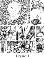
Figure 1.1 represents a cross section of a root of an unidentified plant where the central pit is not preserved and the majority of the remaining cortical cells appear highly distorted and filled with thin- and thick-walled sporangia, and zoospores of different sizes. The additional thin- and thick-walled sporangia and zoospores shown in the remaining figures co-occur on poorly preserved fragments of sections of leaves, roots, and stems of unidentifiable plants. Sporangia occur one per host cell. Zoospores occur within host cells and in thin- and thick-walled sporangia. The infected tissues show signs of hypertrophy.
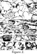 Thin-walled sporangia. Sporangia are nearly spherical and formed by numerous polygonal segments of approximately equal size (Figure 2.1). Other sporangia of broad ellipsoid shape are built of several transversely arranged segments (Figure 2.2, 2.3, 2.4). These segments have a globose aspect, are unequal in size and completely or partially fill the sporangial lumen (Figure 2.2, 2.3, 2.4). In several specimens it is possible to delimit a single-layered wall (up to 2 µm thick) (Figure 2.4). Ruptured individuals are present in some cells (Figure 2.5). An acute papilla-like body projection is present in some individuals (Figure 2.4). Thin-walled sporangia. Sporangia are nearly spherical and formed by numerous polygonal segments of approximately equal size (Figure 2.1). Other sporangia of broad ellipsoid shape are built of several transversely arranged segments (Figure 2.2, 2.3, 2.4). These segments have a globose aspect, are unequal in size and completely or partially fill the sporangial lumen (Figure 2.2, 2.3, 2.4). In several specimens it is possible to delimit a single-layered wall (up to 2 µm thick) (Figure 2.4). Ruptured individuals are present in some cells (Figure 2.5). An acute papilla-like body projection is present in some individuals (Figure 2.4).
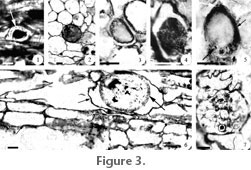
Thick-walled sporangia. Sporangia are ovoid, pyriform, or ellipsoid to curved-ellipsoid in shape (Figure 3.3, 3.4, 3.5, 3.6). Sporangia have a wall that is of different thicknesses (up to 4 µm thick) (Figure 3.5). Numerous small pits ornament the surface of the smallest sporangia (Figure 3.4). A series of ridges are present at the perimeter of larger sporangia (Figure 3.6). Several ruptured sporangia are present and contain zoospores (Figure 3.7). Some sporangia display one small slit at the end of the body (Figure 3.5). A few zoospores are distinguishable in the pyriform sporangia (Figure 3.5).
 Zoospores. Zoospores, including zoospore-like bodies (larger zoospores), (Figure 1.3, 1.4,
Figure 3.1, 3.2) range from 5 to 41 µm in diameter, are spherical or subspherical to sometimes irregularly elongated in shape, and each possesses a large, centrally located, opaque body, although some individuals do not exhibit internal contents (Figure 1.3). Other zoospores are hemispherical (Figure 1.6). Some zoospores have a thick, opaque, short appendage protruding from their body (Figure 1.4). One or more short opaque threads radiate from the opaque body in the center of some zoospores (Figure 1.5). A number of the larger zoospores (more than 10 µm), with the lumen completely or partially empty of opaque contents, or with an extended perimeter and a partially disaggregated opaque central body are present (Figure 1.8, 1.9). In addition, either an irregular scar-like aperture (up to 24
µm) or a neck-shaped wall projection (4-7
µm) opening to the exterior can be distinguished on the surface of the empty larger individuals (Figure 1.8). A number of similarly sized small zoospores (up to 10 µm) occur close to each other (Figure 2.6). Other similarly sized zoospores are attached by their sides, some having individual borders, and others having a common margin (Figure 2.7, 2.8). Zoospores with a common margin exhibit individual or fused opaque contents (Figure 2.8). A few individuals have a thin flagellum (up to 21 µm in length;
Figure 2.9). Opaque particles of dense material are present in the periphery of the opaque central body in some of the larger zoospores (Figure 3.1). A few of the larger zoospores exhibit a lumen homogeneously filled with opaque particles (Figure 3.2). Zoospores. Zoospores, including zoospore-like bodies (larger zoospores), (Figure 1.3, 1.4,
Figure 3.1, 3.2) range from 5 to 41 µm in diameter, are spherical or subspherical to sometimes irregularly elongated in shape, and each possesses a large, centrally located, opaque body, although some individuals do not exhibit internal contents (Figure 1.3). Other zoospores are hemispherical (Figure 1.6). Some zoospores have a thick, opaque, short appendage protruding from their body (Figure 1.4). One or more short opaque threads radiate from the opaque body in the center of some zoospores (Figure 1.5). A number of the larger zoospores (more than 10 µm), with the lumen completely or partially empty of opaque contents, or with an extended perimeter and a partially disaggregated opaque central body are present (Figure 1.8, 1.9). In addition, either an irregular scar-like aperture (up to 24
µm) or a neck-shaped wall projection (4-7
µm) opening to the exterior can be distinguished on the surface of the empty larger individuals (Figure 1.8). A number of similarly sized small zoospores (up to 10 µm) occur close to each other (Figure 2.6). Other similarly sized zoospores are attached by their sides, some having individual borders, and others having a common margin (Figure 2.7, 2.8). Zoospores with a common margin exhibit individual or fused opaque contents (Figure 2.8). A few individuals have a thin flagellum (up to 21 µm in length;
Figure 2.9). Opaque particles of dense material are present in the periphery of the opaque central body in some of the larger zoospores (Figure 3.1). A few of the larger zoospores exhibit a lumen homogeneously filled with opaque particles (Figure 3.2).
Host reaction. Host cells infected with thin-walled sporangia appear enlarged two to five times (Figure 2.1, 2.2) whereas cells hosting thick-walled sporangia are often enlarged three to five times (Figure 2.2,
Figure 3.6). Between zones where host cells are common, there are spaces with sporangia only (Figure 1.2). There are also zones with no sporangia but cells are enlarged and distorted (Figure 2.1). A few host cells have their lumen completely filled with two similarly sized and rather elongated diffuse bodies, one containing opaque inclusions (Figure 1.10).
|