|
|
NOTE ADDED IN PROOF
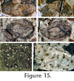
USNM 536213 (Fig. 15.1, 15.2, part and counterpart). Icacinaceae cf. Palaeophytocrene (Reid, E.M., and Chandler, M.E.J. 1933. The London Clay Flora. British Museum [Natural History], London, England, 561 p.). Oval, tuberculate endocarp. Tubercules extend towards center of fruit, tubercles in vertical rows. Dimensions preserved: 2.7 mm length, 1.2 mm width. USNM 536214 (Fig. 15.3, 15.4, part and counterpart). Juglandaceae sp., endocarp. The single specimen is ovoid and basally lobed internally. The endocarp is apparently without a wing and two-lobed, but more specimens would be needed to determine these characters and better assign this fruit taxonomically. Dimensions preserved: 1.3 mm length, 0.8 mm width. USNM 536215 (Fig. 15.6; Fig. 15.5 shows a separated fragment of the same morphotype, on the same rock). Dicot cuticle morphotype with densely spaced, diffuse paracytic stomata and trichome bases, visible under fluorescence microscopy with long-pass green filter. Spherical resin bodies are also visible in the carbon film between cuticle layers (Fig. 15.5), and these are probably organically preserved oil/mucilage idioblasts common in tissues of Laurales, Magnoliales and basal ‘ANITA’ grade angiosperms. These resin bodies are also visible, under fluorescence, in the Lauraceae leaf morphotype exemplar (Fig. 2.3), further confirming that specimen as Lauraceae. |
|