|
TECHNIQUE
X-ray CT is a non-destructive technique for visualizing structures in the interior of opaque objects that enables paleontologists to acquire digital information about the 3-D structural geometry of specimens. Its ability to resolve details as fine as a few tens of microns in objects made of high density material distinguishes this technique from traditional medical CAT-scanning. Complete details of the technique have been published and are available online
at the
High Resolution X-ray CT Facility (Ketcham
and Carlson 2001).
The spatial resolution of a CT image is determined principally by the size and number of detector elements, the size of the X-ray focal spot, and the source-object-detector distances. As a rule of thumb, a CT image should have about as many pixels in each dimension as there are detector channels.
 Thus a 1024-channel linear detector array justifies a 1024x1024 pixel reconstructed image. Resolution in the third dimension is governed by detector aperture or thickness (for single-slice scanners) or vertical spacing (for multi-slice scanners). Thus a 1024-channel linear detector array justifies a 1024x1024 pixel reconstructed image. Resolution in the third dimension is governed by detector aperture or thickness (for single-slice scanners) or vertical spacing (for multi-slice scanners).
No specimen preparation is required prior to scanning, other than the need for the specimen to fit in the field of view. Because the full scan field is a cylinder, the most efficient geometry to scan is a cylinder. Commonly specimens are placed inside a cylindrical container with appropriate filler.
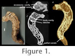
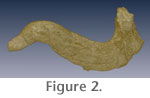 This technique in many cases cannot be used successfully if the specimen contains matrix that has a density similar to the specimen. The rudist specimen scanned here is silicified, and its matrix is carbonate mud, providing an excellent contrast (Figure 1.1, see
Figure 2, 3-D movie). This technique in many cases cannot be used successfully if the specimen contains matrix that has a density similar to the specimen. The rudist specimen scanned here is silicified, and its matrix is carbonate mud, providing an excellent contrast (Figure 1.1, see
Figure 2, 3-D movie).
Scanning was done by Richard Ketcham at the University of Texas High-Resolution X-ray CT Facility.
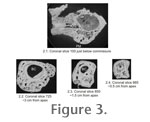 The specimen was first scanned with the high-energy 420-kV scanner subsystem in its longitudinal direction (Figure 1.2) to test for the presence of differentiable details. Following this successful test, the specimen was scanned perpendicular to its long axis (Figure 1.3) on 19 August 2005 using the microfocal subsystem, with X-rays set at 180 kV and 0.133 mA to provide a focal spot of 30 µm. A total of 930 1024x1024 slices were obtained with a slice thickness and inter-slice spacing of 0.1433 mm and a field of reconstruction of 66 mm (Figure 1.3,
Figure 3). Image processing and visualization (Figure 1.4)
was completed by Jessica A. Maisano. The scan can be examined on the DigiMorph
site, an
NSF Digital Library at The University of Texas at Austin. The specimen was first scanned with the high-energy 420-kV scanner subsystem in its longitudinal direction (Figure 1.2) to test for the presence of differentiable details. Following this successful test, the specimen was scanned perpendicular to its long axis (Figure 1.3) on 19 August 2005 using the microfocal subsystem, with X-rays set at 180 kV and 0.133 mA to provide a focal spot of 30 µm. A total of 930 1024x1024 slices were obtained with a slice thickness and inter-slice spacing of 0.1433 mm and a field of reconstruction of 66 mm (Figure 1.3,
Figure 3). Image processing and visualization (Figure 1.4)
was completed by Jessica A. Maisano. The scan can be examined on the DigiMorph
site, an
NSF Digital Library at The University of Texas at Austin.
|