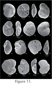
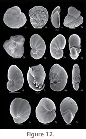
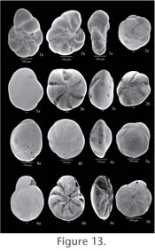
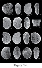
|
|
|
SYSTEMATIC PALEONTOLOGYSuprageneric classification follows that of Loeblich and Tappan (1987). Illustrated specimens are housed in the Department of Earth Sciences, Carleton University, Ottawa, Ontario.
Order FORAMINIFERIDA Eichwald, 1830 1944 Proteonina atlantica Cushman, p. 5, pl. 1, fig. 4. 1980 Saccammina atlantica (Cushman 1944). Rodrigues, p. 69, pl. 3-1, fig. 11. 1995 Saccammina cf. atlantica (Cushman 1944). Blais, p. 92, pl. 2.3, fig. 4. Description. Test free, single elongate, oval, or pyriform chamber; wall agglutinated, fairly coarse; aperture very small, at tapered end of chamber; no distinct neck, but gradually contracted toward apertural end; circular in section.
Saccammina sphaerica Sars, 1872 Description. Test free, single globular chamber, spherical or pyriform; wall agglutinated, fine, smooth; aperture rounded, may be nearly flush or on short neck; inner organic wall layer modified in living specimens to an oral apparatus or entosolenian tube around opening.
Superfamily AMMODISCACEA Reuss, 1862
1948 Ammodiscus gullmarensis Höglund, p. 45. Description. Test free, small, flattened but tending to irregular coiling in last whorls; wall agglutinated with fairly large amount of cement; 7 to 9 whorls; sutures distinct, proloculum central and subspherical.
Superfamily RZEHAKINACEA
Cushman, 1933a 1870 Quinqueloculina fusca Brady, in Brady and Robertson, p. 286, pl. 11, fig. 2. 1972 Miliammina fusca (Brady, 1870) Murray, p. 21, pl. 3, figs. 1-6; Patterson, 1990a, p. 240, pl. 1, fig. 4. Description. Test elongate ovate; narrow chambers one-half coil in length, quinqueloculine arrangement; wall thick, very finely agglutinated; aperture at end of chamber, large and conspicuous, equal in size to transverse section of chamber.
Superfamily LITUOLACEA
de Blainville, 1827 1892 Haplophragmium crassimargo Norman, p. 17, pl. 7-8. 1947 Labrospira crassimargo (Norman 1892). Höglund, p. 141, pl. 11, fig. 1. 1953 Alveolophragmium crassimargo (Norman 1892). Loeblich and Tappan, p. 29, pl. 3, figs. 1-3. 1967 Cribrostomoides crassimargo (Norman 1892). Todd and Low, p. 15, pl. 1, fig. 24. Description. Test free, planispiral, partially involute; wall agglutinated, thick, coarse, with large sand grains, sometimes of orange color; 2 to 4 whorls, 8 to 10 chambers in last whorl; sutures straight, slightly depressed; umbilicus depressed; periphery rounded, lobulate; aperture interio-areal, forming an oblong, curved slit, upper and lower lip well developed.
Cribrostomoides jeffreysii (Williamson 1858) 1858 Nonionina jeffreysii Williamson, p. 34, pl. 3, figs. 72,73. 1953 Alveolophragmium jeffreysii Williamson, 1858. Loeblich and Tappam, p. 31, pl. 3, figs. 4-7. 1972 Cribrostomoides jeffreysii (Williamson 1858). Murray, p. 23, pl. 4, figs. 1-5. Description. Test free, planispiral, almost involute, compressed laterally; wall finely agglutinated, yellow to brown; 6 to 8 chambers in last whorl, with their lateral surface triangular in shape; periphery rounded, slightly lobulate; sutures depressed and radial; aperture a lipped slit near base of apertural face.
Cribrostomoides cf.
subglobosum (Cushman 1910) 1910 Haplophragmoides subglobosum Cushman, p. 105, figs. 162-164. 1947 Labrospira subglobosa (Cushman, 1910). Höglund, p. 144, pl. 11, fig. 2. 1986 Cribrostomoides subglobosus (Cushman, 1910). Schröder, p. 48, pl. 17, figs. 15, 16; Jonasson, 1994, p. 53, pl. 5, fig. 8. 1981 Cribrostomoides subglobosum (Cushman 1910). Poag, p. 57, pl. 11, fig. 2. Description. Test free, planispiral, involute, depressed at the umbilicus; wall agglutinated, somewhat roughened but variable; chambers broad and low, usually 4 to 8 in last whorl, making test as whole subglobular; aperture interio-areal, forming oblong, very narrow curved slit immediately above inner margin of apertural face, with well developed lips. Remarks. Although C. subglobosum is reported to be of a bigger size than the specimens present on the SBIC (J.P. Guilbault, oral communication, 2006), the general shape of these specimens agrees well with the description of C. subglobosum. However, the apertural face in most specimens was covered by debris, making it impossible to recognize the shape of the aperture. They are therefore named as C. cf. subglobosum, until a better designation is found.
Cribrostomoides wiesneri (Parr 1950)
1986 Cribrostomoides wiesneri (Parr 1950). Schröder, p. 48, pl. 17, figs. 10, 12; Poag, 1981, p. 57, pl. 9, fig. 1. Description. Test free, planispiral, not completely involute; wall agglutinated, fine, with excess of yellow to reddish brown cement, smooth; umbilical region depressed, with chambers of earlier whorls visible; about 3 whorls; periphery slightly lobulate; 7 to 9 chambers in last whorl; sutures distinct, slightly depressed; aperture a short narrow slit slightly above base of apertural face.
Cribrostomoides sp. A Description. Test free, planispiral, involute, depressed at the umbilicus; wall agglutinated, somewhat roughened but variable; chambers broad and low, usually 5 to 7 in last whorl; last chamber much inflated and wider, protruding strongly on both sides on apertural view, rest of the chambers more compressed; periphery rounded; aperture interio-areal, forming a curved slit immediately above of inner margin of apertural face. Remarks. This species resembles C. subglobosum, but the width of the last chamber, very prominent in all specimens, sets it apart.
Genus Haplophragmoides
Cushman, 1910 1887 Trochammina robertsoni Brady, p. 893, pl, 20, fig. 4. 1891 Trochammina bradyi Robertson, p. 388. 1947 Haplophragmoides bradyi (Brady 1887). Höglund, p. 134, pl. 10, fig. 1; Murray, 1972, p. 24, pl, 5, figs. 1,2; Schröder, 1986, p. 46, pl. 17, fig. 8. Description. Test free, planispiral, partly evolute, discoidal or compressed, nearly symmetrical bilaterally; wall agglutinated, very fine, brown, very smooth; 5 chambers in outer whorl, somewhat inflated; sutures distinct, depressed; aperture interiomarginal, lipped.
Superfamily HAPLOPHRAGMIACEA
Eimer and Fickert, 1899 1881 Lituola (Haplophragmium) turbinatum Brady, p. 50, pl. 35, fig. 9. 1953 Recurvoides turbinatus (Brady 1881). Loeblich and Tappan, p. 27, pl. 2, fig. 11; Rodrigues, 1980, p. 66, pl. 3-3, fig. 8; Jonasson, 1994, p. 58, pl. 7, fig. 2. Description. Test free, streptospiral, involute on one side and evolute on the other; wall agglutinated, somewhat roughened but variable; chambers not inflated but somewhat irregular in shape; periphery rounded; aperture an elongated areal slit set at an angle on periphery.
Superfamily TROCHAMMINACEA
Schwager, 1877
1986 Trochammina cf. globigeriniformis (Parker and Jones 1865). Schröder, p. 52, pl. 19, figs. 5-8. Description. Test free, trochospiral, varying from depressed to slightly conical; wall medium to coarsely agglutinated with finer grained matrix; 2 or 3 whorls; chambers rapidly increasing in size; periphery rounded; aperture simple at inner margin on umbilical side of last chamber opening into narrow umbilicus.
Genus Deuterammina
Brönnimann 1976 1934 Trochammina discorbis Earland, p. 104, pl. 3, figs. 28-31; Blais, 1995, p. 93, pl. 2-3, figs. 7, 8. 1988 Deuterammina discorbis (EARLAND) Brönnimann and Whittaker, p. 101, fig. 36A-I. Description. Test free, minute, trochospiral; wall agglutinated, very fine with much cement; surface smooth but not very highly polished; 3 or 4 whorls visible on spiral side, 4 or 5 chambers from final whorl visible on dorsal side; dorsal side highly convex, umbilical side nearly flat but with deeply sunk umbilicus; sutures recurved on dorsal side, straight on ventral; periphery subacute; aperture small slit on inner edge of final chamber on ventral side. Deuterammina grisea (Earland 1934) 1934 Trochammina grisea Earland, p. 100, pl. 3, figs. 35-37. 1988 Deuterammina (Deuterammina) grisea (Earland) Brönnimann and Whittaker, p. 107-110, fig. 39D-I. Description. Test free, large, trochospiral; wall agglutinated, fine, smooth, unpolished; 3 whorls with 6 chambers each on dorsal side, only 6 last chambers visible on umbilical side, very slightly inflated; dorsal side flattened, umbilical side deeply depressed at umbilicus; sutures on dorsal side straight and distinct, slightly depressed, sutures more depressed on umbilical side; periphery round, slightly lobulate at last few chambers; aperture a narrow slit on inner apertural face of last chamber.
Deuterammina rotaliformis (Heron-Allen and Earland, 1911) 1911 Trochammina rotaliformis Heron-Allen and Earland, p. 309; Loeblich and Tappan, 1953, p. 51, pl. 8, figs. 6-9; Murray, 1972, p. 39, pl. 12, figs. 1-5; Blais, 1995, p. 93, pl. 2-3, figs. 1-3. Description. Test free, trochospiral; wall agglutinated, imperforate; 20 elongated chambers arranged in 3 whorls on dorsal side, 5 chambers broadly triangular on umbilical side; strongly convex on dorsal side, shallow-concave on umbilical side; periphery rounded, slightly lobulate; sutures not well defined, more depressed on umbilical side; umbilical depression star-shaped, open and deep with 5 arms; primary aperture interiomarginal extraumbilical, secondary posterior aperture umbilical-sutural.
Genus Lepidodeuterammina
Brönnimann and Whittaker 1983 1858 Rotalina ochracea Williamson, p. 55, pl. 5, fig. 113. 1987 Lepidodeuterammina ochracea (Williamson 1858). Loeblich and Tappan, pl. 135, figs. 10-14. 1972 Trochammina ochracea (Williamson 1858). Murray, p. 37, pl. 11, figs. 1-5; Alves Martins and Ruivo Dragão Gomes, 2004, p. 29, fig. 2.12. Description. Test free, trochospiral, depressed; wall agglutinated; two-and-one-half whorls in dorsal side, with 8 or 9 chambers each, filled with dark organic matter on umbilical side; slightly convex on dorsal side, correspondingly concave on ventral side; periphery round, very slightly lobulate at last chambers; sutures sometimes raised, arcuate, flexuous and very prominent; aperture a peripheral interiomarginal arch, secondary umbilical apertures at umbilical inflated portions of chambers.
Genus Lepidoparatrochammina
Brönnimann and Whittaker 1986 1925 Trochammina charlottensis Cushman, p. 39, pl. 6, fig. 4a, b. 1995 Trochammina charlottensis Cushman, 1925. Blais, p. 92, pl. 2-3, fig. 1; Patterson, Burbidge and Luternauer, 1998, p. 4, pl. 26, figs. 5, 6. Description. Test free, compressed, very flat trochospiral; wall coarsely agglutinated, opaque; sutures radiate and depressed, especially on the umbilical side; all chambers visible on spiral side, last 6 chambers visible on the umbilical side; aperture a low interiomarginal arch opening into the open umbilicus. Remarks. This species is easily distinguishable by the very big, inflated last chamber.
Genus Portatrochammina
Echols, 1971 1980 Portatrochammina bipolaris Brönnimann and Whittaker, pp. 181, 183, figs. 15, 16, 18, 19, 20-31. Description. Test free, trochospiral; wall agglutinated, imperforate; about 14 chambers, 6 to 8 chambers in the final whorl, increasing rapidly in size; shallow umbilicus covered by flap from each successive chamber; elongate-ovate in outline, final portion somewhat lobulate; compressed, almost flat spirally, concave umbilically; periphery rounded to sub-acute; final chamber characteristically flat on spiral side and strongly convex on umbilical side; radial sutures straight to sinuous on umbilical side and slightly curved on spiral side; color dark brow grading into light brown; aperture begins as low interiomarginal opening near periphery, extends as slit around umbilical flap.
Genus Polystomammina
Seiglie, 1965 1881 Trochammina nitida Brady, p. 52, Figure in Brady, 1884, pl. 41, figs. 5, 6; Snyder, Hale and Kontrovitz, 1990, p. 277, pl. 7, fig. 3. Description. Test free, low trochospiral, compressed; wall agglutinated, thin and fragile; 2 or 3 whorls on dorsal side, final whorl with 9 chambers, increasing rapidly in size as added; dorsal side flat, umbilical side convex, somewhat excavated at umbilicus; periphery rounded, slightly depressed at sutures; aperture a curved or angled slit, beginning at base of apertural face in equatorial position and curving openly upward onto umbilical side of chamber.
Genus Trochammina
Parker and Jones, 1859 1808 Nautilus inflatus Montagu, p. 81, pl. 18, fig. 3. 1972 Trochammina inflata (Montagu, 1808). Murray, p. 35, pl. 10, figs. 3-6; Patterson, 1990a, p. 241, pl. 1, figs. 8-10. Description. Test free, trochospiral; wall very finely agglutinated, very smoothly finished; 2 or 3 whorls, with 4 to 5 chambers in last whorl; chambers extremely inflated; periphery rounded; sutures distinct but only slightly depressed, nearly radial; aperture a narrow slit on ventral side, at base of last-formed chamber.
Trochammina pacifica
Cushman, 1925 1925 Trochammina pacifica Cushman, p. 39, pl. 6, fig. 3; Patterson, 1990a, p. 240, pl. 1, figs. 5-7; Snyder, Hale and Kontrovitz, 1990, p. 273, pl. 5, fig. 1; Blais, 1995, p. 93, pl. 2.2, figs. 7-8. Description. Test free, trochospiral; wall coarsely agglutinated, smoothly finished; 2 or 3 whorls, with 4 to 5 chambers in last whorl; chambers extremely inflated; periphery rounded; sutures distinct but only slightly depressed, nearly radial; aperture a narrow slit on ventral side, at base of last-formed chamber.
Trochammina squamata
Jones and Parker, 1860 1860 Trochammina squamata Jones and Parker, fig. in Hedley, Hurdle and Burdett, 1964, p. 422, fig. 1, p. 424, figs. 1-3. 1947 Trochammina astrifica Rhumbler, 1938. Höglund, p. 206, pl. 15, fig. 2. 1990 Trochammina squamata Jones and Parker, 1860. Snyder, Hale and Kontrovitz, p. 279, pl. 8, fig. 1. Description. Test free, small, low trochoidal, excavated on umbilical side; wall finely agglutinated, with occasional larger grains, smoothly finished; periphery angular in outline, slightly lobulate; chambers not inflated, lunate shaped; 2 whorls visible on spiral side; sutures distinct, apparently flush with surface of test, visible on spiral side as dark brown curved backwards lines; aperture a narrow slit at inner margin of umbilical side of last chamber.
Genus Zavodovskina
Brönnimann and Whittaker 1988 1881 Haplophragmium nana Brady, p. 50; Brady, 1884, p. 311, pl. 35, figs. 6-8; 1953 Trochammina nana (Brady 1881). Loeblich and Tappan, p. 50, pl. 8, fig. 5; Patterson, Burbidge and Luternauer, 1998, p. 4, pl. 1, figs. 1-3. Description. Test free, trochospiral, with spiral side almost flattened and umbilical side more convex; wall agglutinated, opaque, of medium-size grains, with some larger clear quartz grains, quite smooth; lobulate periphery; two-and-one-half whorls, all chambers visible on spiral side, only final 6 or 7 visible on umbilical side; chambers somewhat inflated; sutures distinct, nearly straight and radiate ventrally, curving backwards to periphery on dorsal side; aperture low interiomarginal arch extending into very open umbilicus.
Superfamily HORMOSINACEA Haeckel, 1894
1995 Leptohalysis catella (Höglund 1947). Blais, p. 91, pl. 2-1, fig. 6. Description. Test free, elongate, slender, tapering, circular in section; wall agglutinated, thin; numerous (up to 20) chambers, increasing in size, arranged in straight line or slightly curved; chambers short and broad, height equaling or slightly exceeding the breadth, chamber sides convex, slightly converging upward, contact surface between chambers broad; aperture a simple rounded opening at top of chamber. Leptohalysis gracilis (Kiaer 1900) 1900 Nodulina gracilis Kiaer, p. 24, figs. 1, 2. 1990 Reophax gracilis (Kiaer, 1900). Alve, p. 693, pl. 1, fig. 17. Description. Test free, elongate, composed of about 19 loosely sutured segments, at the outset short and wide, becoming elongated and pointed towards aperture, with almost horizontally sectioned basal parts; wall agglutinated, smooth. Remarks. R. gracilis differs from R. catella and R. scottii in basal part of its chambers, which look almost horizontally sectioned. Leptohalysis scottii (Chaster 1892) 1892 Reophax scottii Chaster, p. 57, pl.1, fig. 1; Murray, 1972, p. 17, pl. 1, figs. 6-8; Alve, 1990, p. 693, pl. 1, fig. 19. Description. Test free, elongate, narrow; wall agglutinated, very delicate, transparent, flexible when moist; 10-20 chambers arranged in a line, gradually increasing in size; chambers somewhat pyriform, abruptly truncated below; aperture small.
Superfamily SPIROPLECTAMMINACEA
Cushman, 1927 1865 Textularia agglutinans d'Orbigny var. biformis Parker and Jones, p. 370, pl. 15, figs. 23, 24. 1947 Spiroplectammina biformis (Parker and Jones 1865). Höglund, p. 163, pl. 12, fig. 1; Loeblich and Tappan, 1953, p. 34, pl. 4, figs. 1-6; Schröder, 1986, p. 51, pl. 21, fig. 14. Description. Test free, elongate, narrow, ovoid in section, margins broadly rounded, sides nearly parallel; wall agglutinated, solid; large early planispiral coil of few chambers followed by biserially arranged chambers, coil commonly of greater breadth than first few parts of biserial chambers; chambers very slightly inflated; sutures somewhat indistinct, only slightly depressed; aperture a low arch at inner margin of final chamber.
Superfamily VERNEULINACEA
Cushman, 1911 1927 Gaudryina arenaria Galloway and Wissler, p. 68, pl. 11, fig. 5; Patterson, Burbidge and Luternauer, 1998, p. 5, pl. 1, figs. 6, 7. Description. Test free, elongate, of equal with for much of the test length, tapers to base; wall agglutinated, finely perforate, opaque, surface roughened; chambers initially triserial and triangular in cross-section, then become biserial and slightly inflated; sutures indistinct in triserial section, become depressed in biserial portion; aperture arched at base of final chamber.
Superfamily TEXTULARIACEA
Ehrenberg, 1838 1922 Verneuilina advena Cushman, p.141. 1953 Eggerella advena (Cushman 1922). Loeblich and Tappan, p. 36, pl. 3, figs. 8-10; Patterson, Burbidge and Luternauer, 1998, p. 5, pl. 28, fig. 6. Description. Test free, elongate, sharply tapering, early portion with 4 to 5 chambers in a whorl, later portion triserial; wall finely agglutinated with occasional larger grains; chambers numerous, low and broad in early portion, increase in relative height as added, those of final whorl approximately equal in height and breadth; sutures distinct and depressed; aperture small, central, low arch at base of final chamber.
Eggerella sp. 1963 Eggerella advena (Cushman, 1922). Resig, p. 125, fig. 2. Description. Test free, elongate, sharply tapering, early portion with 4 to 5 chambers in a whorl, later portion triserial, triangular in cross-section; wall finely agglutinated with occasional larger grains; normally 4 to 5 whorls; chambers numerous, low and broad in early portion, increasing slowly in relative height as added, normally three chambers in final whorl, very inflated and extending outwards of the axis of the test, giving the test a triangular outline in apertural view and an almost flat apertural face; sides straight, except for sutures, increasing at 25° from the axis; sutures distinct and depressed; aperture small, central, low arch at base of final chamber, sometimes with a narrow lip. Remarks. This species is typically found with E. advena (Cushman), although the tapering of the final chambers and the more elongated test easily differentiates the latter.
Family TEXTULARIIDAE Ehrenberg, 1838 1952 Textularia earlandi Parker, new name for Textularia tenuissima Earland, 1933, p. 95, pl. 3, figs. 21-40; Murray, 1972, p. 33, pl. 9, figs. 1-5; Blais, 1995, p. 92, pl. 2-2, fig. 1. Description. Test minute, very elongate, biserial, straight or slightly curved, oval in section, tapering very gradually to the oral, thickest portion; wall agglutinated, thin, smooth, with little sign of cement; periphery straight in first half of test, then slightly lobulate, rounded throughout; chambers very numerous, distinct, regularly increasing in size and thickness, finally becoming slightly inflated; sutures distinct, depressed; aperture an interiomarginal arch, distinct.
Suborder MILIOLINA
Delage and Hérouard, 1896
Description. Test free, narrow, elongate, arcuate; 2 to 6 elliptical chambers, nearly equal in diameter, slightly overlapping; sutures distinct, constricted, straight; wall calcareous, finely perforate, translucent, so that neck and aperture of earlier chambers may be seen through wall of following chamber, surface smooth and unornamented; aperture radiate, terminal and central in position, very slightly produced.
Suborder LAGENINA
Delage and Hérouard, 1896 1912 Lagena dentaliformis Bagg, p. 45, pl. 13, figs. 1, 2. 1998 Hyalinonetrion dentaliforme (Bagg, 1912). Patterson, Burbidge and Luternauer, p. 8, pl. 2, fig. 9. Description. Test free, large, elongate, circular in cross-section, tapers at ends; wall calcareous, hyaline, smooth, finely perforate, although pores do not penetrate outer wall; aperture small and circular with phialine lip.
Genus Lagena
Walker and Jacob, 1798 1803 Vermiculum leave Montagu, p. 524. 1953 Lagena laevis (Montagu 1803). Loeblich and Tappan, p. 61, pl. 11, figs. 5-8; Murray, 1972, p. 83, pl. 32, figs. 6, 7. Description. Test free, unilocular, flask-shaped, somewhat elongate, widest slightly below midportion of test, base rounded; wall calcareous, hyaline, finely perforate, surface smooth except occasionally very slightly and finely hispid at base, but without distinct spines; upper portion of test tapering gradually to very elongate and slender neck; rounded aperture at top of neck.
Lagena meridionalis
Wiesner, 1931 1931 Lagena gracilis var. meridionalis Wiesner, p. 117, pl. 18, fig. 211 1967 Lagena meridionalis Wiesner, 1931. Todd and Low, p. 24, pl. 3, fig. 21. Description. Test free, unilocular, minute, flask-shaped, wider near rounded base, tapering into long, slender neck; wall calcareous, hyaline, finely perforate, surface ornamented with longitudinal costae, of which every second one is shorter, stopping at the base of the neck, the long costae extend onto neck; aperture simple, terminal, rounded. Lagena parri Loeblich and Tappan, 1953 1946 Lagena laevis (Montagu) var. baggi Cushman and Gray, p. 18, pl. 3, figs. 26, 27. 1953 Lagena parri Loeblich and Tappan, p. 64, pl. 11, figs. 11-13. Description. Test free, unilocular, flask-shaped to ovate, widest in lower half of test, with single distinct basal spine; long, very slender neck; wall calcareous, hyaline, finely perforate, smooth; aperture terminal at top of delicate neck.
Lagena semilineata Wright, 1886 1886 Lagena semilineata Wright, p. 320, pl. 26, fig. 7; Loeblich and Tappan, 1953, p. 65, pl. 11, figs. 14-22. Description. Test free, unilocular, flask-shaped, widest near apiculate base, somewhat tapered at upper portion and grading into very long, slender, delicate neck; wall calcareous, hyaline, finely perforate, upper one-half to two-thirds of test is smooth, lower portion ornamented with fine costae, which double in number by intercalation a short distance from the base, in occasional specimens intercalary costae do not develop; delicate basal spine of about one-half the diameter of neck, sometimes broken; neck long, slender, length about two-thirds of chamber; aperture terminal at end of neck, surrounded by slight lip.
Genus Procerolagena Puri, 1953 1862 Lagena amphora Reuss, p. 330, pl. 4, fig. 57; Todd and Low, 1967, p. 23. Description. Test free, unilocular, big, amphora-shaped, elongate, tapering into long, slender neck; wall calcareous, hyaline, finely perforate, surface ornamented with thick, longitudinal, plate-like costae that extend onto neck; aperture simple, terminal, rounded, with phialine lip.
Procerolagena gracilis (Williamson, 1848) 1848 Lagena gracilis Williamson, p. 13, pl. 1, fig. 5 1998 Procerolagena gracilis (Williamson, 1848). Patterson, Burbidge and Luternauer, p. 9, pl. 6, fig. 1. Description. Test free, unilocular, elongate, circular in cross-section, widest near midpoint; wall calcareous, hyaline; 16 to 20 discontinuous and occasionally anastomosing longitudinal costae that extend from base to neck, in some specimens costae unite to form an elongate process in aboral region; neck narrow and elongate, comprises less than one-half of test length; aperture small and circular with phialine lip.
Procerolagena mollis (Cushman 1944) 1944 Lagena gracillima (Seguenza) var. mollis Cushman, p. 21, pl. 3, fig. 3. 1953 Lagena mollis (Cushman 1944). Loeblich and Tappan, p. 63, pl. 11, figs. 25-27. Description. Test free, unilocular, elongate-fusiform, with basal spine and extremely long and slender neck at opposite end, sides nearly parallel over central portion of test; wall calcareous, hyaline, finely perforate, ornamented with numerous very fine longitudinal ribs which die out at beginning of neck; aperture terminal, surrounded by flared lip, at end of long, slender and smooth neck.
Procerolagena wiesneri (Parr 1950) 1950 Lagena striata (Montagu) var. wiesneri Parr, p. 301. 1998 Procerolagena wiesneri (Parr, 1950). Patterson, Burbidge and Luternauer, p. 10, pl. 5, figs. 5, 6. Description. Test free, unilocular, subglobular, broadest near midpoint; wall calcareous, hyaline, surface smooth, finely perforated between costae; on average 28 delicate costae extending from base, every sixth terminates at aperture, three intervening costae terminate at base of neck, separated by two costae of intermediate length which terminate halfway up to neck; no tendency for costae to form an elongate aboral process; aperture round with a phialine lip.
Genus Pygmaeoseistron
Patterson and Richardson, 1988 1913 Lagena hispidula Cushman, p. 14, pl. 5, figs. 2, 3. 1988 Pygmaeoseistron hispidulum (Cushman, 1913). Patterson and Richardson, p. 243, 245, figs. 7-10; Patterson, Burbidge and Luternauer, 1998, p. 10, pl. 6, figs. 2, 3. Description. Test free, unilocular, elongate, circular in section; wall calcareous, translucent, surface coarse, imperforate; aperture small, circular, with phialine lip, terminal on narrow neck.
Family ELLIPSOLAGENIDAE Silvestri, 1923
Description. Test free, unilocular, subspherical; wall calcareous, hyaline; surface ornamented with 8 to 10 high, stout longitudinal costae, and discontinuous transverse costae, about one-half the height of longidudinal costae, so pattern on test is rectangular; longitudinal costae bifurcate near apex of text to form a single row of hexagonal reticulations; aperture small, round, with phialine lip, may have entosolenian tube.
Favulina melo (d'Orbigny 1839) 1839 Oolina melo d'Orbigny, p. 20, pl. 5, fig. 9; Loeblich and Tappan, 1953, p. 71, pl. 12, figs. 8-15; Murray, 1972, p. 93, pl. 37, figs. 4-6. 1998 Favulina melo (d'Orbigny 1839). Patterson, Burbidge and Luternauer, p. 11, pl. 6, figs. 6-9. Description. Test free, unilocular, globular, circular in section; wall calcareous, hyaline, fine perforations do not penetrate outer wall; 9 to 14 longitudinal costae extend from base to aperture; numerous downward curved cross struts unite longitudinal costae giving squamiform appearance to test surface; aperture small and circular, phialine lip; entosolenian tube short and straight.
Genus Homalohedra
Patterson and Richardson, 1988 1953 Lagena apiopleura Loeblich and Tappan, p. 59, pl. 10, figs. 14, 15. 1998 Homalohedra apiopleura (Loeblich and Tappan, 1953). Patterson, Burbidge and Luternauer, p. 11, pl. 28, figs. 4, 5. Description. Test free, unilocular, rare twins occur, ovate to pyriform, circular in section, base rounded; wall calcareous, hyaline, finely perforate, translucent; 14 longitudinal costae merge to 7 on upper part of test; collar terminates at base of apertural neck, surface smooth or may have costae with slight bifurcations; aperture small, circular, at end of short smooth neck.
Homalohedra borealis (Loeblich and Tappan 1954) 1953 Oolina costata (Williamson 1858). Loeblich and Tappan, p. 68, pl. 13, figs. 4-6, new name for Entosolenia costata Williamson, 1858, p. 9, pl. 1, fig. 18. 1954 Homalohedra borealis (Loeblich and Tappan). Patterson, Burbidge and Luternauer, 1998, p. 11, pl. 7, figs. 1, 2. Description. Test free, unilocular, globular, circular in section; wall calcareous, hyaline, finely perforate but pores do not penetrate outer surface; 11 to 17 narrow longitudinal ribs extend from circular basal ring and grade into smooth area surrounding aperture; aperture terminal and circular with slightly produced rim; entosolenian tube short and straight.
Homalohedra guntheri (Earland 1934) 1934 Lagena guntheri Earland, p. 151, pl. 6, figs. 53, 54. 1998 Homalohedra guntheri (Earland 1934). Patterson, Burbidge and Luternauer, p. 11, pl. 28, figs. 1, 2. Description. Test free, unilocular, pyriform, broadest near midpoint, circular in section; wall calcareous, translucent, imperforate; 6 to 9 longitudinal costae extend from basal ring, then bifurcate and rejoin forming hexagonal pits encircling upper part of test; aperture terminal, small, and round; entosolenian tube short and straight. Remarks. There is a variation in the number of costae present in this species, going from 9 to 14.
Subfamily ELLIPSOLAGENINAE
Silvestri, 1923
Description. Test free, unilocular, ovate, depressed in cross-section, with almond-like outline, occasionally very slightly produced at the base; wall calcareous, translucent, finely perforate, with thin marginal keel, surface smooth; aperture terminal, ovate, with slight lip, entosolenian tube extending about one-half the length of the test.
Fissurina
eburnea (Buchner 1940) 1940 Lagena eburnea Buchner, p. 458, pl. 9, figs. 146, 147. 1998 Fissurina eburnea (Buchner 1940). Patterson, Burbidge and Luternauer, p. 12, pl. 8, figs. 3, 4. Description. Test free, unilocular, compressed, circular in side view; marginal carina completely encircling test; wall calcareous, hyaline, smooth, finely perforate; aperture circular at centre of fissurine opening; entosolenian tube short and straight.
Fissurina lucida (Williamson 1848) 1953 Fissurina lucida (Williamson, 1848). Loeblich and Tappan, , p. 76, pl. 14, fig. 4; Murray, 1972, p. 97, pl. 39, figs. 1-3; Patterson, Burbidge and Luternauer, 1998, p. 12, pl. 8, figs. 5, 6. Description. Test free, unilocular, slightly elongated, compressed in section; wall calcareous, hyaline, smooth; broad horseshoe-shaped white band follows the margin except in the upper part of test; test entirely encircled by broad and thick marginal carina; aperture slightly produced, fissurine and very elongate, within marginal carina; entosolenian tube short and straight.
Fissurina quadrata (Williamson 1858) 1858 Entosolenia marginata (Montagu) var. quadrata Williamson, p. 11, pl. 1, figs. 27, 28. 1994 Fissurina quadrata (Williamson 1858). Loeblich and Tappan, p. 90, pl. 155, figs. 1-6 Description. Test free, unilocular, compressed in section, with the shape of a parallelogram; wall calcareous, hyaline, smooth; test encircled by thin marginal carina; aperture slightly produced, fissurine and very elongate, within marginal carina; entosolenian tube short and straight. Fissurina vitreola (Buchner 1940) 1940 Lagena vitreola Buchner, p. 477, pl. 13, figs. 256-258. 1998 Fissurina vitreola (Buchner 1940). Patterson, Burbidge and Luternauer, p. 12, pl. 9, figs. 1-5. Description. Test free, unilocular, oval in side view, compressed; wall calcareous, hyaline, smooth, fine pores do not penetrate outer wall; 2 wide longitudinal bands near margins, may be connected at base forming horseshoe-shaped structure; aperture an elongate fissurine slit on slight extension of test; entosolenian tube short and straight.
Genus Palliolatella
Patterson and Richardson, 1988 1940 Lagena frangens Buchner, p. 504, pl. 19, figs. 407-409. 1998 Palliolatella frangens (Buchner 1940). Patterson, Burbidge and Luternauer, p. 12, pl. 10, figs. 3, 4. Description. Test free, unilocular, oblong, compressed in cross-section; wall calcareous, hyaline, smooth, finely perforate; wide marginal carina completely encircles test forming a very slight recurved lip around the aperture; 2 high, thin longitudinal costae nearly encircle each test face; aperture a fissurine slit, entosolenian tube attached to one wall and terminating halfway down test wall.
Palliolatella immemora
Patterson, 1990b 1940 Lagena neglecta Buchner, p. 503, pl. 19, fig. 405 (not Lagena neglecta Buchner, 1940, p. 463, pl. 11, figs. 173-178). 1990b Palliolatella immemora Patterson, p. 686, fig. 5; Patterson, Burbidge and Luternauer, 1998, p. 13, pl. 10, figs. 1, 2. Description. Test free, unilocular, compressed, oblong in side view; wall calcareous, hyaline, smooth, finely perforate; thin marginal carina completely encircles test, becomes much wider on short neck; outer margin of carina also thickens on neck, forms recurved area around aperture; aperture small and circular within narrow fissurine slit; entosolenian tube attached to one wall and terminates near test base.
Subfamily PARAFISSURININAE
Jones, 1984 1940 Parafissurina lateralis (Cushman) forma semicarinata Buchner, p. 520, pl. 23, figs. 493-494. 1998 Parafissurina semicarinata (Buchner, 1940). Patterson, Burbidge and Luternauer, p. 13, pl. 10, figs. 5-8. Description. Test free, unilocular, oblong, compressed in section; wall calcareous, hyaline, smooth, finely perforate; narrow lateral carina encircles test; aperture arched; subterminal slit at one side of test with overhanging hood-like extension of opposite wall; entosolenian tube attached to overhanging wall and terminates halfway down test.
Family GLANDULINIDAE Reuss, 1860 1912 Polymorphina trilocularis Bagg, p. 75, pl. 20, figs. 15-18. 1998 Laryngosigma trilocularis (Bagg, 1912). Patterson, Burbidge and Luternauer, p. 13, pl. 12, figs. 7, 8. Description. Test free, elongate, compressed; wall calcareous, hyaline, smooth, finely perforate; chambers 3 to 4 pairs, high, narrow, biserial, added slightly less than 180° apart, form a sigmoid series; aperture terminal and radiate; entosolenian tube short and straight.
Suborder ROTALIINA
Delage and Hérouard, 1896
1998 Bolivina decussata Brady, 1881. Patterson, Burbidge and Luternauer, p. 14, pl. 11, figs. 5, 6. Description. Test free, elongate, very compressed, of nearly equal thickness throughout; wall calcareous, translucent, finely perforate although perforations commonly obscured by coarse reticulations formed by secondary calcification; chambers 8 to 10 pairs, rapidly expand from subacute initial end; sutures distinct, slightly curved at about 45° to longitudinal axis; aperture loop-shaped at top of final formed chamber, with internal toothplate attached to one side.
Bolivina minuta Natland, 1938 1938 Bolivina minuta Natland, p. 146, pl. 5, fig. 10. Description. Test free, very compressed, sides nearly parallel, periphery angled, flattened, forming a slightly concave side; wall calcareous, thin, finely perforated; 8 to 10 pairs of chambers biserially arranged, rapidly increasing in size; sutures distinct, curved, quite oblique; aperture a loop-shaped opening in top of last chamber.
Genus Bolivinellina
Saidova, 1975 1942 Bolivina acerosa Cushman var. pacifica Cushman and McCulloch, p. 185, pl. 21, figs. 2, 3. 1998 Bolivinellina pacifica (Cushman and McCulloch, 1942). Patterson, Burbidge and Luternauer, p. 14, pl. 13, figs. 1,2. Description. Test free, elongate, compressed, broadest near aperture and tapers to base; wall calcareous, hyaline, smooth, finely perforate; chambers 10 to 13 pairs, biserially arranged, slightly inflated and wider than high, gradually increase in size as added; sutures slightly curved and depressed, about 60° to longitudinal axis; aperture lipped, loop-shaped at base of final formed chamber, with internal toothplate attached to one side.
Superfamily CASSIDULINACEA
d'Orbigny, 1939 1839 Cassidulina crassa d'Orbigny, p. 56, pl. 7, figs. 18-20. 2004 Cassidulina crassa d'Orbigny, 1939. Alves Martins and Ruivo Dragão Gomes, p. 118, fig. 2.67. Description. Test free, circular to oval, biconvex; periphery slightly rounded; chambers slightly inflated, last chamber rectangular to trapezoidal; sutures slightly depressed; wall calcareous, delicate, translucent, shiny, finely perforated; aperture a simple slit in the middle of a central, triangular depression of the basal suture of the last chamber, partially closed by an apertural plate. Remarks. C. crassa differs from C. reniforme Nørvang in the more globular test, slightly inflated chambers and depressed sutures.
Genus Islandiella Nørvang, 1959 1976 Islandiella helenae Feyling-Hanssen and Buzas, p. 155, figs. 1-4; Patterson, Burbidge and Luternauer, 1998, p. 15, pl. 31, figs. 1-3. Description. Test free, biconvex with a sub acutely thickened peripheral margin, lenticular; wall calcareous, perforate, translucent to hyaline, very distinctly radial when viewed in polarized light; about 5 pairs of chambers in the final whorl, biserially arranged, evolutely coiled so that the previous whorls are seen through the thick, clear shell material of the umbilical region, chambers bean-shaped, oriented at 45° to umbilicus giving them the appearance of leaning into side of previous chamber; sutures distinct, thickened, flush with the surface, outlining the chambers; aperture a broad short slit paralleling the periphery and with a free apertural tongue projecting out of it.
Superfamily TURRILINACEA Cushman, 1927 1994 Stainforthia feylingi Knudsen and Seidenkrantz, ol. 1, figs. 1-32; pl. 2, figs. 1-6, 8; Patterson, Burbidge and Luternauer, 1998, p. 16, pl. 14, figs. 5, 6. Description. Test free, elongate, streamlined, fusiform, compressed and ovate in cross-section, broadest often near the middle; wall calcareous, hyaline, smooth, finely perforate; chambers slightly inflated, approximately 7 to 11 pairs, triserially arranged in early stages, later biserial and often slightly twisted; sutures depressed and slightly curved at 45° to 50° to longitudinal axis; aperture depressed and loop-shaped at base of final chamber, with narrow incurved lip at one side and broad toothplate at opposite side bending under lip and partially closing opening; toothplate with serrated free folded portion, lower portion attached to preceding chamber wall.
Superfamily BULIMINACEA Jones, 1875
1998 Protoglobobulimina pupoides (d'Orbigny 1846). Patterson, Burbidge and Luternauer, p. 17, pl. 17, figs. 7, 8. Description. Test free, elongate, broadest near base, almost circular in cross-section; wall calcareous, hyaline, smooth, finely perforate; chambers inflated, much higher than wide and strongly overlapping, triserially arranged, in 2 or 3 whorls; sutures depressed; aperture elongate, extends up from base of final chamber; successive chambers connected by an internal toothplate; toothplate final tip can be seen in aperture.
Family BULIMINELLA Cushman, 1911 1839 Bulimina elegantissima d'Orbigny, p. 51, pl. 7, figs. 13, 14. 1972 Buliminella elegantissima (d'Orbigny 1839). Murray, p. 105, pl. 42, figs. 1-4; Patterson, Burbidge and Luternauer, 1998, p. 17, pl. 16, figs. 6, 7. Description. Test free, elongate, with a high and close spiral formed by numerous high narrow chambers in 3 to 4 whorls; wall calcareous, hyaline, smooth, finely perforate; aperture loop-shaped with internal toothplate connecting aperture with foramen of previous chamber.
Subfamily UVIGERININAE Haeckel, 1894 1846 Uvigerina aculeata d'Orbigny, p. 191, pl. 11, figs. 27, 28. 1998 Euuvigerina aculeata (d'Orbigny 1846). Patterson, Burbidge and Luternauer, p. 17, pl. 16, figs. 1-3. Description. Test free, elongated, rounded in section; wall calcareous, hyaline, surface finely perforate and covered with numerous cone-shaped spines occasionally uniting to form discontinuous rib-like elements; 4 to 5 whorls of inflated, triserially arranged chambers become cuneate in later whorls; aperture small and circular within a phialine lip at top of tubular neck; toothplate a narrow twisted ribbon extending from the aperture to fasten against the previous foramen.
Euuvigerina peregrina (Cushman 1923) 1923 Uvigerina peregrina Cushman, p. 166, pl. 42, figs. 7-10; Murray, 1972, p. 121, pl. 50, figs. 1-7. 2004 Euuvigerina peregrina (Cushman 1923). Alves Martins and Ruivo Dragão Gomes, p. 162, figs. 2.93, 2.94. Description. Test free, elongate, triserial to biserial in last chambers; chambers numerous, distinct, inflated; sutures depressed; wall calcareous, perforate, surface with numerous longitudinal costae, generally discontinous, disrupted by sutures; space between costae presents little pustules; aperture terminal, rounded, on tubular neck, bordered with phialine lip, narrow ribbon like and twisted toothplate extends within from aperture to fasten against previous foramen.
Genus Neouvigerina
Thalmann, 1952 1866 Uvigerina proboscidea Schwager, p. 250, pl. 7, fig. 96. 2005 Neouvigerina proboscidea (Schwager 1866). Narayan, Barnes and Johns, p. 137, pl. 5., figs. 20, 21. Description. Test small to medium, stout, 1.5 times as long as wide; 3 to 4 whorls; chambers in early portion triserially arranged, small, closely packed, later portion biserial, much inflated, globular; periphery lobulate; sutures distinct, depressed; wall calcareous, finely perforate, ornamented with numerous fine papillae, appearing slightly spinose or granular; aperture terminal, rounded, at end of elongate, papillose neck with phialine lip. Remarks. The ornamentation in these specimens is very damaged, making it difficult to ascribe them with certainty to this species. However, the overall shape and fainted papillae seem to belong to N. proboscidea.
Subfamily ANGULOGERININAE
Galloway, 1933 1948 Angulogerina fluens Todd, in Cushman and McCulloch, 1948, p. 288, pl. 36, fig. 1; Loeblich and Tappan, 1953, p. 112, pl. 20, figs. 10-12; Patterson, Burbidge and Luternauer, 1998, p. 18, pl. 16, figs. 4, 5. Description. Test free, elongate, trigonal in cross-section, angles subrounded and carinate; wall calcareous, hyaline, smooth except numerous discontinuous longitudinal costae cross suture lines and extend from base to aperture; chambers arranged in 4 to 5 whorls, initially triserial, become cuneate; sutures depressed and curved; aperture terminal, reniform, produced on very short neck and bordered by pronounced lip; internal toothplate extends from foramen to foramen, externally visible as narrow projection in aperture.
Superfamily DISCORBACEA
Ehrenberg, 1838
1998 Gavelinopsis campanulata (Galloway and Wissler 1927). Patterson, Burbidge and Luternauer, p. 18, pl. 18, figs. 5-7. Description. Test free, plano-convex; wall calcareous, hyaline, smooth; all chambers visible on convex spiral side, only final 6 to 7 of final whorl visible on ventral side; sutures smooth and radiating on spiral side, depressed and slightly curved on ventral side, radiating from open umbilicus with central umbilical plug, often invisible, partially filling gap; aperture an interiomarginal extra umbilical arch with secondary sutural openings at margins of previous chambers.
Genus Rosalina d'Orbigny, 1926 1925 Discorbis columbiensis Cushman, p. 43, pl. 6, fig. 13. 1998 Rosalina columbiensis (Cushman 1925). Patterson, Burbidge and Luternauer, p. 18, pl. 19, figs. 1-3. Description. Test free or attached, plano-convex, trochospiral; wall calcareous, hyaline, smooth, finely perforate; chambers irregular in shape and gradually increase in size as added, all visible on spiral side, and only 6 to 7 visible on final whorl around open umbilicus on partially evolute umbilical side; sutures depressed and slightly curved; aperture an interiomarginal arch at base of final chamber, near periphery on umbilical side extending into umbilicus; planktic-stage specimens have large subglobular float chamber completely covering umbilical side.
Superfamily DISCORBINELLACEA Sigal, 1952 1953 Epistominella vitrea Parker, in Parker, Phleger and Peirson, 1953, p. 9, pl. 4, figs. 34-36, 40, 41; Murray, 1972, p. 131 pl. 54, figs. 1-6; Patterson, Burbidge and Luternauer, 1998, p. 19, pl. 20, figs. 3-5. Description. Test free, trochospiral, biconvex, periphery rounded, and slightly lobulate; wall calcareous, hyaline, smooth, finely perforate; spiral side with all 3 whorls and chambers visible and sutures straight, depressed, and oblique; only final 6 chambers visible on umbilical side with sutures radial and depressed; aperture narrow lipped slit oriented slightly oblique to peripheral margin.
Superfamily PLANORBULINACEA
Schwager, 1877 1927 Cibicides fletcheri Galloway and Wissler, p. 64, pl. 10, figs. 8, 9. 1998 Lobatula fletcheri (Galloway and Wissler 1927). Patterson, Burbidge and Luternauer, p. 19, pl. 19, figs. 4-6. Description. Test free, plano-convex, trochospiral with spiral side flattened and umbilical side rounded and convex; wall calcareous, translucent, smooth, coarsely perforate on spiral side; 8 to 9 slightly inflated chambers visible on concave umbilical side; all chambers visible on spiral side with a well developed umbilical boss; sutures slightly curved and depressed; aperture low interiomarginal lipped, may extend along spiral suture on spiral side.
Lobatula lobatula (Walker and Jacob 1798) 1798 Nautilus lobatulus Walker and Jacob, p. 20, pl. 3, fig. 71. 1972 Cibicides lobatulus (Walker and Jacob, 1798). Murray, p. 175, pl. 73, figs. 1-7. 1987 Lobatula lobatula (Walker and Jacob, 1798). Loeblich and Tappan, p. 168, pl. 637, figs. 10-13; Villanueva Guimerans and Cervera Currado, 1999, p. 186, fig. 2.3; Alves Martins and Ruivo Dragão Gomes, 2004, p. 211, fig. 2.126. Description. Test trochospiral, ovate; wall calcareous, coarsely perforate or lacking pores on umbilical side, on spiral side with pores of intermediate size and uniformly distributed; spiral side flat or irregular, umbilical side convex; chambers inflated, 7 or 8 in final whorl, gradually increasing in size as added; periphery acute, carinate; sutures on spiral side slightly raised and imperforate, depressed and slightly backwards on umbilical side; aperture interiomarginal, extraumbilical-equatorial, bordered by protruding rim continuing into spiral supplementary aperture, open in last 1 to 3 chambers.
Lobatula mckannai (Galloway and Wissler 1927)
1998 Lobatula mckannai (Galloway and Wissler 1927). Patterson, Burbidge and Luternauer, p. 19, pl. 19, figs. 7-9; Patterson and Kumar, 2002, p. 121, pl. 1, fig. 5. Description. Test free, plano-convex, trochospiral with flattened spiral side and high convex umbilical side; wall calcareous, translucent, coarsely perforate, especially on umbilical side which has a roughened appearance; only final 9 slightly inflated chambers of umbilical side visible; all chambers visible on spiral side; sutures slightly depressed and radiate; aperture low interiomarginal lipped slit which may extend back several chambers on spiral side.
Subfamily STICHOCIBICIDINAE
Saidova, 1981 1930 Dyocibicides biserialis Cushman and Valentine, p. 31, pl. 10, figs. 1, 2; Patterson, Burbidge and Luternauer, 1998, p. 20, pl. 20, figs. 1, 2. Description. Test attached, elongate, trochospiral, with attachment area on spiral side; wall calcareous, translucent, smooth, coarsely perforate; all chambers visible on flattened spiral side; only 7 to 8 slightly inflated chambers visible in final whorl of umbilical side, gradually increasing in size as added; later chambers uncoiled and irregularly biserial, increasing greatly in size as added; sutures depressed and curved; aperture terminal and lipped.
Superfamily NONIONACEA Schultze, 1854 1945 Nonionella turgida (Williamson) var. digitata Nørvang, p. 29, fig. 4. 1998 Nonionella digitata Nørvang, 1945. Patterson, Burbidge and Luternauer, p. 20, pl. 21, figs. 1-3. Description. Test free, compressed, general outline elongate, elliptical, in low trochospiral coil, periphery rounded; wall calcareous, hyaline, smooth, finely perforate without pustules; spiral side partially evolute around umbonal boss with all chambers visible (about 10), rapidly increasing in size; sutures strongly depressed and slightly curved; umbilical side involute with only 5 to 6 chambers of final whorl visible, flap-like extensions of final chambers subdivided in finger-like projections that cross umbilical regions, obscuring it; aperture small interiomarginal and nearly equatorial arch, extending onto umbilical side.
Nonionella stella
Cushman and Moyer, 1930 1930 Nonionella miocenica Cushman var. stella Cushman and Moyer, p. 56, pl. 7, fig. 17. 1998 Nonionella stella Cushman and Moyer, 1930. Patterson, Burbidge and Luternauer, p. 20, pl. 22, figs. 1-3. Description. Test free, trochospiral, slightly compressed; wall calcareous, translucent, smooth, finely perforate; 7 to 10 inflated low chambers rapidly increase in size as added; large umbilical flap extends from last chamber and covers umbilical region; all chambers visible on spiral side; aperture low arch extending somewhat onto umbilical side, at base of large flat apertural face.
Nonionella cf.
turgida (Williamson 1858) 1858 Rotalina turgida Williamson, p. 50, pl. 9, figs. 95-97. 1972 Nonionella turgida (Williamson, 1858). Murray, p. 193, pl. 81, figs. 1-5. 1998 Nonionella cf. N. turgida (Williamson 1858). Patterson, Burbidge and Luternauer, p. 20, pl. 23, fig. 8. Description. Test free, compressed, slightly elongated in low trochospiral coil, periphery rounded; all chambers visible on spiral side, partially evolute around umbonate boss; only 9 final chambers visible on umbilical side, extension of final chambers into umbilicus, partially obscuring it; sutures depressed and slightly curved on both sides; wall calcareous, hyaline, smooth, finely perforate; aperture low, interiomarginal, a nearly equatorial arch, extending slightly onto umbilical side. Remarks. The few specimens recovered were broken in the last chamber; therefore, the species is named N. cf. turgida.
Genus Nonionellina
Voloshinova, 1958 1860 Nonionica scapha var. labradorica Dawson, p. 191, fig. 4. 1953 Nonion labradoricum (Dawson, 1860). Loeblich and Tappan, p. 86, pl. 17, figs. 1, 2. 1998 Nonionellina labradorica (Dawson 1860). Patterson, Burbidge and Luternauer, p. 20, pl. 23, figs. 1, 2. Description. Test free, trochospiral in early stage, later nearly planispiral and involute, periphery rounded and slightly lobulate; wall calcareous, hyaline, smooth other than fine pustules clustered in sutural depression and filling umbilicus, finely perforate; 14 chambers visible on both umbilical and spiral sides; sutures strongly depressed and curved; aperture low arched slit at base of final chamber.
Genus Pseudononion Asano, 1936 1930 Nonion pizarrensis Berry var. basispinata Cushman and Moyer, p. 54, pl. 7, fig. 18. 1998 Pseudononion basispinata (Cushman and Moyer 1930). Patterson, Burbidge and Luternauer, p. 21, pl. 23, figs. 3-5. Description. Test free, asymmetric planispiral and involute, compressed; wall calcareous, hyaline, smooth, finely perforate; 10 to 16 slightly inflated low chambers rapidly increasing in size as added; sutures slightly depressed and curved, hispid material found in open umbilicus and along lower parts of sutures on one side, or with an umbilical knob on the other side; aperture narrow, interiomarginal with equatorial opening.
Subfamily ASTRONONIONINAE
Saidova, 1981
Description. Test free, planispiral and involute, compressed, periphery rounded and lobulate; wall calcareous, hyaline, with medium sized perforations; 7 to 8 strongly inflated chambers of final whorl visible, increase gradually in size as added; wedge-shaped supplementary chambers surround umbilicus on each side, taper outward to suture about two-thirds the distance to periphery; sutures slightly curved and depressed; aperture low arch at base of final chamber extending on each side toward umbilicus with supplementary opening at outer posterior margin or each of the supplementary chambers.
Family TRICHOHYALIDAE Saidova, 1981 1952 Buccella depressa Andersen, p. 145, 146, figs. 7, 8; Patterson, Burbidge and Luternauer, 1998, p. 22, pl. 24, figs. 1-3. Description. Test free, trochospiral, roughly biconvex, periphery rounded, lobulate; wall calcareous, hyaline, smooth except concentrations of fine pustules in umbilicus and in narrow bands along sutures, coarsely perforate; two-and-one-half whorls and all chambers visible on slightly convex spiral side; spiral side sutures slightly curved, oblique, and very slightly depressed; only highly inflated final 7 to 9 chambers of final whorl visible on umbilical side and gradually increase in size as added; umbilical side sutures slightly curved and radial; aperture interiomarginal with slit-like supplementary apertures found along posterior margins of chambers on umbilical side.
Buccella frigida (Cushman
1922) 1922 Pulvinulina frigida CUSHMAN, p. 144; Loeblich and Tappan, 1953, p. 115, pl. 22, figs. 2, 3; Murray, 1972, p. 129, pl. 53, figs. 1-5; Reinhardt, Easton, and Patterson, 1996, p. 41, fig. 3; Patterson, Burbidge and Luternauer, 1998, p. 22, pl. 25 figs. 6-8. Description. Test free, trochospiral, plano-convex; wall calcareous, hyaline, smooth, finely perforate; all chambers visible on convex, spiral side; only 6 to 7 slightly inflated chambers gradually increase in size as added and visible on flattened umbilical side; sutures oblique on spiral side, slightly curved and radial and depressed on umbilical side; pustulose material most concentrated on the umbilicus partially obscures sutures; aperture interiomarginal with supplementary sutural apertures at posterior margin or each chamber.
Buccella hannai (Phleger and Parker 1951), emend.
Andersen, 1952 1951 Eponides hannai Phleger and Parker, p. 21, pl. 10, figs. 11-14. Buccella hannai (Phleger and Parker 1951). Andersen, 1952, p. 147, fig. 3. Description. Test free, trochospiral, biconvex, ranging from specimens with equal convexity on both sides to extremely convex on dorsal side and flat on ventral side; wall calcareous, hyaline, smooth, finely perforate; 7 to 9 chambers, slightly inflated; three to three-and-one-half whorls; sutures curved, limbate on spiral side, depressed and radial on umbilical side; periphery distinctly lobulate, typically acute and limbate; pustulose material most concentrated on the umbilicus partially obscures sutures; aperture interiomarginal with supplementary sutural apertures at posterior margin or each chamber.
Buccella inusitata
Andersen, 1952 1952 Buccella inusitata Andersen, p. 148, figs. 10a-c, 11a-c; Loeblich and Tappan, 1953, p. 116, pl. 22, fig. 1. Description. Test free, trochospiral, dorsal and ventral sides equally biconvex in the microspheric form, ventral side nearly flat and dorsal side extremely convex in the megalospheric form; wall calcareous, hyaline, smooth, finely perforated; all chambers visible on convex, spiral side; only 7 to 9 slightly inflated chambers visible on flattened umbilical side; sutures slightly limbate, strongly oblique to the peripheral margin on dorsal side, depresses and radial on ventral side; umbilicus, sutures, and basal margin of last chamber with thick coating of pustules; periphery acute and limbate, last 2 or 3 chambers usually lobate; sutural apertures at posterior margin or each chamber.
Buccella tenerrima (Bandy 1950) 1950 Rotalia tenerrima Bandy, p. 278, pl. 42, fig. 3. 1996 Buccella tenerrima (Bandy 1950). Reinhardt, Easton and Patterson, p. 41, fig. 3. Description. Test free, trochospiral, biconvex; wall calcareous, hyaline, smooth, finely perforated; periphery slightly lobulate, carinate; umbilicus pustulose; three-and-one-half whorls, 8 to 10 chambers in last whorl; dorsal sutures somewhat oblique and slightly curved, ventral sutures depressed, nearly radial; pustulose material covering umbilicus and sutures; aperture a series of pores at base of last septal face.
Superfamily ROTALIACEA
Ehrenberg, 1839
1972 Elphidium excavatum (Terquem, 1876). Murray, p. 159, pl. 66, figs. 1-7. 1998 Cribroelphidium excavatum (Terquem 1876). Patterson, Burbidge and Luternauer, p. 22, pl. 25, figs. 4, 5; pl. 26, figs. 1, 2. Description. Test free, planispiral, involute, biumbonate, periphery rounded; wall calcareous, thin, smooth except concentrations of pustules found along sutures, in umbilicus, and around aperture; fine circular pores less concentrated along septa and on apertural face; no pores in central extensions of chamber walls; usually 9 to 11 gradually enlarging chambers in last whorl; sutures depressed, backward-curved, usually closed before reaching the umbilical region, with single row of large sutural pores, and few 2 to 7 short sutural bridges which may not be visible in smaller specimens; aperture multiple interiomarginal. Remarks. Five different morphotypes of C. excavatum are differentiated, following the work of Miller et al. (1982). They are grouped on five formae: C. excavatum forma excavata (Terquem 1876), C. excavatum forma selseyensis (Heron-Allen and Earland 1911), C. excavatum forma clavata (Cushman 1930), C. excavatum forma lidoensis (Cushman 1936), and C. excavatum forma magna (Miller et al. 1982). For more information on the taxonomy and ecology of these formae, the reader is referred to Miller et al. 1982.
Cribroelphidium foraminosum (Cushman, 1927)
1996 Cribroelphidium foraminosum (Cushman, 1927). Reinhardt, Easton and Patterson, p. 41, fig. 3; Patterson, Burbidge and Luternauer, 1998, p. 22, pl. 24, figs. 6-8. Description. Test free, planispiral, involute, biumbonate, periphery rounded; wall calcareous, hyaline, smooth, coarsely perforate except on imperforate apertural face; 10 to 11 chambers of last whorl visible and gradually increase in size as added; sutures depressed, backward-curved, with single row of large oblong sutural pores separated by short sutural bridges; aperture multiple interiomarginal.
Cribroelphidium hallandense (Brotzen 1943) 1943 Elphidium hallandense BROTZEN, p. 268, fig. 109. 1998 Cribroelphidium hallandense (Brotzen, 1943). Patterson, Burbidge and Luternauer, p. 22, pl. 25, figs. 1-3. Description. Test free, planispiral, involute, sides flat, periphery broadly rounded, slightly lobulate margin; wall calcareous, hyaline, smooth except bands of granular material found in umbilicus along sutures and near aperture; 7 to 9 slightly inflated and gradually enlarging chambers in final whorl; sutures slightly depressed and curved; aperture a low interiomarginal equatorial arch often obscured by granular material covering apertural face.
Cribroelphidium magellanicum (Heron-Allen and Earland 1932) 1932 Elphidium (Polystomella) magellanicum Heron-Allen and Earland, p. 440, pl. 16, figs. 26-28. 1972 Elphidium magellanicum (Heron-Allen and Earland 1932). Murray, p. 163, pl. 68, figs. 1-7; Murray, 2002, p. 21, figs. 7.9, 7.10. Description. Test free, planispiral, involute, biumbonate, compressed; 5 to 6 chambers in the last whorl; sutures backward curved and strongly depressed, chambers slightly inflated; periphery rounded, markedly lobulate; sutures filled with very finely granular matter, contrasting with glassy chambers; fossettes concealed under granules; aperture a series of interiomarginal pores.
Cribroelphidium microgranulosum (Galloway and Wissler 1927) 1927 Themeon decipiens Galloway and Wissler, p. 83, pl. 12, figs. 15, 16. 1990 Cribroelphidium microgranulosum (Galloway and Wissler 1927). Patterson, Brunner, Capo, and Dahl, p. 11, fig. 13; Reinhardt, Easton, and Patterson, 1996, p. 41, fig. 3. Description. Test free, planispiral, involute, biumbonate, compressed; 5 to 6 chambers in the last whorl; periphery very slightly lobulate; sutures slightly curved, slightly depressed and each provided with 8 to 10 small, indistinct, round pores; wall calcareous, granular, completely covered with pustulose material; aperture a very narrow curved slit.
Genus Elphidiella Cushman, 1936 1927 Elphidium hannai Cushman and Grant, p. 77, pl. 7, fig. 1 1996 Elphidiella hannai (Cushman and Grant 1927). Reinhardt, Easton, and Patterson, p. 41, fig. 3; Patterson, Burbidge and Luternauer, 1998, p. 22, pl. 26, figs. 3, 4. Description. Test free, lenticular, planispiral, involute, bilaterally symmetrical, periphery rounded; wall calcareous, hyaline, smooth except concentration of granular material near aperture; 13 to 15 chambers of the last whorl visible and increase gradually in size as added; sutures distinct, thickened but not raised, slightly curved, bordered by double row of fine sutural pores that extend to smooth umbilical region; aperture a row of pores at base of apertural face of final formed chamber.
Suborder GLOBIGERININA
Delage and Hérouard, 1896
1981 Globigerinita uvula (Ehrenberg 1861). Saito, Thompson and Breger, p. 81, pl. 24.2, fig. 3. Description. Test free, very small, high trochospiral; 3 or 4 whorls with 15 to 20 chambers, 3 or 4 chambers in last whorl; chambers spherical, increasing slowly in size as added, much embracing; sutures distinct; wall calcareous, fragile, translucent, very smooth, finely perforate, non-spinose, slightly pustulate near aperture; aperture interiomarginal, umbilical, a small, low arch.
Superfamily GLOBIGERINACEA
Carpenter, Parker and Jones, 1862 1826 Globigerina bulloides d'Orbigny, p. 277; Murray, 1972, p. 211, pl. 87, figs. 1-5; Saito, Thompson and Breger, 1981, p. 40, pl. 7.1, fig. 1. Description. Test free, size variable, low to medium trochospiral, strongly lobulate; 2 to two-and-one-half whorls with 8 to 10 chambers, 3 to 5 globular chambers in the last whorl; chambers spherical to slightly ovoid, well separated, size increasing slowly; sutures deep; wall calcareous, uniformly and densely perforated, spines simple and with circular cross-sections; aperture umbilical, interiomarginal, a high, symmetrical arch occasionally with thin rim-like lip.
Globigerina quinqueloba
Natland, 1938 1938 Globigerina quinqueloba Natland, p. 149, pl. 6, fig. 7; Murray, 1972, p. 217, pl. 90, figs. 1-5; Saito, Thompson and Breger, 1981, p. 48, pl. 10, fig. 1. Description. Test free, small, low trochospiral; two-and-one-half whorls with 15 chambers, 5 chambers in final whorl; chambers subglobular, slightly flattened radially, final chamber of mature specimens typically partially or completely covers umbilicus like a bulla; sutures distinct, slightly incised; wall calcareous, thick, finely perforated, hispid, spinose; aperture umbilical-extraumbilical, interiomarginal, frequently obscured by final chamber with its rim-like basal lip.
Genus Globigerinoides
Cushman, 1927 1927 Globigerina cyclostoma Galloway and Wissler, p. 42, pl. 7, figs. 8, 9. Globigerinoides cyclostomus (Galloway and Wissler 1927). Saito, Thompson and Breger, 1981, p. 60, pl. 15, fig. 2. Description. Test free, small, low to medium trochospiral; 3 whorls with 11 chambers, 3 chambers on final whorl; chambers subspherical to ovoid, increasing moderately in size but closely packed giving the test a rectangular outline; wall calcareous, coarsely perforated, spinose; spines simple rounded cross sections, in simple raised bases; primary aperture umbilical, small oval opening, secondary aperture(s) on spiral side at intersection of spiral and intercameral sutures.
Subphylum SARCODINA Schmarda, 1871 1832 Arcella aculeata Ehrenberg, p. 91. 1983 Centropyxis aculeata (Ehrenberg, 1832). Medioli and Scott, p. 39, figs. 11.7-11.16, pl. 7, figs. 10-12, 18, 19. 1998 Centropyxis aculeata (Ehrenberg, 1832) strain aculeata Reinhardt, Dalby, Kumar and Patterson, pl. 1, fig. 1. Description. Test depressed; in dorsal view, usually large and more or less circular; anterior slope large, with small (15° to 45°) anterior angle; posterior slope ill-defined, practically absent, fusing into fundus; fundus quite posterior; 1 to 8 spines in postero-lateral margin; wall basically organic, agglutinated, with siliceous particles completely covering the membrane; aperture subcentral, usually slightly anterior, invaginated.
Centropyxis constricta (Ehrenberg 1843) 1843 Arcella constricta Ehrenberg, p. 410, pl. 4, fig. 35, pl. 5, fig. 1. 1983 Centropyxis constricta (Ehrenberg 1843). Medioli and Scott, p. 41, figs. 12.7, 12.14, 12.16, 12.17, pl. 7, figs. 1-4, 6-9. 1998 Centropyxis constricta (Ehrenberg 1843) strain constricta Reinhardt, Dalby, Kumar, and Patterson, pl. 1, fig. 4. Description. Test much less depressed than in C. aculeata, usually elliptical on dorsal view, with profile usually raised posteriorly; large (40° to 65°) anterior angle; posterior angle well-defined; fundus raised in uppermost position; ventral side often small; three or fewer spines on fundus; wall agglutinated, completely covered with mineral particles of various nature; aperture antero-marginal, with variable degree of invagination.
Centropyxis constricta (Ehrenberg, 1843) 1929 Centropyxis aerophila Deflandre. 1980 Centropyxis aerophila Deflandre, 1929. Ogden and Hedley, p. 48-49. 1983 Centropyxis constricta (Ehrenberg, 1843). Medioli and Scott, p. 41, figs. 12.6, 12.8-12.12, 12.15, 12.18-12.26, pl. 7, fig. 5. 1998 Cucurbitella [sic.] constricta Reinhardt, Dalby, Kumar, and Patterson, pl. 1, fig. 6. 1998 Centropyxis constricta (Ehrenberg, 1843) strain aerophila Kuman and Dalby, fig. 5.1. Description. Test much less depressed than in C. aculeata, usually elliptical on dorsal view, with profile usually raised posteriorly; large (40° to 65°) anterior angle; posterior angle well-defined; fundus raised in uppermost position; ventral side often small; spines absent; wall agglutinated, completely covered with mineral particles of various nature; aperture antero-marginal, with variable degree of invagination.
Genus
Cyclopyxis Deflandre, 1929 1929 Centropyxis kahli Deflandre, p. 330. 1980 Cyclopyxis kahli (Deflandre, 1929). Ogden and Hedley, p. 70-71, pl. 24, figs. a-e; Roe and Patterson, 2006, p. 29, pl. 1, fig. 7, p. 33, pl. 3, figs. 1-6. Description. Test plano-convex, radially symmetrical, hemispherical; wall agglutinated; aperture circular, large, centered.
Family HYALOSPHENIIDAE Schulze, 1877 1874 Difflugia sphagni Leidy, p. 157 1879 Heleopera picta Leidy. 1983 Heleopera sphagni (Leidy, 1879). Medioli and Scott, p. 37-38, fig. 9, pl. 6, figs. 15-18. Description. Test strongly compressed, ovoid; oral pole narrower in broadside view; test composed of siliceous idiosomes, substituted more or less extensively by xenosomes; aperture at narrow end of test, an elongated, narrow ellipse with acute commissures.
Genus Nebela
(Leidy 1874) 1848 Nebela collaris Ehrenberg; Ogden and Hedley 1980, p. 94-95; Kumar and Dalby, 1998, fig. 9.2. Description. Test strongly compressed, ovoid; oral pole narrower in broadside view; test composed of siliceous plates; collar showing at narrow end of test; aperture elongate, narrow, at end of collar.
Family DIFFLUGIDAE Stein, 1859 1832 Difflugia oblonga Ehrenberg, p. 90; Ogden and Hedley, 1980, p. 148, pl. 63, figs. a-c; Haman, 1982, p. 397, pl. 3, figs. 19-25; Scott and Medioli, 1983, p. 818, fig. 6, pl. 2, figs. 1-17. Description. Test variable in shape and size, pyriform, rounded in cross-section; fundus rounded to subacute; neck subcylindrical, elongated, gradually narrowed toward oral end; test agglutinated, comprised of detrital particles, sometimes mixed with diatom frustules; aperture terminal, circular to slightly oval.
Difflugia protaeiformis
Lamarck, 1816 1816 Difflugia protaeiformis Lamarck, p. 95; Kumar and Dalby, 1998, figs. 14.1, 15.1, 15.2. Description. Test pyriform, rounded in cross-section; fundus tapering, acuminate with one or more spines; oral end sometimes narrower; test agglutinated, fine to coarse grains; aperture terminal, circular.
Difflugia urceolata
Carter, 1864 1864 Difflugia urceolata Carter, p. 27, pl. 1, fig. 7; Medioli and Scott, 1983, p. 31, figs. 1-23, pl. 4, figs. 1-4; Kumar and Dalby, 1998, figs. 22.1, 22.2. Description. Test spheroid to acutely ovate, amphora-like to cauldron-like; fundus rounded to acuminate; neck short, terminating in evaginated, sometimes recurved or straight collar of variable shape and size; wall agglutinated, composed of detrital grains of variable coarseness; aperture wide, circular, terminal.
Genus Lagenodifflugia
Medioli and Scott, 1983 1874 Difflugia vas Leidy, p. 155. 1983 Lagenodifflugia vas (Leidy, 1874). Medioli and Scott, p. 33, pl. 2, figs. 18-23, 27, 28; Reinhardt, Dalby, Kumar, and Patterson 1998, pl. 1, fig. 8. Description. Main part of test usually ovoid, with neck arising from its narrower extremity; wide fundus; neck usually slightly higher than wide, coniform, truncated at aperture and constriction; constriction between body and neck not always conspicuous, normally visible in wet specimens; wall agglutinated; aperture rounded, terminal. |
|
