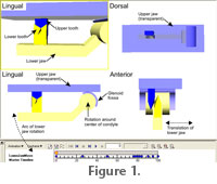
Issue
Table of Contents

Carnivoran Chewing:
EVANS & FORTELIUS

Plain-Language &
Multilingual  Abstracts

Abstract
Introduction
Methods
Results
Discussion
Acknowledgements
References

Test

Print article


METHODS

Eight carnivoran species were chosen to include a range of dental and jaw joint morphologies. They fall into four basic morphologies:

Group 1) upper – well-developed carnassial with a very small first molar; lower – only a well-developed carnassial (Acinonyx jubatus and Crocuta crocuta);

Group 2) upper – scimitar-shaped carnassial and a single molar; lower – carnassial and a single posterior molar (Mustela lutreola and Vormela peregusna);

Group 3) both upper and lower have a carnassial with two posterior molars (Canis aureus and Alopex lagopus); and

Group 4) upper – carnassial and two molars; lower – carnassial and a single posterior molar (Genetta genetta and Herpestes ichneumon).

Carnassial and post-carnassial upper and lower tooth rows of one side were scanned using a Nextec Hawk 3D laser scanner (Evans et al. 2007). These scans are viewable in the MorphoBrowser database, an online dental morphology database. Left and right condyle and post-glenoid surfaces and incisor positions were also scanned, maintaining correct relative position and orientation between all features. Tooth surfaces were exported as text point files, interpolated using GIS software (Surfer for Windows v. 8.0, Golden Software, Colorado, USA), and then exported as VRML (Virtual Reality Modelling Language) files. Scans were exported as STL files and converted to VRML using Rhino for Windows v. 3.0 (McNeel, Washington, USA). Upper and lower teeth and joint surfaces were aligned in CosmoWorlds for Windows v. 2.0 (Silicon Graphics, Inc., California, USA), and CosmoWorlds was used for the jaw rotation and translation movements in the occlusal reconstructions.

To simulate the chewing cycle, the lower jaw was first placed at the end of the closing stroke, i.e., centric occlusion, with the condyles placed within the glenoid fossae, protocones within talonid basins (where present) and incisors in a closed position. Figure 1 shows the general setup for the simulations using model upper and lower jaws and teeth at Time 0 (centric occlusion), with four views of the occluding jaws. The right tooth row was used in the simulation, and so the right side was the working side. The presence of the soft tissue of the joint capsule was accommodated by leaving a small space between the bone joint surfaces. The sagittal cross-section of the condyle was estimated as a circle, and the axis of rotation of the lower jaw passed through the centres of the left and right circles (Figure 1). As the first step in reconstructing the movement of the jaw, the lower jaw was moved in the opposite direction to the normal chewing stroke to allow easier manual alignment of the teeth and jaws. The jaw was rotated downwards to the position where the teeth would first come close to contacting each other. The condyles are held within the glenoid fossae. This leaves a lateral space between the teeth, and so the jaw was translated laterally until the teeth make contact (Figure 1, bottom right). Tooth-tooth contact was estimated to occur when small amounts of one surface passed through the contacting surface – automatic collision detection was not used. The jaw was rotated one or two degrees towards the closed position, and the jaw moved laterally again to maintain very slight contact between the teeth. This was repeated until centric occlusion was achieved. Small necessary adjustments were made to maintain contact between the occluding teeth as the rotation and translation progressed. Additional jaw positions were added to approximate the remaining portions of the chewing cycle, including jaw opening and the initial phase of jaw closing, where there is no tooth-tooth contact. Figure 2 shows the complete chewing cycle for the model jaws and teeth.

Two main measurements of the jaw movement were taken: the angle between initial tooth contact and centric occlusion, and the lateral movement of the jaw between initial tooth contact and centric occlusion. The second of these was standardised by the distance between the outer edges of the two glenoid fossae, termed the outer glenoid fossae distance.

The shape and curvature of the large attrition facets of Acinonyx and Crocuta were examined by comparing them to a plane in 3D space. Species in Group 1 were examined as they have the largest attrition facets and are the easiest to examine for this part of the study, but this means that the results may not be applicable to the other groups.


Next Section

Carnivoran Chewing
Plain-Language & Multilingual  Abstracts | Abstract | Introduction | Methods
Results | Discussion | Acknowledgements | References
Print article