|
|
|
Material and methods
Composite imaging:
Specifically, we used three different types of light:
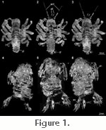
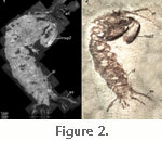
Ultraviolet: The smaller of the two specimens of Cancrinos claviger and additionally a specimen of Sculda spinosa (part and counterpart) were documented under UV light (358 nm) on an Axio Scope 2 with a mounted Axiocam. Green light: A specimen of Sculda pennata and a specimen of ?Sculda sp. were both documented under green light (546 nm). All other settings were the same as for the documentation with UV light. Combining UV-fluorescence images of part and counterpart: Part and counterpart of the specimen of Sculda spinosa (see above) were digitally combined. All black areas on the counterpart were set as transparent using the magic wand tool in Adobe Photoshop (CS3). The composite image of the counterpart was then mirrored and placed above the composite image of the part (terms 'part' and 'counterpart' are exchangeable in this special specimen as it is extremely flattened and both parts contain a lot of substance of the fossil; in general it would be useful to leave the part with more substance unaltered while processing the other one with transparency). Combining normal light images with a UV-fluorescence image of the same specimen: The composite images of the smaller of the two specimens of Cancrinos claviger, one under normal light, one under UV light, were combined into a single image. For this purpose, the image under normal light was inverted, and all parts not assignable to the fossil were set transparent. The resulting image was placed on the UV image. 3D attempts:
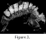
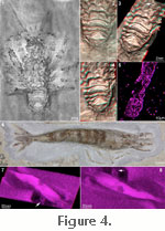
Confocal laser scanning microscopy (cLSM): Details of the specimen of ?Sculda sp. were documented under a Leica cLSM. The excitation wavelength was set to a range of 488–543 nm. In total the stack was made up of 43 images. The resulting stack of images was further processed using the freely available software program ImageJ, where the 'find edges' function was used to enhance contrast. The resulting stack was 3D-projected (Maximum Intensity Projection, MIP) using the freely available DICOM-viewer OsiriX. Stereo images and 3D models via Structure from Motion (SFM): The pleon of the larger possible specimen of Cancrinos claviger was documented as a stereo image, produced with a tiltable Zeiss Stemi 1000 stereomicroscope with a mounted DCM 500 ocular camera (tilting of about 6°). The images were then loaded into the freely available test version software "Structure from Motion" (MeeSoft) (for principles of this method see Dellaert et al. 2000). Five reference points were assigned, i.e., corresponding structures on the two single images of the stereo image were marked. Based on this, a 3D model was calculated. Computer tomography (CT) scans: A large uncompressed specimen of Antrimpos sp. was scanned in a medical CT-scanner (Philips Brilliance iCT 256) under the setting for small structures (resolution of 0.667 mm) for very heavy patients to enhance the emitted energy. The resulting stack of 402 images was processed using the freely available DICOM-viewer OsiriX (volume rendering). SEM-EDX analysis:
The specimen of ?Sculda sp. and the specimen of an undetermined caridean shrimp were put into the SEM (ZEISS DSM 962) and their elemental composition analysed using a mounted energy dispersive X-ray spectroscopy (EDX) unit. The acceleration voltage used was 25 kV. |
|