|
|
|
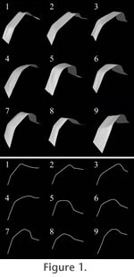
INTRODUCTIONIn the present-day study of palaeontology, new technology, such as 3D modelling, is increasingly utilized. Powerful computers and digital 3D models of fossils enable many new research projects. However, 3D scanners tend to be rather expensive and may not be accessible to all researchers. Also, unless the 3D scanner is very portable, it may be difficult to use one in field conditions or during museum visits. The purpose of this work is to test how well a method, in which 3D models are produced from ordinary digital photographs, works for palaeontological studies. The great advantage of this kind of method is that all that is needed for generating 3D models is an ordinary digital camera and a computer for post-processing. Hence, the method can be used whenever it is possible to take photographs, because no special 3D scanners are needed. In this work we studied the dietary regimes of Pleistocene rhinoceros species as a test case. We analysed dental fossils of four Pleistocene rhinoceros species from North-Western Europe, Stephanorhinus kirchbergensis (Jäger, 1839), S. hemitoechus (Falconer, 1868), S. hundsheimensis (Toula, 1902), and Coelodonta antiquitatis (Blumenbach, 1799). In the last couple of years these species have been the subject of a vivid discussion (among others Lacombat 2006a, 2006b, 2007; Billia 2007, 2008a, 2008b; von Koenigswald et al. 2007; Kahlke and Lacombat 2008; Tong and Wu 2010; Kahlke and Kaiser 2010). Our intention was to find out whether wear patterns of lower molars of rhinoceroses could be used for mesowear analysis. We compared the reconstructed palaeodiets of the Pleistocene species to the dietary regimes of the five Recent species, Diceros bicornis (Linnaeus, 1758), Ceratotherium simum (Burchell, 1817), Rhinoceros unicornis (Linnaeus, 1758), R. sondaicus (Desmarest, 1822) and Dicerorhinus sumatrensis (Fischer, 1814). Mesowear analysis is a method, which can be used to reconstruct the diets of ungulate species (Fortelius and Solounias 2000). The method is based on facet development on the occlusal surfaces of upper cheek teeth. Mesowear is recorded by examining the shape of adequately worn cusp apices macroscopically to estimate the relative amount of abrasive (food on tooth) and attritive (tooth on tooth) wear. Leaf browsing tends to result in an attritive wear pattern, where the cusp apices are sharp, whereas more abrasive grazing tends to result in blunt cusp apices. Originally Fortelius and Solounias (2000) employed only second upper molars for mesowear analysis, but later the method was also extended to the analysis of other upper cheek teeth of ruminants, horses and rhinoceroses, as well as to the analysis of the lower cheek teeth of horses and ruminants (see Franz-Odentaal and Kaiser 2003; Kaiser and Fortelius 2003; Kaiser and Solounias 2003; Kaiser and Rössner 2007; Kahlke and Kaiser 2010). However, the classical mesowear analysis does not lend itself to the study of lower cheek teeth of rhinoceros species. Restricting analyses only to the upper teeth of rhinoceroses limits the use of the method, as the fossil assemblages contain both upper and lower teeth.
The dietary regimes of the Pleistocene rhinoceros species have raised questions for a long time. Especially the diet of the species Stephanorhinus hemitoechus has been a controversial issue. Earlier S. hemitoechus has been seen as a grass consuming steppe rhino (see for example Zeuner 1934; Loose 1975), whereas in later studies a grazing diet has been considered as highly improbable (Fortelius 1982; Fortelius et al. 1993). Since all the Stephanorhinus Kretzoi, 1942 species studied here seem to occasionally have occupied the same areas (for example, fossils of all the three species have been found at Mosbach, see Fortelius et al. 1993), the possible differences in their dietary regimes are very interesting. The woolly rhino, Coelodonta antiquitatis, has for a long time been considered a cold adapted grazing animal, whose winter diet might have been different from its summer diet (Loose 1975).
Institutional abbreviations |
|
