

Article Search
Volume 27.1
January–April 2024
Full table of contents
ISSN: 1094-8074, web version;
1935-3952, print version
Recent Research Articles
See all articles in 27.1 January-April 2024
See all articles in 26.3 September-December 2023
See all articles in 26.2 May-August 2023
See all articles in 26.1 January-April 2023
 Emiliano Bruner. Grupo de Paleobiología, Centro Nacional de Investigación sobre la Evolución Humana, Paseo Sierra de Atapuerca 3, 09002 Burgos, Spain. emiliano.bruner@cenieh.es
Emiliano Bruner is Research Group Leader in Paleoneurology at the National Research Centre for Human Evolution in Burgos, Spain. He is a Ph.D. in Animal Biology for the University La Sapienza, Rome, Italy. He applies digital anatomy and computed morphometrics in functional craniology and evolutionary neuroanatomy. His main research lines deal with paleoneurology, parietal lobe evolution, brain-braincase integration and craniovascular morphology. He also works in cognitive archaeology, particularly on those functions associated with parietal cortex and visuospatial integration. He writes for many dissemination journals and blogs, on topics concerning evolutionary anthropology. Main webpage: https://paleoneurology.wordpress.com.

 Naomichi Ogihara. Faculty of Science and Technology, Department of Mechanical Engineering, Keio University, Yokohama, Kanagawa 223-8522, Japan. ogihara@mech.keio.ac.jp
Naomichi Ogihara received a B.S. degree in Mechanical Engineering, in 1995, and M.S. and Ph.D. degrees in Biomedical Engineering, in 1997 and 2000, respectively, all from Keio University, Japan. From 2000 to 2009, he was an assistant professor of Department of Zoology, Kyoto University, Japan. He joined the Faculty of Science and Technology of the Keio University in 2009, where he is currently professor in Mechanical Engineering.
FIGURE 1. During registration, sliding landmarks are allowed to move tangentially according to their neighborhood landmarks in order to minimize differences in terms of spatial distances or bending energy (1). Surface analysis can be useful to quantify 3D differences on smooth objects, like endocasts: frontal lobe differences between a modern human and an archaic human (2; after figure 3 of Beaudet and Bruner [2017]), and endocranial differences between modern human and chimpanzee (3; after figure 3 of Dupej et al. [2018]). In both cases, maps show, in red, regions of relative dilation in modern humans.
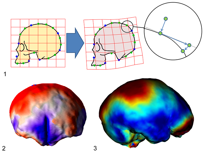
FIGURE 2. In anatomical regions which are not delimitated with homologous landmarks, surface analyses or sliding landmarks are not able to detect local differences, distributing equally the differences over the whole area. Apart from the lack of information, this can introduce a bias on the final output. The 3D form is interpreted as an undifferentiated object, and not as a specific anatomical system.
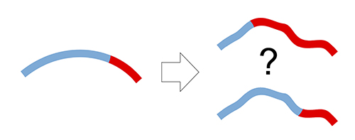
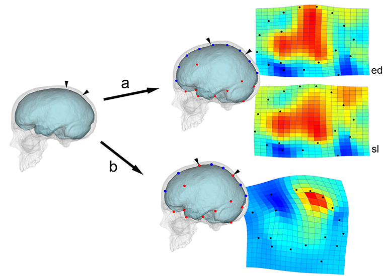
FIGURE 3. A digital model (skull and endocast) has been graphically deformed at the parietal surface, generating a flatter braincase (on the left). The differences from the original image is analyzed through thin-plate spline deformation grids, and expansion maps showing the areas undergoing dilation (red) and compression (blue). Knowing that the actual difference is due only to a marked parietal enlargement (small arrows), we used equally-spaced semi-landmarks (blue points) for the vault profile (a), without (ed) or after (sl) sliding according to a bending energy minimization criterion. The other landmarks have no specific meaning. The shape comparison gives the same importance to every landmark, “spreading” differences throughout the configuration. The parietal difference is interpreted as a more extended effect (mostly when landmarks can slide). The same comparison was computed using distinct spaced landmarks for the frontal, parietal, and occipital outlines separately, that is including the boundaries between the three districts in the model (b). In this case, the shape comparison is able to properly describe the geometrical expansion of the parietal morphology. For this visual example, we used an interpolant function, but a similar effect can be found in every registration procedure (such as Procrustes superimposition or point-to-point minimization in surface analysis) when information on anatomical boundaries and local proportions is not included. Comparisons were computed with PAST 2.17c (Hammer et al., 2001).
Surfin’ endocasts: The good and the bad on brain form
Plain Language Abstract
Computed methods have deeply improved morphometrics. Nonetheless, these techniques require a specific expertise and are based on complex numerical transformations. Appealing graphic outputs or numerical results can easily introduce biased information, if the limitations of these tools are not taken into account. Methods based on rough geometry instead of actual anatomical elements are particularly sensitive to these kinds of drawbacks. Numerical and spatial models in morphometrics refer to specific geometrical or physical properties of the organisms and not to the organisms themselves.
Resumen en Español
Surfear endocráneos: lo bueno y lo malo en morfología cerebral
La anatomía digital y la morfometría computada actualmente representan herramientas básicas en antropología, zoología y paleontología. A pesar de las interfaces fáciles de usar de los programas, estos métodos requieren una sólida experiencia en estadística, imágenes biomédicas y gráficos por computadora. El modelado geométrico tiene como objetivo normalizar la variación de forma y comparar formas dentro de un espacio de referencia compartido. Como cualquier otro enfoque de modelado, se puede usar para probar hipótesis o para investigar la estructura de una variación de muestra. En ambos casos, los modelos se refieren a variables y parámetros específicos, y siguen criterios numéricos que se basan en reglas algebraicas y convencionales. Si los modelos se interpretan de manera demasiado amplia y se confunden con los elementos anatómicos reales, las conclusiones pueden estar muy sesgadas. Este riesgo puede ser particularmente relevante cuando se trata de métodos morfométricos que no utilizan referencias anatómicas, como análisis de puntos homólogos deslizantes, análisis de superficie o morfometría basada en reconocimiento de voxel. Todas estas técnicas se emplean principalmente en craneología, paleoneurología y neuroanatomía evolutiva. Siguiendo estos enfoques, los elementos se analizan como "objetos" y no como "elementos anatómicos", introduciendo ruido e inconvenientes debido a los procesos de registro y a la ausencia de restricciones asociadas con los límites anatómicos. Las desventajas pueden evitarse interpretando modelos geométricos como representaciones específicas de un conjunto de propiedades de los sistemas anatómicos originales y no como mera efigie generalizada de elementos biológicos.
Palabras clave: paleoneurología; morfometría; análisis de forma; análisis de superficie
Traducción: Enrique Peñalver (Sociedad Española de Paleontología)
Résumé en Français
Surfer sur les endocrânes : le bon et le mauvais sur la forme du cerveau
L’anatomie numérique et la morphométrie assistée par ordinateur représentent actuellement les outils basiques en anthropologie, zoologie, et paléontologie. Malgré les interfaces ergonomiques des programmes, ces méthodes nécessitent une expertise robuste en statistiques, imagerie biomédicale, et infographie. La modélisation géométrique vise à normaliser la variation de conformation pour comparer les formes au sein d’un espace de référence partagé. Comme avec toute autre approche de modélisation, cela peut être utilisé pour tester des hypothèses ou pour étudier la structure de la variation d’un échantillon. Dans les deux cas, les modèles s’appuient sur des variables et des paramètres spécifiques, et ils suivent des critères numériques qui sont basés sur des règles conventionnelles d’algèbre. Si les modèles sont interprétés de manière trop large et confondus avec les vrais éléments anatomiques, les conclusions peuvent être sérieusement biaisées. Ce risque peut être particulièrement pertinent pour les méthodes morphométriques qui n’utilisent pas de points de référence anatomiques, comme les points de repère glissants (« sliding landmarks »), les analyses de surface, ou la morphométrie basée sur les voxels. Toutes ces techniques sont fréquemment utilisées en craniologie, paléoneurologie, et neuroanatomie évolutive. Dans ces approches, les éléments sont analysés comme des « objets » et non comme des « éléments anatomiques », ce qui introduit du bruit et des limitations liées aux processus de saisie des données et à l’absence de contraintes associées avec les limites anatomiques. Ces limitations peuvent être évitées en interprétant les modèles géométriques comme des représentations spécifiques d’un jeu de propriétés des systèmes anatomiques d’origine et non comme une effigie généralisée des éléments biologiques.
Mots-clés : paléoneurologie ; morphométrique ; analyse des conformations ; analyse de surface
Translator: Antoine Souron
Deutsche Zusammenfassung
Surfin’ endocasts: Das gute und schlechte über Hirnformen
Digitale Anatomie und Computer-Morphometrie sind derzeit die Basiswerkzeuge in der Anthropologie, Zoologie und Paläontologie. Trotz der bedienerfreundlichen Oberflächen und Programme erfordern sie eine gute Expertise in Statistik, biomedizinischer Bildgebung und anderen Computergrafiken. Geometrisches Modellieren ist bestrebt Formavarianten zu normalisieren im Vergleich mit Formen innerhalb einer gemeinsamen Referenzfläche. Wie jeder andere Modellierungsansatz kann es zum Testen von Hypothesen oder zur Strukturuntersuchung einer Probenvariante genutzt werden. In beiden Fällen beziehen sich die Modelle auf spezifische Variablen und Parameter und sie folgen numerischen Kriterien die auf mathematischen und konventionellen Regeln basieren. Wenn Modelle zu weit ausgelegt werden und mit den echten anatomischen Elementen durcheinandergebracht werden, können die Schlussfolgerungen erheblich verzerrt werden. Das Risiko kann besonders relevant sein im Umgang mit morphometrischen Methoden wie verschiebbare Orientierungspunkte, Oberflächenanalyse oder voxel-basierter Morphometrie. All diese Techniken werden häufig in der Kraniologie, Paläoneurologie und der evolutionären Neuroanatomie eingesetzt. Gemäß diesen Ansätzen werden Elemente als „Objekte“ analysiert und nicht als „anatomische Elemente“ was ein Rauschen mit sich bringt und nachteilig ist wegen der Registrierungsverfahren und dem Fehlen von Constraints, die mit den anatomischen Grenzen zusammenhängen. Nachteile können vermieden werden, wenn geometrische Modelle als konkrete Darstellungen eines Satzes von Eigenschaften des originalen anatomischen Systems interpretiert werden und nicht als generalisierte Abbildungen eines biologischen Systems.
Schlüsselwörter: Paläoneurologie; Morphometrik; Formanalyse; Oberflächenanalyse
Translator: Eva Gebauer
Arabic
Translator: Ashraf M.T. Elewa
-
-
-
Review: The Princeton Field Guide to Mesozoic Sea Reptiles
 The Princeton Field Guide to Mesozoic Sea Reptiles
Article number: 26.1.1R
April 2023

 Poster Winners 2024