 FIGURE
4.61-80.
FIGURE
4.61-80.Cibicides inagawaensis Matsunaga (1963, p. 116, pl. 51, figure 5a-5c).
4.61. Dorsal view, 22.5x (LD = .98 millimeters).
4.62. Ventral view, 22.5x (LD = .98 millimeters).
4.63. Edge view, 22.5x (LD = .98 millimeters).
Photographs of holotype (#85377) in Tohoku University collections. This species is not a Cibicides species; it may fit into the Cibicidoides group and appears to be a distinct species.
Cibicides kamadai Asano (1951a, p. 17, figures 33-35).
4.64. Dorsal view, 37.5x (LD = .56 millimeters).
4.65. Ventral view, 37.5x (LD = .56 millimeters).
Photographs of holotype (no number) in Tohoku University collections. It is difficult to say what this specimen is because it appears deformed, but similar to C. lobatulus.
Cibicides malloryi Matsunaga (1963, P. 116, pl. 51, figures 7 and 8).
4.66. Ventral view, 22.5x (LD = .57 millimeters).
4.67. Dorsal view, 22.5x (LD = .57 millimeters).
Photograph of holotype (no number) in Tohoku University collections. This is not similar to C. lobatulus, but may be close to C. mundulus (Brady et al. 1890). However, this specimen has a sugary outside texture, which is probably a postmortem feature.
Cibicides (?) omurai Asano and Inomata (in Asano 1952, p. 17, figures 97-99).
4.68. Dorsal view, 13.5x (LD = .74 millimeters).
4.69. Ventral view, 13.5x (LD = .74 millimeters).
Photographs of holotype (#75254) in Tohoku University collections. It is easy to see why Asano and Inomata had a questionable generic designation, but the species is distinct. Takayanagi feels this species may have some relationship to the genus Parrelloides.
Cibicides yoitaensis Matsunaga (1963, p. 117, pl. 52, figure 3a-3c).
4.70. Ventral view, 22.5x (LD = .71 millimeters).
4.71. Dorsal view, 22.5x (LD = .71 millimeters).
Photographs of the holotype (#85381) in the Tohoku University collections. This specimen is similar to C. malloryi (4.66-4.67).
Cribroelphidium aomoriense Asano (1950c, p. 11, figures 60 and 61).
4.72. Side view, 13.5x (LD = 1.26 millimeters).
4.73. Edge view showing aperture, 13.5x (LD = 1.18 millimeters).
Photographs of holotype? (not labeled as holotype, but numbered #66185) in Tohoku University collections. This very badly etched specimen could be any one of a number of elphidiid species and therefore could not be designated as a new species.
Cribroelphidium cribrojenseni Matsunaga (1963, p. 108, pl. 35, figure 11a-11b).
4.74. Side view, 13.5x (LD = 1.11 millimeters).
4.75. Edge view showing aperture, 13.5x (LD = 1.11 millimeters).
Photographs of holotype (#85182) in Tohoku University collections. This species is very similar to the original Polystella jenseni Cushman (1924) that itself was put into synonymy with Elphidium fichtellianum (dOrbigny 1846) by Hayward et al. (1997).
Cribroelphidium imanishii Asano (1953b, p. 52, figure 11a-11b).
4.76. Side view, 13.5x (LD = 1.18 millimeters).
4.77. Edge view showing aperture, 13.5x (LD = 1.18 millimeters).
Photographs of holotype (#75287) in Tohoku University collections. This species, similar to C. aomoriense (4.72 and 4.73), is badly etched, but is probably not a distinct species.
Cribroelphidium kannonjiense Matsunaga (1963, p. 108, pl. 35 figure 12a-12b).
4.78. Side view, 13.5x (LD = 1.33 millimeters).
4.79. Edge view showing aperture, 13.5x (LD = 1.26 millimeters).
Photographs of holotype (#85184) in the Tohoku University collections. This type is broken, but it is believed that this species is probably one of the Elphidium excavatum (Terquem 1876) group, not a distinct species.
Cribroelphidium nishiyamaense Matsunaga (1963, pl. 35, figure 13a-13b).
4.80. Side view, 13.5x (LD = 1.41 millimeters).
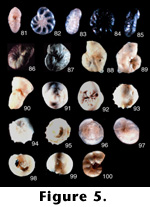
Photographs of the holotype (#85185) in the Tohoku University collections. Although this specimen is badly broken and etched, it is probably part of the E. excavatum group (forma selseyensis Heron-Allen and Earland 1911). (See also 5.81.)
Cribroelphidium nishiyamaense Matsunaga (continued from 4.80).
5.81. Edge view showing aperture, 13.5x (LD = 1.33 millimeters).
Cribrononion miyakoens Ujiie and Kusukawa (1969, p. 766, pl. 1, figures 3-7).
5.82. Side view, 45x (LD = .38 millimeters).
5.83. Edge view showing aperture, 45x (LD = .38 millimeters).
Photographs of holotype (#244, 245) in the Tokyo National Museum. This species is identical to Elphidium poeyanum (d'Orbigny 1839) and therefore a junior synonym of that species.
Cribrononion multicameratum Ujiie (1977, p. 96, pl. 19, figures 1-3, pl. 20, figures 4 and 5).
5.84. Side view, 45x (LD = .29 millimeters).
5.85. Edge view showing aperture 45x (LD = .31 millimeters).
Photographs of holotype (#973-975) in the Tokyo National Museum. These photographs turned out poorly even after several retakes because the specimens were stained. The color figure in this case is quite good, but it was impossible to get a good electronic scan for the plate here. The specimen appears to be part of the E. excavatum group, although it is difficult to say which forma it is.
Cyclammina ezoensis Asano (1951b, p. 5, figures 16 and 17).
5.86. Slanted side view, 7x (LD = 2.86 millimeters).
Photograph of hypotype (holotype was broken during photography, #67075) in the Tohoku University collections. This species is identical to C. cancellata (Brady 1879) and hence is a junior synonym of that species.
Cyclammina japonica Asano (1950d, p.78, pl. 11, figures 3-8).
5.87. Side view, 3x (LD = 6.67 millimeters).
Photograph of holotype (#66193) in the Tohoku University collections. This species is also a junior synonym to C. cancellata. No edge view is shown, but it is identical to C. cancellata.
Discanomalina japonica Asano (1951a, p. 13, figures 3-5).
5.88. Side view, 13.5x (LD = 1.18 millimeters).
5.89. Opposite side view, 13.5x (LD = 1.18 millimeters).
5.90. Edge view, 13.5x (LD = 1.04 millimeters).
Photographs of holotype (#67129) in Tohoku University collections. Although this species has been shown already to be the junior synonym of D. semipunctata (Bailey 1851) by Medioli and Scott (1978), it is still the type species for the genus. It is an important forma in the lineage of semipunctata because it represents the extreme end member of the attached forms.
Discopulvinulina hofkeri Asano (1951c, p. 5, figures 30 and 31).
5.91. Dorsal view, 13.5x (LD = 1.31 millimeters).
5.92. Ventral view, 13.5x (LD = 1.33 millimeters).
Photographs of holotype (#67172) in the Tohoku University collections. This looks like a distinct species.
Discorbis nakamurai Asano (1951c, p. 2, figures 8-10).
5.93. Dorsal view of first specimen, 30x (LD = .63 millimeters).
5.94. Ventral view of first specimen, 30x (LD = .50 millimeters).
5.95. Ventral view of second specimen. 30x (LD = .63 millimeters).
All photographs from specimens on the holotype slide (#67164) in the Tohoku University collections. This was a very interesting group because it had three specimens in the holotype slide: two specimens joined in plastogamy (which was broken apart; Figures 93 and 94); the other is a single specimen where only the ventral side was photographed (5.95).
Discorbis ozawai Asano (1951c, p. 3, figures 14-16).
5.96. Dorsal view, 25x (LD = .80 millimeters).
5.97. Ventral view, 25x (LD = .80 millimeters).
Photographs of holotype (#67163) in the Tohoku University collections. This appears to be a distinct species.
Discorbis subopercularis Asano (1951c p. 3, figures 17-19).
5.98. Ventral view, 13.5x (LD = 1.63 millimeters).
5.99. Dorsal view, 13.5x (LD = 1.70 millimeters).
Photographs of holotype (#67167) in the Tohoku University collections. This looks like a distinct species. This particular specimen is damaged on the ventral side; however, the radial structure on the ventral chambers can still be seen, which may give the species affinities to genus Glabratella.
Echigoina hataii Matsunaga (1963, p. 115, pl. 50, figure 4a-4b).
5.100. Side view, 45x (LD = .56 millimeters).
Photographs of holotype (#85368) in the Tohoku University collections. This species may be distinct, but the genus is probably Astrononion and not the genus described by Matsunaga. Holotype of the genus does fit with the type description, except for the trochospiral chamber arrangement. Another species in the same paper placed in this genus (for which Scott did not find the specimen, E. furutsuensis) does show some trace of a trochospiral arrangement. This species was so rare in the type material that we are treating them as aberrant specimens of the genus Astrononion. However, the description of Astrononion does not mention any trace of trochospiral arrangement and emendation of the genus would require finding that characteristic in the type species of Astrononion. (See also 6.101)
Echigoina hataii Matsunaga (continued from 5.100).
6.101. Tilted edge view, 45x (LD = .49 millimeters).
Eggerella matsunoi Takayanagi n. sp.
6.102. Side view showing aperture, 45x (LD = .67 millimeters).
6.103. Side view, 45x (LD = .67 millimeters).
6.104. Side view showing aperture, 45x (LD = .67 millimeters).Holotype: Specimen number 75023 in Tohoku University collections.
Type locality: Lat. 44o, 41' 54.6"N, long. 141o, 59' 03.2". Location no. EN-84, cliff of the upper stream of the Rubeshube River, a branch of the Ebetsu River, about 6,600m ENE of the Chuo Post office, Embetsu-machi, Teshio-gun, Teshio Province, Hokkaido.
Type formation: East Chikubetsu Formation.
Type level: Miocene.
Description: Test free, small elongate, moderately tapering in early portion, nearly cylindrical in later triserial portion, frequently compressed; chambers numerous, low and broad, increasing rather rapidly in size in early portion with four to five chambers to a whorl, later becoming inflated and increasing gradually in size as added; sutures distinct and depressed; wall finely agglutinated, with much cement, surface smoothly finished; aperture an indistinct low arch at the base of the last -formed chamber; white in color.
Remarks: This species is similar to Eggerella karamatensis (Brönnimann 1953) from the Oligo-Miocene of Trinidad, but is easily distinguished from that species by a more elongate test.
Specific name is given in honor of Dr. Kyuya Matsuno, formerly of the Geological Survey of Japan.
Elphidiella nagaoi Asano (1938a, p. 590, pl. 14 (3), figure 8a-8b).
6.105. Side view, 10x (LD = 2.00 millimeters).
6.106. Slanted edge view, 10x (LD = 2.00 millimeters).
Photograph of holotype (#21421) in the Tohoku University collections. This species is identical in every respect to E. arctica (Parker and Jones in Brady 1864), including characters such as a large test, thick shell, and the typical double rows of sutural pores with associated striations. It is therefore a junior synonym of the Parker and Jones species.
Elphidium asanoi Matsunaga (1963, p. 109, pl. 36, figure 6a-6b).
6.107. Side view, 45x (LD = .47 millimeters).
6.108. Edge apertural view, 45x (LD = .47 millimeters).
Photographs of holotype (#85190) in the Tohoku University collections. This specimen is in poor condition, but could be a distinct species. It does not appear to be a member of the E. excavatum group.
Elphidium advena Cushman gorokuense Takayanagi (1950, p. 27, figure 4).
6.109. Side view, 13.5x (LD = .89 millimeters).
6.110. Edge view showing aperture, 13.5x (LD = .89 millimeters).
Photographs of holotype (#66105) in the Tohoku University collections. According to Scott, this species is an ecophenotype of E. advena (Cushman 1921), not a subspecies.
Elphidium etigoense Husezima and Maruhasi (1944, p. 392, pl. 34, figure 1a-1b.
6.111. Side view, 37.5x (LD = .59 millimeters).
6.112. Edge view showing aperture, 37.5x (LD = .59 millimeters).
Photographs of hypotype (no number) in the Tohoku University collections. This particular specimen is badly etched, but we agree with a later paper by Ishiwada (1964) that is referable to E. bartletti Cushman (1933) and is not a distinct species.
Elphidium ezoense Asano (1937a, p. 787. pl. 24 (12), figures 1 and 2, text figure 1a-1b).
6.113. Side view, 7.5x (LD = 3.06 millimeters).
6.114. Slanted side view, 7.5x (LD = 2.93 millimeters).
Photographs from holotype slide (#21433) in Tohoku University collections with about 50 specimens. None of the specimens was designated as the holotype, so a representative specimen was selected. This could be a distinct species, but Scott thought it very similar to E. galvestonense (Kornfeld 1931), for which this would be a junior synonym. However, unlike galvestonense, this is a cold water species. Takayanagi believes this species may be closer to E. oregonense (Cushman and Grant 1927), which is a colder water species. This hypothesis is probably correct because E. ezoense has cribrate apertures similar to those of E. oregonense. Asano (1950a) assigned this species to the genus Cribroelphidium.
Elphidium hanzawai Asano (1939, p. 426, text figures 3, 4, and 6).
6.115. Side view, 13.5x (LD = 1.26 millimeters).
6.116. Edge view, 13.5x (LD = 1.26 millimeters).
Photographs of holotype (#62910) in the Tohoku University collections. This is another junior synonym of Cushmans 1921 species, E. advena.
Elphidium hokkaidoense Asano (1950a, p. 8, figures 44 and 45).
6.117. Side view, 37.5x (LD = .53 millimeters).
6.118. Edge view, 37.5x (LD = .53 millimeters).
Photographs of paratype (#66178) in the Tohoku University collections. This poorly preserved specimen could be a variant of Elphidium advena.
Elphidium kusiroense Asano (1938a, p. 590, pl. 14 (3), figure 2).
6.119. Side view, 30x (LD = .73 millimeters).
6.120. Edge view, 30x (LD = .67 millimeters).
Photographs of holotype (#21420) in the Tohoku University collections. This species is close to E. margaritaceum Cushman (1930), but this specimen has a large umbilical boss that is not present in margaritaceum while having the same sugary texture common to E. margaritaceum.
NOTE: LD (longest dimension).
![]()
 FIGURE
5.81-100.
FIGURE
5.81-100.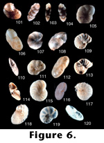 FIGURE
6.101-120.
FIGURE
6.101-120.