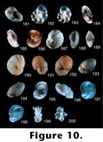
 FIGURE
10.181-200.
FIGURE
10.181-200.Haplophragmoides taiwanensis Nakamura (continued from 9.180).
10.181. Edge view showing aperture, 13.5x (LD = 1.85 millimeters).
Hyalinea inflata Ujiie and Kusukawa (1969, p. 767, pl. 2, figures 1-3).
10.182. Side view, 45x (LD = .38 millimeters).
10.183. Ventral view, 45x (LD = .38 millimeters).
Photographs of holotype (specimen no longer exists) from the National Science Museum (Tokyo, Shinjuku branch). The holotype was crushed during photography and the photographs of this species did not turn out well. They are included here because they are the only light photographic record of this species. This appears to be a distinct species; in the absence of a holotypic specimen, a lectotype should be designated to replace the holotype from the collections of the National Science Museum.
Miliammina echigoensis Asano and Inomata (in Asano 1952, p. 5, figures 21-24).
10.184. Side view of 4 chambered side, 45x (LD = .51 millimeters).
10.185. Side view showing aperture, 45x (LD = .44 millimeters).
Photographs of Holotype (#75251) in the Tohoku University collections. This looks distinct from M. fusca (Brady 1870) with no apparent inner lining.
Nonion aimonoi Matsunaga (1963, p. 109, pl. 37, figures 2a and 2b).
10.186. Side view, 45x (LD = .49 millimeters).
Photograph of holotype (#85387) in the Tohoku University collections. There is no question that this is a junior synonym of Nonion affinis (Reuss 1851) and not a distinct species.
Nonion nagasawaense Matsunaga (1963, p. 109, pl. 37, figures 7a and 7b).
10.187. Side view 13.5x (LD = 1.18 millimeters).
10.188. Edge view, 13.5x (LD = 1.18 millimeters).
Photographs of holotype (#85201) in the Tohoku University collections. This looks like a distinct species.
Nonion nakosoense Asano (1949, p. 428, figure 2, nos. 14-17).
10.189. Edge view showing aperture, 13.5x (LD = .67 millimeters).
10.190. Side view, 13.5x (LD = .96 millimeters).
Photographs of holotype (#67044) in the Tohoku University collections. This looks like a distinct species, although it is similar to Nonionellina labraodorica (Dawson 1860) in the North Atlantic, except that the sutures of this species are slightly filled with a line of clear calcite.
Nonionella hanzawai Asano (1953b, p. 52, figure 4a-4c).
10.191. Slanted dorsal view, 13.5x (LD = .74 millimeters).
10.192. Ventral view, 13.5x (LD = .67 millimeters).
Photographs of holotype (#75286) in the Tohoku University collections. This very odd species is definitely a distinct species.
Nonionella higashiyamaensis Matsunaga (1963, p. 110, pl. 38, figure 3a-3c).
10.193. Side view, 45x (LD = .47 millimeters).
10.194. Edge view showing aperture, 45x (LD = .47 millimeters).
Photographs of holotype (#85206) in the Tohoku University collections. This specimen is highly recrystallized, but very similar to Nonionella turgida (Williamson 1858) and hence a possible junior synonym of that species.
Paracassidulina nabetaensis Nomura (1983a, p. 98, pl. 2, figure 16a and b; pl. 5, figure 5; pl. 25, figure 7).
10.195. Side view showing aperture, 45x (LD = .58 millimeters).
10.196. Edge view showing aperture, 45x (LD = .58 millimeters).
Photographs of paratype (#97406) in the Tohoku University collections. This species is similar in some ways to Cassidulina reniforme (Nörvang 1945), but its shape is much more elongate and chambers are elongated, so it appears to be a distinct species.
Paracassidulina quasicarinata Nomura (1983a, p. 100, pl. 2, figure 19a-c; pl. 25, figures 9-11).
10.197. Side view showing aperture, 45x (LD = .35 millimeters).
10.198. Slanted edge view showing aperture, 45x (LD = .35 millimeters).
Photographs of paratype (#97254) in the Tohoku University collections. Scott suggests this is similar to Islandiella teretis (Tappan 1951), and hence is a junior synonym; Takayanagi indicates it is close to I. Teretis, but differs in umbilical and apertural features.
Pararotalia? takayanagii Matoba (1970, p. 63, pl. 6, figures 9 and 10).
10.199. Edge view, 45x (LD = .20 millimeters).
10.200. Ventral view, 45x (LD = .20 millimeters).
Photographs of paratype (#91312C) in the Tohoku University collections. This very unusual species is certainly distinct. This species was subsequently placed in the genus Murrayinella by Takayanagi and Hasegawa (1987). (See also 11.201.)
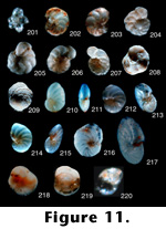
Pararotalia? takayanagii Matoba (continued from 10.200).
11.201. Dorsal view, 45x (LD = .20 millimeters).
Planulina convexa Takayanagi (1953, p. 34, pl. 4, figure 14a-14c).
11.202. Ventral view, 45x (LD = .51 millimeters).
11.203. Dorsal view, 45x (LD = .53 millimeters).
Photographs of holotype (#67145) in the Tohoku University collections. This species appears to be distinct.
Planulina nipponica Asano (1953a, p. 13, pl. 3, figure 13a-13c).
11.204. Ventral view, 13.5x (LD = 1.18 millimeters).
11.205. Dorsal view, 13.5x (LD = 1.26 millimeters).
Photographs of holotype (#75269) in the Tohoku University collections. This looks like a distinct species because of the presence of a strong keel around its outer margin.
Planulina subdepressa Asano (1951a, p. 15, figures 16-18).
11.206. Dorsal view, 13.5x (LD = 1.63 millimeters).
11.207. Ventral view, 13.5x (LD = 1.63 millimeters).
Photographs of holotype (no number on slide) in the Tohoku University collections. Scott considers this species to be a junior synonym of Planulina wuellerstorfi (Schwager 1866); Takayanagi finds that it is different because it has moderately curved and depressed sutures on the umbilical side.
Poroeponides cribroconcameratus Asano and Uchio (in Asano 1951c, p. 18, figures 132 and 133).
11.208. Ventral view, 7.5x (LD = 2.80 millimeters).
11.209. Dorsal view, 7.5x (LD = 2.80 millimeters).
Photographs of holotype (#74703) in Tohoku University collections. This appears to be a distinct species.
Pseudononion japonicum Asano (1936b, p. 347, text figures a-c).
11.210. Side view, 13.5x (LD = .81 millimeters).
11.211. Edge view showing aperture, 13.5x (LD = .81 millimeters).
Photographs of holotype (#21362) in the Tohoku University collections. This specimen appears to be the same as Nonionella atlantica Cushman (1947). Pseudononion japonicum is therefore the valid name for this well known Atlantic species.
Pseudononion kanbaraense Matsunaga (1963, p. 110, pl. 38, figure 8a-8c).
11.212. Side view, 45x (LD = .44 millimeters).
11.213. Edge view, 45x (LD = .42 millimeters).
Photographs of holotype (#85211) in the Tohoku University collections. This species is very close to P. japonicum.
Pseudononion oinomikadoi Matsunaga (1963, p. 110, pl. 39, figure 1a-1c).
11.214. Side view, 45x (LD = .49 millimeters).
11.215. Edge view showing aperture, 45x (LD = .49 millimeters).
Photographs of holotype (#85212) in the Tohoku University collections. This species is very similar to the previous two Pseudononion species (11.21011.213).
Pseudononion trececum Asano (1936c, p. 622, pl. 33, figure 7a-7c).
11.216. Side view, 13.5x (LD = 1.11 millimeters).
11.217. Slanted edge view, 13.5x (LD = 1.11 millimeters).
Photographs of lectotype (#21375) in the Tohoku University collections. This may be a distinct species or it could be a variation of the other three Pseudononion species.
Pseudoparella japonica Asano (1949, p. 430, figure 2, nos. 2-4).
11.218. Ventral view, 13.5x (LD = .89 millimeters).
11.219. Dorsal view, 13.5x (LD = .89 millimeters).
Photographs of holotype (#67045) in the Tohoku University collections. This appears to be a distinct species. The genus for this species was changed to Alabamina (Takayanagi and Hasegawa 1987).
Pseudoparella tamana Kuwano (1950, p. 317, figure 5a-5c).
11.220. Ventral view, 45x (LD = .13 millimeters).
Photographs of ideotype (no number) in the National Science Museum (Tokyo, Shinjuku branch). The photographs here are not high quality because the specimen was disintegrating as it was being photographed. In spite of the poor quality of the photographs, the form of the species appears to have a thinner test wall than E. takayanagii Iwasa and slightly less distinct sutures. This is also similar to Stetsonia arctica (Green 1960), which has a thin test wall and less distinct sutures. Unfortunately, this is a problem that will not be solved easily in the absence of the author of the species. This may be another species that should have its type figures designated as lectotypes. (See also 2.221.)
Pseudoparella tamana Kuwano (continued from 11.220).
12.221. Dorsal view, 45x (LD = .11 millimeters).
Pseudorotalia borneensis Ujiie (1977, p. 95, pl. 18, figures 1-3, pl. 21, figures 7 and 8).
12.222. Dorsal view, 13.5x (LD = .89 millimeters).
12.223. Ventral view, 13.5x (LD = .89 millimeters).
Photographs of holotype (#1008) in the National Science Museum (Tokyo, Shinjuku branch). This is a very distinctive species with incised ventral sutures and a well developed keel on the periphery.
Rotalia? minuta Takayanagi (1955, p. 52, figure 29a-29c).
12.224. Ventral view, 45x (LD = .24 millimeters).
12.225. Dorsal view, 45x (LD = .26 millimeters).
Photographs of the holotype (#67152) in the Tohoku University collections. This species is remarkably similar to Pararotalia takayanagii Matoba, especially in the texture of the test. If these are the same, then Matoba's species is a junior synonym. Hasegawa notes that minuta is different in the periphery of the test and other features, but Scott still believes this one is a variation of Takayanagis species. The genus for this species was changed to Murrayinella (Takayanagi and Hasegawa 1987).
Rotalia sadoensis Asano (1951c, p. 16, figures 120 and 121).
12.226. Ventral view, 10x (LD = 1.40 millimeters).
12.227. Dorsal view, 10x (LD = 1.40 millimeters).
Photographs of holotype (#67198) in the Tohoku University collections. This is an interesting species because of the slits on the dorsal sutures and truncated ventral sutures. However, it could also be an extreme variation of Ammonia beccarii (Linne 1758).
Siphotextularia masudai Asano (1953a, p. 17, pl. 1, figure 8a and 8b).
12.228. Side view, 13.5x (LD = .96 millimeters).
12.229. Edge view showing aperture, 13.5x (LD = .96 millimeters).
Photographs of holotype (#75267) in the Tohoku University collections. This species is identical to S. rolhauseri (Phleger and Parker 1951) and is a junior synonym of that species.
Spiroplectammina higuchii Takayanagi (1953, p. 27, pl. 4, figures 1a and 1b).
12.230. Side view, 45x (LD = .42 millimeters).
12.231. Apertural view, 45x (LD = .42 millimeters).
Photographs of holotype (#67134) in the Tohoku University collections. This species is certainly different from any other Neogene species of this genus.
Spiroplectammina niigataensis Asano and Inomata (in Asano 1952, p. 4, figures 15-17).
12.232. Side view, 13.5x (LD = 1.56 millimeters).
12.233. Edge view showing aperture, 13.5x (LD = 1.56 millimeters).
Photographs of holotype (#75252) in the Tohoku University collections. This is also a different species from any other Neogene form.
Spiroplectammina shibataensis Matsunaga (1963, p. 106, pl. 25, figure 2).
12.234. Side view, 22.5x (LD = .93 millimeters).
Photograph of holotype (#85040) in the Tohoku University collections. This is a very interesting species, quite distinct from the other Japanese species of this genus as well as those from anywhere else, with a large coil and tapering biserial chambers.
Textularia andenensis Asano (1950c, p. 2, figures 5 and 6).
12.235. Side view, 13.5x (LD = 2.07 millimeters).
12.236. Edge view showing aperture, 13.5x (LD = 2.07 millimeters).
Photographs of a specimen that is not the holotype, but was the only specimen of this species (#66190) in the Tohoku University collections. Scott places this species with T. conica (d'Orbigny 1839), making this species a junior synonym.
Textularia aokii Asano (1936d, p. 325, pl. 36, figure 1a and 1b).
12.237. Side view, 13.5x (LD = .96 millimeters).
12.238. Edge view showing aperture, 13.5x (LD = .96 millimeters).
Photographs of holotype (#21330) in the Tohoku University collections. This species resembles a number of different species from the Mediterranean, but may be a distinct species.
Textularia hoppoensis Nakamura (1937, p. 134, pl. 10, figure 4a and 4b).
12.239. Side view, 13.5x (LD = 2.29 millimeters).
Photograph of holotype (#60855) in the Tohoku University collections. The very coarse grains making up the specimen obscure the sutures, but it is probably a distinct species.
Textularia intosiana Nakamura (1937, p. 134, pl. 10, figures 5 and 6).
12.240. Side view, 7.5x (LD = 3.06 millimeters).
Photographs of paratype (#60857) because the holotype in the Tohoku University collections was broken. This is a very distinctive species; the sutures and aperture are very easy to see, unlike most species of this genus. (See also 13.241.)
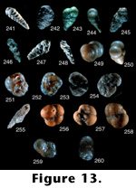
NOTE: LD (longest dimension).
![]()
 FIGURE
11.201-220.
FIGURE
11.201-220.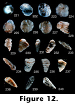 FIGURE
12.221-240.
FIGURE
12.221-240.