METHODS
 The
PTM images can be acquired with a single light source that is manually
positioned for each exposure, but it is more practical to use a set of
computer-controlled light sources in fixed positions.
The
PTM images can be acquired with a single light source that is manually
positioned for each exposure, but it is more practical to use a set of
computer-controlled light sources in fixed positions. 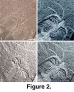 At
the time of writing there are four such machines available, two of which have
been used for this article. The original "flash dome" was described by
Malzbender
et al. (2001). A dome built at the Geological Museum in Oslo was based on
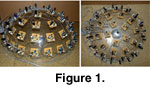
the same design, with minor modifications. For the Oslo version (Fig.
1), 50 computer-controlled flashes were mounted on an acrylic dome
(hemisphere) with a diameter of 40 cm.
At
the time of writing there are four such machines available, two of which have
been used for this article. The original "flash dome" was described by
Malzbender
et al. (2001). A dome built at the Geological Museum in Oslo was based on
the same design, with minor modifications. For the Oslo version (Fig.
1), 50 computer-controlled flashes were mounted on an acrylic dome
(hemisphere) with a diameter of 40 cm.  A
Nikon Coolpix 995 digital camera was mounted at the top of the dome, pointing
down onto the specimen, which is placed on a surface in the center of the dome.
The flashes are synchronized with the camera exposures by using the flash
synchronization output ("hot shoe") of the Coolpix camera. Custom-made
software controls the flashes, the camera, and the transfer of images to the
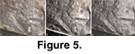
computer in an automated sequence. For the examples shown here (Fig.
2, Fig. 3, Fig.
4, and Fig. 5), a relatively low
resolution of 1600 x 1200 pixels was used (this is cropped to smaller size in
the illustrations), but the system can handle higher resolutions at the cost of
slower image transfer, image processing, and interactive manipulation. The
optical zoom range of the Coolpix 995 camera and the distance from specimen to
the top of the dome allow visualization of specimens ranging in size from 50 to
300 mm across.
A
Nikon Coolpix 995 digital camera was mounted at the top of the dome, pointing
down onto the specimen, which is placed on a surface in the center of the dome.
The flashes are synchronized with the camera exposures by using the flash
synchronization output ("hot shoe") of the Coolpix camera. Custom-made
software controls the flashes, the camera, and the transfer of images to the
computer in an automated sequence. For the examples shown here (Fig.
2, Fig. 3, Fig.
4, and Fig. 5), a relatively low
resolution of 1600 x 1200 pixels was used (this is cropped to smaller size in
the illustrations), but the system can handle higher resolutions at the cost of
slower image transfer, image processing, and interactive manipulation. The
optical zoom range of the Coolpix 995 camera and the distance from specimen to
the top of the dome allow visualization of specimens ranging in size from 50 to
300 mm across. 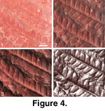 Hence,
this camera is not optimized for very small specimens. The light sensitivity of
the camera chip was set to an equivalent of ISO 200, and the compression to
"fine" (JPEG compression to 25% of original file size; as JPEG is a
somewhat destructive compression, TIFF or equivalent is recommended if storage
space allows).
Hence,
this camera is not optimized for very small specimens. The light sensitivity of
the camera chip was set to an equivalent of ISO 200, and the compression to
"fine" (JPEG compression to 25% of original file size; as JPEG is a
somewhat destructive compression, TIFF or equivalent is recommended if storage
space allows).
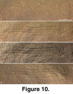
The original "flash dome" (Malzbender
et al. 2001), used for Fig. 6, Fig.
7, Fig. 8, Fig.
9, and Fig. 10, has a diameter of 64
cm and employs a Kodak DCS460 camera with a pixel resolution of 3060 x 2036
pixels and a Nikon ED AF Micro Nikkor 70-180 mm zoom lens. In order to minimize
reflections from external objects, the dome is constructed of acrylic and the
interior painted opaque matte black.
 The
50 images were processed using software developed at HP Labs and available in
the public domain:
The
50 images were processed using software developed at HP Labs and available in
the public domain:
HP Laboratories (latest
version)
PE copy (version at time of writing).
 In
a preliminary step, the program ptmfitter is used to combine the images
into a single, compact PTM file containing the coefficients of the paraboloids
approximating the brightness of each pixel as a function of the direction of
incoming light. These data are then further processed by a viewing program (ptmviewer),
allowing interactive control of image enhancement algorithms and the positions
of virtual light sources.
In
a preliminary step, the program ptmfitter is used to combine the images
into a single, compact PTM file containing the coefficients of the paraboloids
approximating the brightness of each pixel as a function of the direction of
incoming light. These data are then further processed by a viewing program (ptmviewer),
allowing interactive control of image enhancement algorithms and the positions
of virtual light sources.
The results (Fig.
2, Fig. 3, Fig.
4, Fig. 5, Fig.
6, Fig. 7, Fig.
8, Fig. 9, and Fig.
10 PTM images) are presented here as PTM images, allowing the user full
interactivity, including adjustment of the number, intensity, and positions of
virtual light sources as well as manipulations of surface reflectance. For users
who cannot run the ptmviewer software (it is currently only available in
Windows, Linux, and HP-UX versions), simpler interaction can be achieved using
the included QuickTime VR files (see primary figures 210) (cf. Lyons
and Head 1998), which require that the free QuickTime
Player is installed on the computer. An even simpler format, clickable JPEG
images, which only requires a browser, is also included for Figure
2 and Figure 6. The
QTVR and clickable JPEG formats only allow the user to select from a finite
number of positions of a single light source and fail to provide interactive
modification of surface reflectance parameters. However, they do provide a
glimpse of the full interactivity provided by the PTM format. Finally, static
images are given in a composite figure for each object, to illustrate the
effects of a few selected manipulations of light source and reflectance on the
specific object.
The fossils chosen for this study are deposited at
the Palaeontological Museum, Oslo University (PMOU), the Swedish Museum of
Natural History, Stockholm (SMNH), and the South Australian Museum, Adelaide
(SAM). They cover a range of preservational modes. In order to take advantage of
possible variation in surface reflectance with light angle, specimens were not
whitened using ammonium chloride.