 2A
2AThe h parameter of the curve is directly amenable to Fourier methods without transformation, but the non-uniqueness problem requires that theta be transformed. Transforming the angular position to the difference between the observed and expected positions solves the problem of non-unique points along the suture (Foote 1989).
The transformation applied to theta is φ(n) = π n/N - θ(n) θ(n) = π n/N - φ(n)
 2A
2A
and the inverse transformation is
 2B
2B
where n is the index of the point, N is the total number of points along the suture line, from the ventral (or external, n = 0) to the dorsal (or internal, n = N) points of symmetry.
Mirror-plane symmetry of suture lines provides us with opportunities to simplify the analysis. This study uses the point where the suture line crosses the venter as the origin (n = 0, h = 0, theta = 0). Because suture patterns are symmetric about this origin, they can be fully described by cosine and sine series. Note that h is an even function (h(n) = h(-n)) and can be described using cosine series, and that theta and phi are odd functions (theta(n) = -theta(-n)) and can be described using sine series. Because points measured in one half of the suture have counterparts in the other half, any number of points measured in the half suture describes an even number of points in the full suture.
The Fourier description of the coordinates of point S(n) along the suture is
 3A
3A
 3B
3B
where Ah(i), and A phi (i) are the Fourier amplitudes for the frequency i.
Suture patterns of the same individual show a great deal of variation throughout ontogeny. Sutures in an ontogenetic series are treated as different patterns, as if they belonged to different species. Variation can also be found between the left and right sides of the suture of the same individual. This variation? is typically not considered to be important, as shown by the convention of illustrating only half-sutures. There is also variation between individuals of the same species, a common situation in any morphometric study.
A discrete Fourier transform is used to calculate the Fourier series. There are two common algorithms for calculating Fourier series: the fast Fourier transform (FFT) and the discrete Fourier transform (DFT). For sine and cosine series, with no imaginary component to the amplitudes (phase angles), FFT and DFT produce the same results. The FFT is a significantly faster computational method for very large data sets, but is constrained to data consisting of 2n points (where n is any integer). The DFT is slower, but only requires an even number of points (2n). Since the use of half-sutures guarantees that there are an even number of points in the full-suture, this requirement is automatically met. The FFT's requirement of 2n points is not automatically met and would require interpolating points along the suture before the Fourier series could be calculated. Since the data for suture lines is limited to a few thousand points, the constraint on the number of points (2n versus 2n) becomes more significant than any advantage in speed the FFT would provide, and the DFT is preferred. The algorithms for the sine and cosine DFT used in this study are modified from Pachner (1984).
 The DFT returns a description of the suture pattern as two series of amplitudes, each with many elements. The suture pattern can be considered as comprised of two different signals,
h and phi. The number of frequencies needed for the reconstruction of suture patterns depends upon the complexity of the suture and the detail needed
(Figure 4). Simple patterns, such as that of
Agoniatites, require as few as 64 amplitudes (32 frequencies for both phi and
h). More complex suture patterns, such as that of Scaphites, require many more data. The reconstruction of the suture pattern of
Scaphites requires 512 frequencies. More complex suture patterns require even more data.
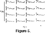
 Much of the visual complexity of a suture pattern appears to be the result of the
phi-frequencies, at least subjectively
(Figure 5). Uniformly reducing all h-frequencies by the same factor while leaving the phi frequencies unchanged produces suture patterns that are vertically shortened, yet still retain their saddles and lobes in recognizable shapes. Uniformly reducing all the
phi-frequencies while leaving the h-frequencies unchanged produces a different result. The number and magnitude of non-unique points along the suture are reduced. The new pattern is no longer recognizable as being the same as the unaltered suture pattern. An ammonitic suture pattern plotted with reduced values for
phi appears to be a less complex structure.