EXAMPLES OF APPLICATION OF SILICON RUBBER CASTING,
EPOXY REPLICATION, AND STEREO IMAGING
Example 1: Silicon Rubber Casting of Acid Prepared Mould of a Natural Assemblage of
Distomodus
Specimen BGS MWL 4702 preserves a partial skeleton of a
conodont, but the identity of the taxon could not be determined because the P elements were exposed on a slab of black shale with their lower surface uppermost. The taxonomically diagnostic features of the elements were facing downwards, embedded in the matrix. After taking a silicon rubber mould to preserve a record of the surface of the specimen, it was prepared by carefully dissolving the phosphatic crown tissue of the elements in 10%
hydrochloric acid. When dry the surface was consolidated with pvb in methanol, and a cast of the external mould of the elements made using RTV 913.
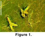 After curing, the rubber cast was removed, mounted and coated (with silver) for scanning electron microscopy. Stereo pairs were prepared by acquiring images of the specimen at approximately -5 and +5 degrees relative to horizontal. Red-green anaglyph stereo images
(Figure 1) were prepared by the method outline above and detailed in
Appendix 2. These images have been used for primary research on the specimen (as the only way of visualising details of the surface of the P elements in 3D), for communication with colleagues and collaborators (email, presentations, and reports), and for publication
(Purnell et al.
2003).
After curing, the rubber cast was removed, mounted and coated (with silver) for scanning electron microscopy. Stereo pairs were prepared by acquiring images of the specimen at approximately -5 and +5 degrees relative to horizontal. Red-green anaglyph stereo images
(Figure 1) were prepared by the method outline above and detailed in
Appendix 2. These images have been used for primary research on the specimen (as the only way of visualising details of the surface of the P elements in 3D), for communication with colleagues and collaborators (email, presentations, and reports), and for publication
(Purnell et al.
2003).
Example 2: Acid Preparation, Consolidation, Silicon Rubber Moulding and Epoxy Casting of the Mouth of
Protopteraspis vogti
Specimen A28720 (Paleontologisk Museum, Oslo) was collected from the Devonian, Ben Nevis Formation during the 1925 expedition to
Spitzbergen. The slab preserves articulated remains of several individuals of Protopteraspis vogti
Kaier, including two with articulated oral plates. Heterostracans preserving articulated oral plates are rare, and the outer surface of one of the specimens on the slab, A28720/2, was mechanically prepared to expose the details of the mouth (by Anatol
Heintz, sometime between 1925 and 1930). The specimen lies at an angle to the surface of the slab, and as a result of the removal of material from the mouth the exposed oral plates now sit within a steep-sided recess, more than 1 cm deep
(Figure 2). 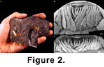 As part of an investigation into the structure of the mouth of pteraspid heterostracans
(Purnell 2002, and ongoing work with D.K. Elliot), detailed images of the mouth were required. The fact that the specimen sits on a recess caused some difficulties, especially in trying to obtain scanning electron photomicrographs.
As part of an investigation into the structure of the mouth of pteraspid heterostracans
(Purnell 2002, and ongoing work with D.K. Elliot), detailed images of the mouth were required. The fact that the specimen sits on a recess caused some difficulties, especially in trying to obtain scanning electron photomicrographs.
Additional acid preparation of the specimen was carried out to remove carbonate rock matrix from around the oral plates of the mouth using ethanoic acid (acetic acid), buffered according to the method of
Jeppsson et al.
(1985). Areas of the specimen which required no further preparation were protected from the acid by a covering of
pvb, and as matrix was removed by acid dissolution, newly exposed areas of the oral plates were also covered with
pvb. Prior to moulding, these protective coatings were removed, and a thin layer of pvb was applied as a precautionary separator over the area to be moulded (following the procedure outlined above).
Figure 2B-2C shows scanning electron images of an epoxy replica of the mouth of
P. vogti specimen A28720/2. The replica was cast using Araldite 2020 in a mould of RTV913. The figure shows the level of detail revealed by the acid preparation and retained through the moulding and casting process. Because the specimen sits in a recess, the anterior view of the mouth
(Figure 2C) could not have been obtained by direct optical or scanning electron microscope imaging.
Example 3: Acid Preparation, Consolidation, Silicon Rubber Moulding and Epoxy Casting of the Apparatus of Conodont Specimen RMS GY 1992.41.2
National Museum of Scotland specimen RMS GY 1992.41.2 is one of the 10 specimens from the Granton Shrimp bed of Edinburgh that preserve traces of soft tissue
(Aldridge et al.
1993). Although the exceptional preservation of body remains has transformed the study of conodont
palaeobiology, studying the conodont elements in some of the specimens has been rather problematic because of the difficulties of preparing elements without damaging soft tissue remains. In some cases it has not been possible to assign a specimen to a
taxon, simply because the identity of the elements could not be determined due to significant morphological details being obscured by rock. Also, scanning electron microscope imaging of elements in some of the specimens has been prevented because the blocks are too large.
Interpretation of specimen RMS GY 1992.41.2 has been hampered by both these problems.
Aldridge et al. (1993 p. 420) noted that in this specimen "The Pa element is not sufficiently exposed to allow the taxon to be positively identified." The conodont lies in the centre of a large block of laminated, organic rich limestone (16-20 cm long, 9-12 cm wide, 8-9 cm thick), thus effectively preventing imaging of the elements in a scanning electron microscope.
In order to overcome these difficulties, acid preparation of the
P1 element ( = Pa, see Purnell et al. 2000 for discussion of element notation) was undertaken, using pvb to coat adjacent areas and limit the effects of the acid to the area of the
P1 element only. Buffered methanoic acid (formic acid), prepared according to the method of
Jeppsson and Anehus
(1995), was applied to the element and its immediate surroundings drop by drop. The reaction was constantly monitored under a binocular microscope to ensure that the specimen was not adversely affected in any way. The area under preparation was washed periodically with
deionised water, and a fine paintbrush (00) and a very fine flexible steel mounted needle (0.25 mm diameter) were used to assist with the removal of the matrix from around the element.
 After several hours, the element was considered sufficiently well exposed and, after washing, drying, and application of a thin layer of pvb as a separator, a rubber mould was made using RTV 913 contained with a wall of Colténe® President following the method detailed above. From this step an epoxy cast was made using Araldite 2020 (see above).
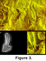
Figure 3A shows an anaglyph stereo image of the epoxy replica of the elements preserved in specimen RMS GY 1992.41.2. The
P1 element, toward the left is now well exposed. The enlargements show the level of detail reproduced by the replica, with incremental growth lamellae evident in the basal cavity of the
P1 (arrowed in
Figure 3B) and along the lower margin of the S4 elements (arrowed in
Figure 3C).
