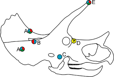
Figure 5. Schematic of Triceratops prorsus skull, showing the approximate locations of cranial lesions observed in actual Triceratops specimens. A) SMNH P1163.4, B) USNM 1201, C) SMM P62/1/1, D) YPM 1823, E) USNM 4708. Locations of lesions from the left side of the skull are mirrored to that of the right side. The colors of the dots, following the convention of Figure 4, indicate horn locking models with which the lesions are consistent. A yellow dot indicates a lesion which does not correspond to the horn locking positions inferred here.