SYSTEMATIC
PALEONTOLOGY
(continued)
Coracoid. The
left coracoid (Mal-235; Figure 19C) is slightly damaged. Typical of titanosaurians, the bone is roughly quadrangular (301 mm by 230 mm) and thickens
towards the glenoid fossa. The glenoid fossa is rugose. The lateral surface is
convex, and the medial surface is concave as in Apatosaurus (Gilmore
1936), Camarasaurus (Osborn and Mook 1921), and in Isisaurus
colberti (Jain and Bandyopadhyay 1997). The coracoid foramen is elliptical
and restricted to the dorsal portion of the element penetrating diagonally from
the lateral surface through the bone.
 Humerus. Four
humeri (left, Mal-221, Figure 20A-D; right, Mal-289,
Figure 20E; right,
Mal-316; and left, Mal-317; Table 7) of possibly three individuals (different
sizes and quarries) are well preserved and complete. The proximal end is
slightly more expanded than the distal end. The head is rugose and placed more
medially than laterally and directed posteriorly as in Alamosaurus
(Gilmore 1946), Opisthocoelicaudia (Borsuk-Bialynicka 1977), and
Saltasaurus (Powell 1986). The anterior surface is hollowed out and has a
strongly marked facet proximally, below which is a well-marked depression for
muscular attachment. The deltopectoral crest overhangs the center of the shaft
on the proximal anterolateral margin. The shaft is straight and rounded. The
ulna and radial condyles are strongly pronounced. The ulnar condyle is more
prominent than the radial condyle.
Humerus. Four
humeri (left, Mal-221, Figure 20A-D; right, Mal-289,
Figure 20E; right,
Mal-316; and left, Mal-317; Table 7) of possibly three individuals (different
sizes and quarries) are well preserved and complete. The proximal end is
slightly more expanded than the distal end. The head is rugose and placed more
medially than laterally and directed posteriorly as in Alamosaurus
(Gilmore 1946), Opisthocoelicaudia (Borsuk-Bialynicka 1977), and
Saltasaurus (Powell 1986). The anterior surface is hollowed out and has a
strongly marked facet proximally, below which is a well-marked depression for
muscular attachment. The deltopectoral crest overhangs the center of the shaft
on the proximal anterolateral margin. The shaft is straight and rounded. The
ulna and radial condyles are strongly pronounced. The ulnar condyle is more
prominent than the radial condyle.
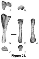 Ulna. Three
ulnae (Mal-190, Mal-218, Figure 21A-C, left; Mal-309;
Table 7) of at least two
different individuals are present. Mal-218 is well preserved, but Mal-190 and
Mal-309 are less well preserved. The olecranon process is strongly developed and
protrudes above the articular surface. The articular surface is concave
medially, convex laterally. The shaft is triangular in section and has a deep
radial fossa proximally, and is quadrangular distally. A strong posterior
tuberosity for muscle attachment emerges about 240 mm down and extends to the
distal end. The distal end has a medially concave reniform outline.
Ulna. Three
ulnae (Mal-190, Mal-218, Figure 21A-C, left; Mal-309;
Table 7) of at least two
different individuals are present. Mal-218 is well preserved, but Mal-190 and
Mal-309 are less well preserved. The olecranon process is strongly developed and
protrudes above the articular surface. The articular surface is concave
medially, convex laterally. The shaft is triangular in section and has a deep
radial fossa proximally, and is quadrangular distally. A strong posterior
tuberosity for muscle attachment emerges about 240 mm down and extends to the
distal end. The distal end has a medially concave reniform outline.
Radius. A
complete right radius (Mal-41; Figure 21D-G,
Table 7) was found with a distal
fragment (Mal-160) of a right humerus. The proximal and distal ends are about
equally expanded and rugose. The proximal end is slightly concave and
semicircular, whereas the distal end is slightly convex and ovoid. A
longitudinal groove occurs on the posterior surface.
Manus. Four
complete, associated metacarpals, and two fragmentary metacarpals are preserved.
Based on comparison with Alamosaurus (Gilmore 1946),
Opisthocoelicaudia (Borsuk-Bialynicka 1977), and Neuquensaurus (von
Huene 1929; Powell 1986) the associated metacarpals pertain to Mc I to IV. The
proximal end of right metacarpal I (Mal-196; Figure 22A-D,
Table 7) is roughly
triangular and rugose.
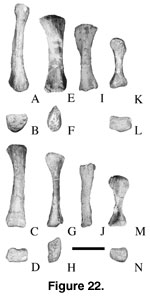 The shaft is rounded proximally and becomes mediolaterally compressed distally. The lateral surface has a triangular
roughened surface proximally to articulate with metacarpal II. A ridge occurs
midshaft on the lateral surface. The distal end is rectangular.
The shaft is rounded proximally and becomes mediolaterally compressed distally. The lateral surface has a triangular
roughened surface proximally to articulate with metacarpal II. A ridge occurs
midshaft on the lateral surface. The distal end is rectangular.
Metacarpal II
(Mal-208-2, right, Mal-214, left distal fragment;
Figure 22E-H,
Table 7) has an
oval proximal end. The proximal surface is rugose and generally convex. The
shaft is transversely compressed and somewhat constricted medially. The medial
surface of the shaft has a well-developed ridge proximally and a depression
distally. The distal surface is subrectangular, convex, and rugose.
Metacarpal III
(Mal-209 right, Figure 22I-J,
Table 7) has a triangular proximal end with a
posterior acute angle. In lateral view, about one third of the proximal portion
is paddle shaped while the distal portion is more rounded. The lateral surface
is flat and rugose, broad proximally and narrow distally. The distal end is
rectangular and is widest anteromedio-posterolaterally. Mal-209 is shorter and
more slender than metacarpal II.
Metacarpal IV
(Mal-208-1, right, Mal-144, left proximal fragment;
Figure 22K-N,
Table 7) has a
strongly twisted shaft. The proximal end is expanded and subrectangular. The
proximal surface is slightly convex. The shaft is medially constricted and
slightly twisted. About 20 mm from the distal end is a well-defined broad
triangular tuberosity on the anterior surface. This is similar to Alamosaurus.
The distal end is expanded mediolaterally, and the articular surface is convex.
In Alamosaurus,
the lengths of the metacarpals decrease laterally (Gilmore 1946). The manus is
reported but not illustrated in Saltasaurus and Aeolosaurus
(Powell 1986). In Opisthocoelicaudia, metacarpal I is the longest,
whereas metacarpals II and III are approximately the same length. In
Camarasaurus (McIntosh et al. 1996a,
1996b) and in Apatosaurus (Gilmore
1936, p. 226, top table) the third metacarpal is the longest and the two lateral
metacarpals are the shortest. In Malawisaurus, the length of metacarpals
decreases laterally.
There is no direct
evidence of phalanges although the distal ends of metacarpals have articular
surfaces that suggest the presence of phalanges. Titanosaurians are said to have
no manual phalanges. If manual phalanges were present in Malawisaurus,
this would suggest that basal titanosaurians had manual phalanges that were lost
in more derived species.
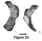 Ischium. Three
ischia (left, Mal-42, length = 400 mm; width = 125 mm at the widest point, and =
90 mm at the narrowest point, Figure 23A, [Jacobs
et al. 1993, figure 1g];
Mal-184; right, Mal-183-1, length = 382 mm; width = 114 mm at the widest point
and = 100 mm at the narrowest point, Figure 23B) have dorsoventrally broad pubic
articular facets. Opposite the pubic peduncle is a small, well-defined tubercle
on the lateral surface for muscle attachment. The posterior blade is plate-like
and short (Jacobs
et al. 1993). The angle between the posterior surface of the ischium and the ischium-ischium articular surface is steep as in Alamosaurus
(Gilmore 1946) and Opisthocoelicaudia (Borsuk-Bialynicka 1977),
suggesting that the cross section of the ischium shaft is nearly coplanar.
Ischium. Three
ischia (left, Mal-42, length = 400 mm; width = 125 mm at the widest point, and =
90 mm at the narrowest point, Figure 23A, [Jacobs
et al. 1993, figure 1g];
Mal-184; right, Mal-183-1, length = 382 mm; width = 114 mm at the widest point
and = 100 mm at the narrowest point, Figure 23B) have dorsoventrally broad pubic
articular facets. Opposite the pubic peduncle is a small, well-defined tubercle
on the lateral surface for muscle attachment. The posterior blade is plate-like
and short (Jacobs
et al. 1993). The angle between the posterior surface of the ischium and the ischium-ischium articular surface is steep as in Alamosaurus
(Gilmore 1946) and Opisthocoelicaudia (Borsuk-Bialynicka 1977),
suggesting that the cross section of the ischium shaft is nearly coplanar.
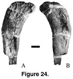 Femur. The
femur (Figure 24,
Table 8) is represented by a right proximal fragment (Mal-201;
length of the preserved portion = 0.745 m). The anterior and posterior surfaces
of the proximal end are damaged. The proximal portion is medially deflected and
has a lateral bulge. The shaft of the femur is flattened anteroposteriorly and
widened transversely so that it has an elliptical cross section with the long
axis oriented mediolaterally. The humerus/femoral ratio is 0.72 in
Opisthocoelicaudia (Borsuk-Bialynicka 1977), 0.74 in Titanosaurus
(McIntosh 1990), 0.78 in Aegyptosaurus (McIntosh 1990), and 0.80 in
Rapetosaurus (Curry Rogers and Forster 2001) with an average of 0.76. The
larger humerus of Malawisaurus (Mal-221) is 0.722 m. Thus, the estimated
length of Mal-201 is 0.950 m.
Femur. The
femur (Figure 24,
Table 8) is represented by a right proximal fragment (Mal-201;
length of the preserved portion = 0.745 m). The anterior and posterior surfaces
of the proximal end are damaged. The proximal portion is medially deflected and
has a lateral bulge. The shaft of the femur is flattened anteroposteriorly and
widened transversely so that it has an elliptical cross section with the long
axis oriented mediolaterally. The humerus/femoral ratio is 0.72 in
Opisthocoelicaudia (Borsuk-Bialynicka 1977), 0.74 in Titanosaurus
(McIntosh 1990), 0.78 in Aegyptosaurus (McIntosh 1990), and 0.80 in
Rapetosaurus (Curry Rogers and Forster 2001) with an average of 0.76. The
larger humerus of Malawisaurus (Mal-221) is 0.722 m. Thus, the estimated
length of Mal-201 is 0.950 m.
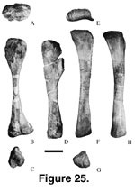 Tibia. The
surface of the right tibia (Mal-207; Figure 25,
Table 8) is weathered. The tibia
has a prominent and anteriorly projecting cnemial crest. The shaft is slightly
twisted and medially constricted. A lateral groove occurs at the distal end of
the shaft. The distal posteroventral process is broad transversely.
Tibia. The
surface of the right tibia (Mal-207; Figure 25,
Table 8) is weathered. The tibia
has a prominent and anteriorly projecting cnemial crest. The shaft is slightly
twisted and medially constricted. A lateral groove occurs at the distal end of
the shaft. The distal posteroventral process is broad transversely.
Fibula. The
proximal end of the right fibula (Mal-189; Figure 25E-H,
Table 8) is expanded anteroposteriorly. The proximal surface is crescent shaped. The proximal tibial
scar on the medial surface is triangular and well marked. The shaft is flattened
medially and convex laterally. About 230 mm downshaft on the lateral surface is
an elongate lateral trochanter for muscle attachment (flexors of digits;
Borsuk-Bialynicka 1977) beyond which the bone becomes more cylindrical. The
distal end is triangular in cross section.
Pes. Metatarsal
III? (Mal-145) is represented by a small fragment. The articular surface is
subrectangular in outline and is concave as in the proximal surface of
metatarsal III of Opisthocoelicaudia that articulates with the
astragalus. Based on these observations, Mal-145 is interpreted as the proximal
fragment of metatarsal III of Malawisaurus.
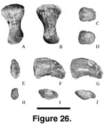 Metatarsal V (right; Mal-210;
Figure 26A-B,
Table 8)
is flattened mediolaterally. The proximal end is semicircular, convex, and
expanded. The shaft is laterally compressed. The medial surface is more concave
proximally than distally. The distal end is convex and ellipsoidal.
Metatarsal V (right; Mal-210;
Figure 26A-B,
Table 8)
is flattened mediolaterally. The proximal end is semicircular, convex, and
expanded. The shaft is laterally compressed. The medial surface is more concave
proximally than distally. The distal end is convex and ellipsoidal.
A proximal right pedal
phalanx, probably of digit I (Mal-213; Figure 26C-D), is conical proximally with
the apex of the cone being placed laterally. The ventral surface is shallowly
depressed medially.
The left ungual
(Mal-211; Figure 26E-G,
Table 8) probably of digit I, is compressed laterally.
The proximal surface is concave while the distal end is pointed. Longitudinal
grooves occur on the interpreted lateral surface as in Apatosaurus
(Gilmore
1936) and in Camarasaurus (BYU 9047;
McIntosh et al. 1996b).
Although shallow grooves occur, the medial surface is relatively smooth compared
to the lateral surface. The ventral surface has a proximal medial tuberosity. In
lateral view, it arcs ventrally to the point.
Another left ungual
(Mal-212; Figure 26H-J) is smaller than Mal-211. In Apatosaurus, the
shapes of the ungual phalanges are similar in the first, second, and third
digits. The sizes decrease laterally. Thus, Mal-212 is likely a more lateral
phalanx than Mal-211, probably from the second digit.
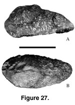 Armor.
Previously it was reported that Malawisaurus lacked direct evidence of
dermal armour, but calcite pseudomorphs shaped like dermal scutes were found
associated with the bones in the same quarry (Jacobs
et al. 1993, figure 2e). A
large dermal scute was more recently discovered (Mal-204;
Figure 27). It is 190
mm long and 95 mm wide. The morphology of this dermal armour is similar to the
large dermal scutes associated with Saltasaurus (PVL 4017-112), whereas
the pseudomorphs are similar to the small dermal scutes of Saltasaurus
(PVL 6017-118) and small dermal scutes of the titanosaurian from Madagascar
(FMNH PR 2021;
Dodson et al. 1998).
Armor.
Previously it was reported that Malawisaurus lacked direct evidence of
dermal armour, but calcite pseudomorphs shaped like dermal scutes were found
associated with the bones in the same quarry (Jacobs
et al. 1993, figure 2e). A
large dermal scute was more recently discovered (Mal-204;
Figure 27). It is 190
mm long and 95 mm wide. The morphology of this dermal armour is similar to the
large dermal scutes associated with Saltasaurus (PVL 4017-112), whereas
the pseudomorphs are similar to the small dermal scutes of Saltasaurus
(PVL 6017-118) and small dermal scutes of the titanosaurian from Madagascar
(FMNH PR 2021;
Dodson et al. 1998).
