SYSTEMATIC PALEONTOLOGY
(continued)
Colubroidea Oppel, 1811
Genus
et species indeterminate
Colubroid morphotype A
Figure 6
Referred specimens. H-GSP 26722, isolated
precloacal vertebra; H-GSP 26666, incomplete, associated, and partially
articulated postcranial skeleton.
Localities and ages.
Y-726 (13.01 Ma), Y-698 (12.94 Ma).
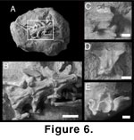 Description. Colubroid morphotype A
consists of approximately 106 partially associated and articulated vertebrae and
ribs representing a single animal from locality Y-726, and an isolated
precloacal vertebra from locality Y-698. The partially articulated specimen
consists of precloacal and a single caudal vertebra embedded in calcareous
siltstone (Figure 6A). In ventral view, precloacal elements are elongate (Figure 6B). Hypapophyses are present on all elements. They originate anteriorly at the
level of the parapophyses and are elongate and posteriorly angled.
Well-developed prezygapophyseal accessory processes are present, and are
anteroposteriorly swollen with sharply pointed distal margins. Prominent
ventrolateral cotylar processes are present on all specimens.
Description. Colubroid morphotype A
consists of approximately 106 partially associated and articulated vertebrae and
ribs representing a single animal from locality Y-726, and an isolated
precloacal vertebra from locality Y-698. The partially articulated specimen
consists of precloacal and a single caudal vertebra embedded in calcareous
siltstone (Figure 6A). In ventral view, precloacal elements are elongate (Figure 6B). Hypapophyses are present on all elements. They originate anteriorly at the
level of the parapophyses and are elongate and posteriorly angled.
Well-developed prezygapophyseal accessory processes are present, and are
anteroposteriorly swollen with sharply pointed distal margins. Prominent
ventrolateral cotylar processes are present on all specimens.
In
lateral view (Figure 6C), the zygosphene is low with anteroposteriorly elongate
articular facets. Ventrally, elongate, pointed parapophyseal processes occur on
all vertebrae. The hypapophysis is low and blade-like. It extends to the
posterior margin of the condyle where it terminates in a sharp point. Dorsally,
the posterior median notch is well developed with straight margins. The neural
spine extends anterior to the dorsal margin of the zygosphene. The spine is tall
and uniformly thick. In anterior view (Figure 6E), the neural canal is rounded
in outline and is capped by a small zygosphene. The prezygapophyseal accessory
facet is slightly dorsolaterally angled. The accessory process is approximately
horizontal. There are no accessory-process foramina.
The
single caudal vertebra is exposed in ventral view (Figure 6D). The pleurapophyses are elongate and ventrally deflected. Their posterior margins
extend to the condyle, giving the element a triangular appearance. Large
subcentral foramina are present on the ventrolateral margins of the centrum.
Anteriorly, the cotylar ventral margin is flush with the prominence, and
posteriorly, the condyle is small and unexpanded. Preserved costal elements
consist of elongate, thin proximal and mid-shaft regions. No complete ribs were
recovered. Proximal regions of the ribs are elongate and thin, and include a
well-developed pseudotuberculum (Figure 6B).
Genus
et species indeterminate
Colubroid morphotype B
Figure 7A
Referred specimens. H-GSP 13970, 24343,
24350, 24352, 24354, 24355, 24356, 24368, 24386, 24418, 24421, 24423, 27061,
27162, 27173, 27177, 50264, 51121, 53305, 53306, 53307, 53308, 53310, 53369,
53370, isolated precloacal vertebrae.
Localities and ages.
Y-367 (8.95 Ma), Y-024 (8.14 Ma), Y-547 (7.93 Ma), Y-457 (7.30 Ma), Y581 (6.97
Ma), Y-908 (6.78 Ma).
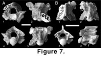 Description. Specimens referred to as
colubroid morphotype B are small, relatively stout elements, all of which
possess well-developed hypapophyses and prezygapophyseal accessory processes. In
anterior view (Figure 7A.1), the neural canal is rounded in outline and is
capped by a zygosphene that possesses a flat dorsal margin. The cotyle is
circular and includes large ventrolateral processes. A large paracotylar foramen
is present on either side of the cotyle. The parapophyseal process is large with
a rounded anterior margin. The prezygapophyseal articular facets are low and
horizontal. Where preserved, the prezygapophyseal accessory processes are
elongate and distally pointed. A large accessory process foramen is present at
the base of the process. In dorsal view (Figure 7A.2), the interzygapophyseal
ridge is smoothly concave. The prezygapophyseal articular facet is
anteroposteriorly wide and approximately subtriangular with an anteromedially
angled apex. The base of the neural spine is elongate and extends onto the
zygosphene. The anterior margin of the zygosphene is approximately straight to
slightly concave. The margins of the posterior median notch are smoothly convex.
In ventral view (Figure 7A.3), the centrum is relatively stout with large
subcentral foramina. Anteriorly the parapophyseal process is rounded.
Hypapophyseal bases indicate an elongate, posteriorly angled hypapophysis. In
posterior view (Figure 7A.4), the dorsal margin of the neural arch is slightly
convex with a relatively steep angle compared to other Siwalik Group taxa. The
posterior margin of the arch is smooth. There is no indication of
epizygapophyseal spines or parazygosphenal foramina. The parapophysis is
elongate and thin in posterior view.
Description. Specimens referred to as
colubroid morphotype B are small, relatively stout elements, all of which
possess well-developed hypapophyses and prezygapophyseal accessory processes. In
anterior view (Figure 7A.1), the neural canal is rounded in outline and is
capped by a zygosphene that possesses a flat dorsal margin. The cotyle is
circular and includes large ventrolateral processes. A large paracotylar foramen
is present on either side of the cotyle. The parapophyseal process is large with
a rounded anterior margin. The prezygapophyseal articular facets are low and
horizontal. Where preserved, the prezygapophyseal accessory processes are
elongate and distally pointed. A large accessory process foramen is present at
the base of the process. In dorsal view (Figure 7A.2), the interzygapophyseal
ridge is smoothly concave. The prezygapophyseal articular facet is
anteroposteriorly wide and approximately subtriangular with an anteromedially
angled apex. The base of the neural spine is elongate and extends onto the
zygosphene. The anterior margin of the zygosphene is approximately straight to
slightly concave. The margins of the posterior median notch are smoothly convex.
In ventral view (Figure 7A.3), the centrum is relatively stout with large
subcentral foramina. Anteriorly the parapophyseal process is rounded.
Hypapophyseal bases indicate an elongate, posteriorly angled hypapophysis. In
posterior view (Figure 7A.4), the dorsal margin of the neural arch is slightly
convex with a relatively steep angle compared to other Siwalik Group taxa. The
posterior margin of the arch is smooth. There is no indication of
epizygapophyseal spines or parazygosphenal foramina. The parapophysis is
elongate and thin in posterior view.
Gansophis gen. nov.
Type species. Gansophis potwarensis
sp. nov.
Etymology. Gans + Ophis (Gr. Masc),
“snake.” “Gans’ snake,” generic nomen in honor of Dr. Carl Gans for his many
contributions to herpetology.
Diagnosis. Colubroid snake of
indeterminate status recognized by a posteriorly projecting accessory
parapophyseal process that forms an elongate articular surface for the costal
tubera.
Gansophis potwarensis
gen. et sp. nov.
Figure 7B
Holotype. H-GSP 49900, isolated
precloacal vertebra.
Type locality and age. Y-908 (6.78 Ma).
Diagnosis. As for genus.
Etymology. Specific name from the Potwar
Plateau in north-central Pakistan.
Description. The single vertebra assigned
to Gansophis is incomplete, missing most of the zygosphene, left
prezygapophysis and synapophysis, right prezygapophyseal accessory process,
distal portion of the hypapophysis, most of the cotyle, and the posterior margin
of the neural spine.
In
dorsal view (Figure 7B.1), the diapophyseal articular facet extends beyond the
base of the synapophysis. The dorsal margin of the neural spine is laterally
expanded with a shallow median groove, producing a slightly bilobate
anterodorsal margin to the spine. Posteriorly, the margins of the posterior
median notch are crenulate. In ventral view (Figure 7B.2), the parapophyseal
accessory process is triangular in outline and is laterally recurved. The
ventral and medial portions of the process are covered by smooth periosteum that
is continuous with the rest of the element and forms an ovoid, elongate
articular surface for the costal tubera. Anteriorly, a single, well-developed
ventrolateral cotylar process is preserved along the right ventral margin of the
cotyle.
In
lateral view (Figure 7B.3), the neural spine is elongate, and the anterior
margin of the spine overhangs the zygosphene. The posterior margin of the neural
arch is elevated, and the posterior margin of the neural spine is shorter than
the anterior margin. The synapophysis is well developed, with a prominent,
laterally extended diapophyseal process. The parapophyseal process is expanded
to include a small, posteriorly oriented accessory process. The process is
triangular in lateral view, and its lateral margin consists of a costal
articular facet continuous with the main body of the synapophysis. The
anteroventral margin of the hypapophysis is steeply angled, and the distance
between the margin and the cotyle indicates that the hypapophysis was broad.
In
posterior view (Figure 7B.4), the lateral margins of the neural arch are
straight and slightly angled at the level of the dorsolateral margin of the
zygantrum. A small prominence is present at the lateral margin of the
postzygapophysis, but it is considerably smaller than the epizygapophyseal
spines of other colubroid taxa. The posterior surface of the neural arch lacks
parazygantral foramina.
Discussion. Of the three indeterminate
colubroids, only Gansophis can be diagnosed by an apomorphic character,
the presence of the posterior accessory parapophyseal process. The process
is not described for any other snake (e.g.,
Rochebrune 1881;
Dowling and Duellman 1978;
Rage 1984;
Holman 2000). It is possible that the accessory
process represents a teratological or pathological abnormality, conditions that
are known to occur in the vertebral column of snakes (e.g.,
Albrecht 1883).
Examination of the process does not demonstrate irregular or abnormal bone
growth. Instead, it is smoothly continuous with the rest of the parapophysis,
suggesting functional articulation with an expanded costal tuberculum. Among the
other two taxa, colubroid morphotype A is unique in the Siwalik Group record in
the absence of large foramina at the anteroventral margins of the
prezygapophyseal articular facets. Colubroid morphotype B can be distinguished
by a comparatively short centrum among colubroid specimens.
The
higher order systematic relationships of these specimens within Colubroidea
cannot be precisely resolved. Morphotypes A and B can be definitively excluded
from Colubrinae and Viperidae on the basis of hypapophyseal morphology, but
could be included in a wide range of colubroid higher-order lineages. Because
Gansophis is represented by only a single incomplete specimen, its
systematic relationships within Colubroidea are more poorly constrained.
Colubroidea indeterminate
Referred specimens. H-GSP 24181, 24342,
24344, 24347, 24349, 24353, 24355, 24366, 24369- 71, 24375, 24376, 24379, 24380,
24386, 24387, 24388, 24390, 24393, 24395, 24397, 24398, 24407, 24420, 24421,
24424, 24425, 26220, 26223, 26245, 26248, 27058, 27060, 27062, 27064, 27074,
27077, 27079, 27080, 27082-6, 27088, 27099, 27100, 27104, 27105, 27150, 27152,
27157, 27160, 27161, 27163, 27165, 27167, 27168, 27172, 27174-6, 27179-85,
27200, 27202, 27204, 27216, 27217, 27219, 27220, 27222, 27233, 27303, 27321,
27322, 46699, 53295, 53311- 4, 53329, 53331, 53337, 53340, 53344, 53348, 53351,
53353-6, 53358-60, 53364-8, 53371, 53373-5, 53381-4, 53387-9, 53399, 53404,
53406, 53415, isolated precloacal and caudal vertebrae.
Localities and ages.
Y-747 (18 Ma), Y-680 (14.10 Ma), Y-491 (13.77 Ma), Y-641 (13.55 Ma), Y-640
(13.55 Ma), Y-668 (13.30 Ma), Y-690 (13.04 Ma), Y-504 (11.52 Ma), Y-809 (11.4
Ma), Y-076 (11.31 Ma), Y-450 (10.16 Ma), Y-311 (10.00 Ma), Y-182 (9.16 Ma),
Y-367 (8.95 Ma), Y-388 (8.68 Ma), Y-387 (8.64 Ma), Y-024 (8.14 Ma), Y-547 (7.93
Ma), Y-906 (7.80 Ma), Y-866 (7.33 Ma), Y-457 (7.30 Ma), Y-931 (7.24 Ma), Y-581
(6.97 Ma).
Description and Discussion.
Indeterminate colubroid specimens consist of fragmentary precloacal and caudal
elements that possess some combination of the following characters: well-developed prezygapophyseal accessory processes and accessory-process foramina;
large ventrolateral cotylar processes; paracotylar foramina; strongly
differentiated diapophyseal and parapophyseal articular facets of the
synapophyses; prominent hypapophyses; elongate centra; subcentral paralymphatic
fossae; elongate pleurapophyses and haemapophyses on caudal elements.
Indeterminate colubroid remains have been recovered from screen-washed
localities throughout the Siwalik Group and undoubtedly include multiple taxa.
