 Holotype. H-GSP 24346, isolated
precloacal vertebra.
Holotype. H-GSP 24346, isolated
precloacal vertebra.
Colubrinae Opell, 1811
Chotaophis gen. nov.
Type species. Chotaophis padhriensis sp. nov.
Etymology. Chota (Urdu) “little” + Ophis (Gr. Masc), “snake” indicating the small size of referred specimens.
Diagnosis. Small colubrine snake possessing narrow zygapophyses, anteriorly positioned lateral foramina, transversely thin neural spine, and parazygantral foramina.
Chotaophis padhriensis sp. nov.
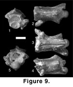
Figure 9
Holotype. H-GSP 24346, isolated
precloacal vertebra.
Paratype. H-GSP 26246, isolated vertebra.
Type locality. Y-367 (8.95 Ma)
Locality and age. Y-457 (7.30 Ma)
Diagnosis. As for genus
Etymology. Specific nomen from Padhri, the local community at locality Y-457. Nomen translates as “little snake from Padhri.”
Description. In anterior view (Figure 9.1), the vertebrae are short and narrow. The neural spine is thin. The zygosphene is transversely wide with a thin anterior margin and small articular facets. The cotyle is relatively small with ventrolateral processes represented only by a slight squaring of the ventral margin of the centrum. The prezygapophyses are low relative to the dorsal margin of the zygosphene and possess horizontal articular facets. There is no indication of elongate prezygapophyseal accessory processes, but large accessory-process foramina are present at the ventrolateral margins of the articular facets. In dorsal view (Figure 9.2), the interzygapophyseal ridge is smoothly concave. The posterior median notch has straight margins. Anteriorly, the prezygapophyseal articular facet is ovoid and elongate. The anterior margin of the zygosphene is broadly convex. The base of the neural spine is transversely uniform in width.
In ventral view (Figure 9.3), the centrum is elongate. The haemal keel is well developed and becomes progressively wider posteriorly, terminating in a blunt, triangular tip. Small subcentral foramina are present on either side of the keel toward the cotyle. The postzygapophyseal articular facet is elongate and anteroposteriorly oriented.
In lateral view (Figure 9.4), the ventral margin of the centrum is concave. The haemal keel possesses a flat ventral margin. Anteriorly, the cotyle is angled more posteroventrally than in other Siwalik taxa. Above the cotyle, the dorsal margin of the zygosphene is elevated. The neural spine extends to the anterior margin of the zygosphene. Although the majority of the spine is not preserved in either specimen, its posterior portion is gradually curved and appears to include a portion of the dorsal margin, suggesting that the spine was low. Small lateral foramina are present on the sides of the centrum. The foramina are located just posterior to the synapophyses and are more anteriorly positioned than in other taxa. In posterior view (Figure 9.5), the neural arch is steeply angled with a concave margin. The posterior margin of the arch includes prominent parazygantral foramina located lateral to the zygantrum. The postzygapophysis is not as expanded laterally as in other Siwalik taxa.
Discussion. Chotaophis is recognized as a distinct taxon based on the unique combination of anteriorly positioned lateral foramina, presence of parazygantral foramina, and elongate centrum. Chotaophis is placed with Colubrinae based on the presence of a well-formed haemal keel. The fossil record of Old World colubrines is extensive; however, vertebral morphology has been considered too ambiguous to define interrelationships and taxonomy within the clade (e.g., Szyndlar 1984, 1987). Old World fossil colubrines include multiple species of the extant genera Coluber, Elaphe, and Coronella, as well as the extinct genera Zelceophis and Texasophis (Rage 1984; Szyndlar 1984; Szyndlar and Schleich 1993; Ivanov 2000). Chotaophis can be differentiated from Coluber, Elaphe, Coronella, and Zelceophis by the absence of prezygapophyseal accessory processes. Chotaophis and Texasophis share relatively unexpanded prezygapophyses and reduced prezygapophyseal accessory processes. However, Chotaophis has more anteriorly positioned lateral foramina, and is relatively more elongate than published specimens of Texasophis. Texasophis is considered a problematic taxon, because vertebral morphology in colubrines is relatively undocumented (Szyndlar 1987), and the genus has not been diagnosed using discrete characters. As a result, there is no evidence to support referral to Texasophis.
Type species. Sivaophis downsi sp. nov.
Etymology. “Siva” (Hindi), “God of death” + Ophis (Gr. Masc), “snake.” Generic nomen continues a secular tradition in naming Siwalik taxa and should not be interpreted as favoring or disparaging a particular religion.
Diagnosis. Colubrine possessing wasp-waisted interzygapophyseal ridges in dorsal and ventral view, large lateral foramina, and an extremely tall neural spine.
Sivaophis downsi sp. nov.
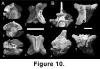
Figure 10
 Holotype. H-GSP 49661, isolated
precloacal vertebra.
Holotype. H-GSP 49661, isolated
precloacal vertebra.
Paratype. BSP 1956 II 2267, isolated precloacal vertebra.
Referred specimens. BSP 1956 II 2267, H-GSP 05063, 09764 17618, 24174, 24339, 24340, 26221, 26367, 27120, 27122, 27315, 31363, 31364, 49898, 50263, 50700, 53302, 53303, 53309, 53418, 53419, BSP 5.2.56, isolated precloacal vertebrae.
Type locality. Y-227 (9.29 Ma).
Localities and ages. Dhok Pathan Formation, Winnewala (BSP collections), Y-059 (13.60 Ma), Y-640 (13.55 Ma), Y-825 (12.55 Ma), Y-504 (11.52 Ma), Y-224 (9.29 Ma), Y-227 (9.29 Ma), Y-260 (9.19 Ma), KL-01 (9.18 Ma), Y-182 (9.16 Ma), Y-367 (8.95 Ma), Y-388 (8.67 Ma), Y-457 (7.30 Ma), Y-908 (6.78 Ma), DP-13 (6.35 Ma).
Etymology. Specific nomen translates as “Downs,” in honor of the contributions of William R. Downs III to the paleontology of the Siwalik Group. Full nomen translates to “Downs’ Siva snake.”
Description. In anterior view (Figure 10A.1, B.1), the zygosphene is thick and has a broadly convex dorsal margin. The cotyle is round with very small ventrolateral processes in all specimens. A single pair of small paracotylar foramina is present at the lateral margins of the cotyle. The prezygapophysis is low slung and approximately horizontal. Small, triangular accessory processes are present below the prezygapophyseal articular facets. The synapophyses are small with low-angled dia- and parapophyseal articular surfaces.
In dorsal view (Figure 10A.2, B.2), the neural spine is elongate and terminates anteriorly between the zygosphenal articular facets. The anterior margin of the zygosphene is highly variable, ranging from straight to broadly concave to strongly crenulate (Figure 10B.2, B.3). The prezygapophyseal articular facets are relatively small, ovoid, and elongate. Between the zygapophyses, the interzygapophyseal ridge is concave and strongly angled toward the apex, giving the element a strongly wasp-waisted appearance in dorsal view. The posterior median notch is well developed and extends anteriorly toward the transverse midline of the element. The margins of the notch and the posterior neural arch are sigmoid in dorsal view from the base of the postzygapophysis to the posterior margin of the neural spine. In ventral view (Figure 10A.3, B.3), the centrum is elongate and triangular. The haemal keel is narrow and well developed, extending from the ventral margin of the cotyle to the anterior margin of the condyle, where it terminates in a blunt, unexpanded point. Deep paralymphatic channels define the keel laterally. The condyle is relatively smaller than in other Siwalik taxa. The postzygapophyseal articular facets are small and restricted to the lateral margins of the neural canal.
In lateral view (Figure 10A.4, B.4), the neural arch is tall. The zygosphenal articular facets are elevated well beyond the level of the prezygapophysis and are anterodorsally angled. The neural spine is partially preserved in only a single specimen (Figure 10B.4). The spine is wide and tall, with a convex dorsal margin. The majority of the anterior margin is not preserved in BSP 5.2.56; however, the angle of the spine at its base suggests that it was slightly angled posteriorly. The posterior margin of the neural arch is elevated with vertical to posteriorly angled margins. A large lateral foramen is present in a shallow fossa just behind the prezygapophysis. The zygosphenal ridge forms the dorsal margin of the fossa, with a sharp angle between the pre- and postzygapophyses. In posterior view (Figure 10A.5), the neural arch is elevated and strongly vaulted, with curved lateral margins. The posterior surface of the neural arch is broad, and the zygantrum is deeply recessed. Medially, the neural canal is tall and narrow.
Discussion. Sivaophis is diagnosed by the presence of wasp-waisted interzygapophyseal ridges, which distinguish it from other Siwalik specimens and all other fossil and extant taxa in which vertebral morphology is described (e.g., Dowling and Duellman 1978; Rage 1984; Szyndlar 1984; Holman 2000). The neural spine in Sivaophis is much taller than seen in many extant and extinct colubroids (e.g., Dowling and Duellman 1978; Holman 2000), although a larger spine is present in some lineages (e.g., Boiga). Among Siwalik specimens, Sivaophis also possesses relatively larger lateral foramina. The presence of a prominent haemal keel and small but well- developed prezygapophyseal accessory processes justifies inclusion in Colubrinae.
Differences in the shape of the zygosphenal anterior margin were previously used as systematically valid characters in diagnosing fossil snakes (e.g., Auffenberg 1963), but examination of complete precloacal vertebral columns demonstrates intracolumnar variability in the shape of the margin from strongly concave to strongly convex (see also Rieppel et al. 2002). Two distinct taxa were previously considered to be present in the collection referred to as Sivaophis (Head 2002) based on variation in the anterior margin of the zygosphene. However, intracolumnar variation in this character prohibits such diagnoses. The original diagnosis for the genus was also based on inferences of missing morphology that have been falsified by examination of BSP 1956 II 2267. As a result, the hypodigm has been recombined, with S. downsi as the type and only species.
Colubrinae indet.
Referred specimens. H-GSP 27076, 27081, 27145, 27149, 27178, 27232, 27301, 30812, 53400, 53410.
Localities and ages. Y-709 (14.30 Ma), Y-641 (13.55 Ma), Y-809 (11.40 Ma), Y-450 (10.16 Ma), Y-311 (10.00 Ma), Y-388 (8.68 Ma), Y-387 (8.64 Ma), Y-547 (7.93 Ma).
Description and Discussion. Specimens referred to Colubrinae indeterminate consist of partial centra and are too incomplete to be assigned to either Chotaophis or Sivaophis, but can be recognized on the basis of the following morphology: in ventral view, the centrum is elongate and the haemal keel is well developed and wide. The keel widens posteriorly with a posterior margin that ranges from sharply pointed to spoon-shaped and rounded. In dorsal view, the posterior median notch is well developed with strongly convex medial margins. There is no indication of epizygapophyseal spines on the postzygapophyses.
![]()