MATERIAL AND METHODS
Remains of diplodocid sauropods from the Late Jurassic (Kimmeridgian) Morrison Formation of Wyoming were provided by the Saurier-Museum Aathal/Switzerland for the
NT and CT analyses. The sample consisted of five cervical vertebrae and one cervical rib of approximately 10 cm in length, with each belonging to juvenile individuals. A 40 cm long midcervical vertebra of an adult specimen did not fit the rotating table for the NT and therefore was only
analyzed using CT. One caudal vertebra of 12 cm length was scanned both with CT and NT to compare with a vertebra without internal cavities.
The fossil bones of all scanned sauropod remains were diagenetically altered (Zocco and Schwartz 1994). With the exception of the caudal vertebra and the cervical rib, all specimens are lateromedially slightly distorted and compressed. The internal cavities, openings and foramina are filled with siliciclastic mud characterized by a calcareous cement (Ayer 2000). The bones are partially badly fractured and most fractures and cracks were filled with quick drying cyano-acrylate resin. The bone surfaces were partially slightly painted and missing parts of the vertebrae have been remodelled with polyester cast resin.
The CT scans were performed with a Multidetector CT-scanner (Sensation 16, Siemens, Erlangen; Germany) at the Department of Medical Radiology of the University Hospital Basel. All vertebrae were scanned with their anteroposterior long axis perpendicular to the scanner rotation axis, so that the resulting image stack runs through the vertebra either from anteriorly to posteriorly or vice versa. Therefore, the maximum path length of X-rays through the largest specimen was 17.9 cm. The parameter setting was 140 kV and 350
MA and a primary collimation of 16 x 0.75 mm. The raw data were reconstructed applying a standard algorithm for human osseous structures using the standard CT imaging processor with the imaging software version VA 70C. The pixel dimensions of the camera were 512 x 512 [pixels], and the resulting field of view was 598 x 323 mm. All CT data were reconstructed in all orthogonal planes at 3 mm thickness and additionally along dedicated planes along anatomical structures. The basic overlapping axial datasets were saved as DICOM files with a defined dimension of 1 x 1 x 1 mm/voxel, and
to allow for further 3-D reconstructions. Scanning and data processing took approximately 10 minutes per specimen.
The NT scans were performed in the Neutron Transmissions Radiography Station NEUTRA at the PSI in Villigen/Switzerland (Vontobel et al. 2003). Samples were positioned on a rotary table at position 3 of NEUTRA, i.e., providing a neutron flux of about 3.6 106 [n/cm2/s] and a collimation ratio L/D = 550. While the samples were rotated over 180o, 240-300 transmission projections were taken. The maximum path length of neutrons through the largest specimen was 12 cm.
 Neutrons were converted into light by a 6Li based neutron scintillator screen with thickness of 0.25 mm, which was imaged with a 1024 X 1024 pixel CCD camera (DV434) from
Andor technology. The resulting field of view was 279 x 279 mm. After exposure normalization and flatfield correction the projection data were reconstructed into slices by filtered backprojection. The voxel data were saved into DICOM-format with a defined dimension of 0.272 x 0.272 x 0.272 mm/voxel. The time needed for the NT scans was 1.5 to 3 hours per specimen, and together with the data processing
was up to 5 hours per specimen.
Neutrons were converted into light by a 6Li based neutron scintillator screen with thickness of 0.25 mm, which was imaged with a 1024 X 1024 pixel CCD camera (DV434) from
Andor technology. The resulting field of view was 279 x 279 mm. After exposure normalization and flatfield correction the projection data were reconstructed into slices by filtered backprojection. The voxel data were saved into DICOM-format with a defined dimension of 0.272 x 0.272 x 0.272 mm/voxel. The time needed for the NT scans was 1.5 to 3 hours per specimen, and together with the data processing
was up to 5 hours per specimen.
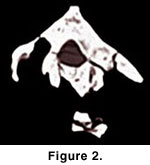
 From both CT and NT scans, resulting tomographic images were edited with ImageJ and OsiriX software for MacIntosh. The quality of the images was improved by individual changes of brightness and contrast, gray levels and sharpening with image processing tools. The image stacks were also exported into movie files (Figure 2,
Figure 3). Three-dimensional polygon-surface models (stl-format) were extracted from the NT data with the help of VG Studio software and from the CT data with the help of Mimics 8.0 software.
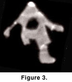
From both CT and NT scans, resulting tomographic images were edited with ImageJ and OsiriX software for MacIntosh. The quality of the images was improved by individual changes of brightness and contrast, gray levels and sharpening with image processing tools. The image stacks were also exported into movie files (Figure 2,
Figure 3). Three-dimensional polygon-surface models (stl-format) were extracted from the NT data with the help of VG Studio software and from the CT data with the help of Mimics 8.0 software.
